Review
. 2021 Feb 1;6(2):145-151.
doi: 10.1302/2058-5241.6.200048. eCollection 2021 Feb.

Subtrochanteric femur fractures: current review of management

Affiliations
Review

Subtrochanteric femur fractures: current review of management

Ian Garrison et al. EFORT Open Rev. .

Abstract

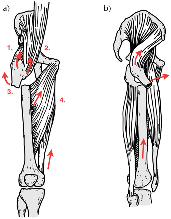
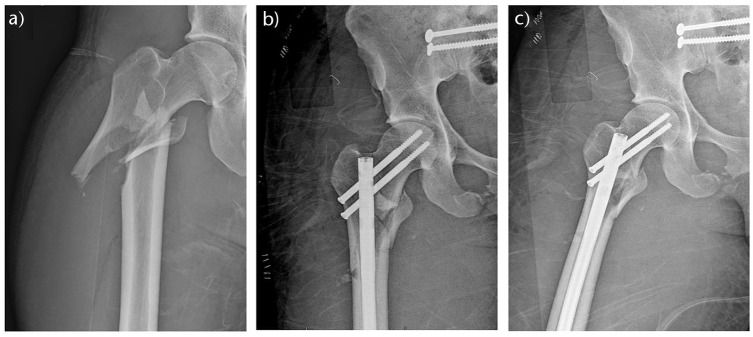
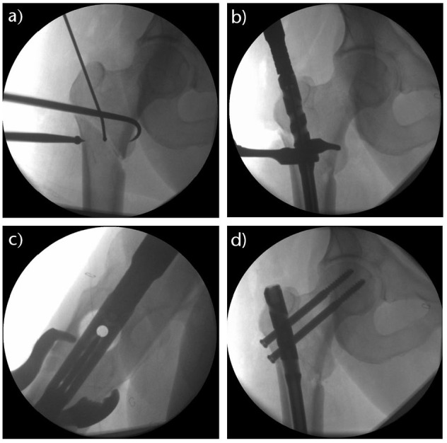
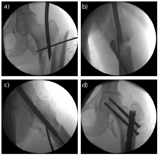
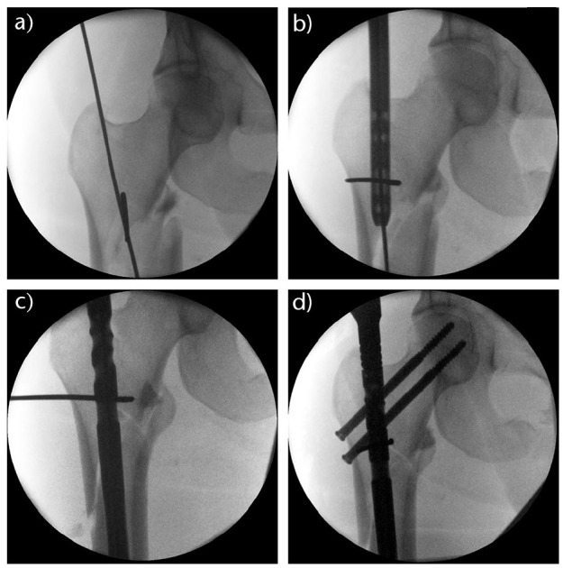
Subtrochanteric (ST) femur fractures are proximal femur fractures, which are often difficult to manage effectively because of their deforming anatomical forces.Operative management of ST fractures is the mainstay of treatment, with the two primary surgical implant options being intramedullary (IM) nails and extramedullary plates.Of these, IM nails have a biologic and biomechanical superiority, and have become the gold standard for ST femur fractures.The orthopaedic surgeon should become familiar and facile with several reduction techniques to create anatomical alignment in all unique ST fracture patterns.This article presents a comprehensive and current review of the epidemiology, anatomy, biomechanics, clinical presentation, diagnosis, and management of subtrochanteric femur fractures. Cite this article: EFORT Open Rev 2021;6:145-151. DOI: 10.1302/2058-5241.6.200048.

Keywords: femur; intramedullary nail; reduction techniques; subtrochanteric fracture.


Conflict of interest statement

ICMJE Conflict of interest statement: The other authors declare no conflict of interest relevant to this work.

Figures

Fig. 1
Fig. 1
The deforming forces (red arrows) of the proximal and distal fragments in subtrochanteric fractures in the coronal (A) and sagittal (B) planes. The proximal fragment is abducted by the gluteus medius and minimus (1), flexed by the iliopsoas (2), and externally rotated by short external rotators (3). The distal fragment is adducted and shortened by the adductors and gracilis (4).
Fig. 2
Fig. 2
Preoperative (A) anteroposterior (AP) and immediate postoperative AP (B) and lateral (C) radiographs of a right subtrochanteric femur fracture with intertrochanteric extension that was treated with a piriformis entry reconstruction nail.
Fig. 3
Fig. 3
Use of a ball spike pusher to medialize the distal fracture fragment while simultaneously pulling the proximal fragment with a bone hook to address the varus fracture deformity (A). After the guidewire was placed, a clamp was utilized to maintain the reduction in the coronal (B) and sagittal (C) planes. Anatomical reduction was achieved and the subtrochanteric femur fracture was fixed with a trochanteric entry reconstruction nail (D).
Fig. 4
Fig. 4
Multiple reduction techniques were used to address this complex subtrochanteric femur fracture with intertrochanteric extension. The finger reduction tool was placed into the piriformis fossa entry portal to gain control of the proximal fragment (A). A Cobb periosteal elevator and a posterior blocking wire were utilized to correct the sagittal plane deformity (B). The finger reduction tool was then passed to the level of the distal fragment to allow for passage of the guidewire (C). Anatomical reduction was achieved and maintained with a piriformis entry reconstruction nail (D).
Fig. 5
Fig. 5
A blocking wire was placed in the concavity of the deformity in the proximal fracture fragment just medial to the guidewire and was left in place during reaming to guide the reaming of the proximal fragment (A). As the nail was passed (B), the blocking wire effectively lateralized the distal segment (C) and created an anatomical reduction that was maintained with a piriformis entry reconstruction nail (D).

References

    1. Joglekar SB, Lindvall EM, Martirosian A. Contemporary management of subtrochanteric fractures. Orthop Clin North Am 2015;46:21–35. - PubMed
    1. Dell RM, Adams AL, Greene DF, et al. . Incidence of atypical nontraumatic diaphyseal fractures of the femur. J Bone Miner Res 2012;27:2544–2550. - PubMed
    1. Ng AC, Drake MT, Clarke BL, et al. . Trends in subtrochanteric, diaphyseal, and distal femur fractures, 1984–2007. Osteoporos Int 2012;23:1721–1726. - PMC - PubMed
    1. Napoli N, Schwartz AV, Palermo L, et al. . Risk factors for subtrochanteric and diaphyseal fractures: the study of osteoporotic fractures. J Clin Endocrinol Metab 2013;98:659–667. - PMC - PubMed
    1. Koch JC. The laws of bone architecture. Am J Anat 1917;21:177–211.

LinkOut - more resources