Multispectral Imaging for Metallic Biopsy Marker Detection During MRI-Guided Breast Biopsy: A Feasibility Study for Clinical Translation
- PMID: 33828972
- PMCID: PMC8020905
- DOI: 10.3389/fonc.2021.605014
Multispectral Imaging for Metallic Biopsy Marker Detection During MRI-Guided Breast Biopsy: A Feasibility Study for Clinical Translation
Abstract
Purpose: To assess the feasibility and diagnostic accuracy of multispectral MRI (MSI) in the detection and localization of biopsy markers during MRI-guided breast biopsy.
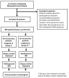
Methods: This prospective study included 20 patients undergoing MR-guided breast biopsy. In 10 patients (Group 1), MSI was acquired following tissue sampling and biopsy marker deployment. In the other 10 patients (Group 2), MSI was acquired following tissue sampling but before biopsy marker deployment (to simulate deployment failure). All patients received post-procedure mammograms. Group 1 and Group 2 designations, in combination with the post-procedure mammogram, served as the reference standard. The diagnostic performance of MSI for biopsy marker identification was independently evaluated by two readers using two-spectral-bin MR and one-spectral-bin MR. The κ statistic was used to assess inter-rater agreement for biopsy marker identification.
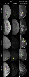
Results: The sensitivity, specificity, and accuracy of biopsy marker detection for readers 1 and 2 using 2-bin MSI were 90.0% (9/10) and 90.0% (9/10), 100.0% (10/10) and 100.0% (10/10), 95.0% (19/20) and 95.0% (19/20); and using 1-bin MSI were 70.0% (7/10) and 80.0% (8/10), 100.0% (8/8) and 100.0% (10/10), 85.0% (17/20) and 90.0% (18/20). Positive predictive value was 100% for both readers for all numbers of bins. Inter-rater agreement was excellent: κ was 1.0 for 2-bin MSI and 0.81 for 1-bin MSI.
Conclusion: MSI is a feasible, diagnostically accurate technique for identifying metallic biopsy markers during MRI-guided breast biopsy and may eliminate the need for a post-procedure mammogram.
Keywords: biopsy marker; breast magnetic resonance imaging (MRI); magnetic resonance imaging (MRI)-guided breast biopsy; mammography; multispectral imaging.
Copyright © 2021 Eskreis-Winkler, Simon, Reichman, Spincemaille, Nguyen, Christos, Drotman, Prince, Pinker, Sutton, Morris and Wang.
Conflict of interest statement
YW and PS are inventors on QSM-related patents issued to Cornell University and hold equity in Medimagemetric LLC. KP received payment for activities not related to the present article including lectures including service on speakers bureaus and for travel/accommodations/meeting expenses unrelated to activities listed from the European Society of Breast Imaging (MRI educational course, annual scientific meeting), and the IDKD 2019 (educational course). EM has received a grant from GRAIL, Inc. for research not related to the present article. The remaining authors declare that the research was conducted in the absence of any commercial or financial relationships that could be construed as a potential conflict of interest.
Figures


Similar articles
-
Dipole modeling of multispectral signal for detecting metallic biopsy markers during MRI-guided breast biopsy: a pilot study.Magn Reson Med. 2020 Apr;83(4):1380-1389. doi: 10.1002/mrm.28017. Epub 2019 Oct 21. Magn Reson Med. 2020. PMID: 31631408 Free PMC article.
-
MRI-guided breast biopsy based on diffusion-weighted imaging: a feasibility study.Eur Radiol. 2021 May;31(5):2645-2656. doi: 10.1007/s00330-020-07396-2. Epub 2020 Oct 30. Eur Radiol. 2021. PMID: 33128183 Free PMC article.
-
Magnetic resonance imaging-guided breast biopsy in lesions not visualized by mammogram or ultrasound.Am Surg. 2012 Oct;78(10):1087-90. Am Surg. 2012. PMID: 23025947
-
Localization and biopsy of breast lesions by magnetic resonance imaging guidance.J Magn Reson Imaging. 2001 Jun;13(6):903-11. doi: 10.1002/jmri.1129. J Magn Reson Imaging. 2001. PMID: 11382951 Review.
-
Multiparametric MRI in detection and staging of prostate cancer.Dan Med J. 2017 Feb;64(2):B5327. Dan Med J. 2017. PMID: 28157066 Review.
References
Grants and funding
LinkOut - more resources
Full Text Sources
Other Literature Sources

