Chemotactic TEG3 Cells' Guiding Platforms Based on PLA Fibers Functionalized With the SDF-1α/CXCL12 Chemokine for Neural Regeneration Therapy
- PMID: 33829009
- PMCID: PMC8019790
- DOI: 10.3389/fbioe.2021.627805
Chemotactic TEG3 Cells' Guiding Platforms Based on PLA Fibers Functionalized With the SDF-1α/CXCL12 Chemokine for Neural Regeneration Therapy
Abstract
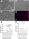
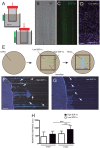
(Following spinal cord injury, olfactory ensheathing cell (OEC) transplantation is a promising therapeutic approach in promoting functional improvement. Some studies report that the migratory properties of OECs are compromised by inhibitory molecules and potentiated by chemical concentration differences. Here we compare the attachment, morphology, and directionality of an OEC-derived cell line, TEG3 cells, seeded on functionalized nanoscale meshes of Poly(l/dl-lactic acid; PLA) nanofibers. The size of the nanofibers has a strong effect on TEG3 cell adhesion and migration, with the PLA nanofibers having a 950 nm diameter being the ones that show the best results. TEG3 cells are capable of adopting a bipolar morphology on 950 nm fiber surfaces, as well as a highly dynamic behavior in migratory terms. Finally, we observe that functionalized nanofibers, with a chemical concentration increment of SDF-1α/CXCL12, strongly enhance the migratory characteristics of TEG3 cells over inhibitory substrates.
Keywords: CXCL12; PLA nanofibers; SDF-1alpha; cell migration; electrospinning; gradients; olfactory ensheathing cells.
Copyright © 2021 Castaño, López-Mengual, Reginensi, Matamoros-Angles, Engel and del Rio.
Conflict of interest statement
The authors declare that the research was conducted in the absence of any commercial or financial relationships that could be construed as a potential conflict of interest.
Figures


Similar articles
-
Biological effects different diameters of Tussah silk fibroin nanofibers on olfactory ensheathing cells.Exp Ther Med. 2019 Jan;17(1):123-130. doi: 10.3892/etm.2018.6933. Epub 2018 Nov 6. Exp Ther Med. 2019. PMID: 30651772 Free PMC article.
-
BDNF production by olfactory ensheathing cells contributes to axonal regeneration of cultured adult CNS neurons.Neurochem Int. 2007 Feb;50(3):491-8. doi: 10.1016/j.neuint.2006.10.004. Epub 2006 Dec 8. Neurochem Int. 2007. PMID: 17157963
-
[Biocompatibility of silk fibroin nanofibers scaffold with olfactory ensheathing cells].Zhongguo Xiu Fu Chong Jian Wai Ke Za Zhi. 2009 Nov;23(11):1365-70. Zhongguo Xiu Fu Chong Jian Wai Ke Za Zhi. 2009. PMID: 19968182 Chinese.
-
The migration of olfactory ensheathing cells during development and regeneration.Neurosignals. 2012;20(3):147-58. doi: 10.1159/000330895. Epub 2012 Mar 27. Neurosignals. 2012. PMID: 22456085 Review.
-
Enhancing the Therapeutic Potential of Olfactory Ensheathing Cells in Spinal Cord Repair Using Neurotrophins.Cell Transplant. 2018 Jun;27(6):867-878. doi: 10.1177/0963689718759472. Epub 2018 May 31. Cell Transplant. 2018. PMID: 29852748 Free PMC article. Review.
Cited by
-
A Dexamethasone-Loaded Polymeric Electrospun Construct as a Tubular Cardiovascular Implant.Polymers (Basel). 2023 Nov 6;15(21):4332. doi: 10.3390/polym15214332. Polymers (Basel). 2023. PMID: 37960012 Free PMC article.
-
A Review on Electrospun Nanofibers Based Advanced Applications: From Health Care to Energy Devices.Polymers (Basel). 2021 Oct 29;13(21):3746. doi: 10.3390/polym13213746. Polymers (Basel). 2021. PMID: 34771302 Free PMC article. Review.
-
Region-specific brain decellularized extracellular matrix promotes cell recovery in an in vitro model of stroke.Sci Rep. 2025 Apr 7;15(1):11921. doi: 10.1038/s41598-025-95656-w. Sci Rep. 2025. PMID: 40195414 Free PMC article.
References
-
- Akter F. (2016). Tissue Engineering Made Easy. Amsterdam: Elsevier Science.
-
- Álvarez Z., Mateos-Timoneda M. A., Hyroššová P., Castaño O., Planell J. A., Perales J. C., et al. (2013). The effect of the composition of PLA films and lactate release on glial and neuronal maturation and the maintenance of the neuronal progenitor niche. Biomaterials 34 2221–2233. 10.1016/j.biomaterials.2012.12.001 - DOI - PubMed
LinkOut - more resources
Full Text Sources
Other Literature Sources

