. 2021 Jun:26:100163.
doi: 10.1016/j.biotri.2021.100163. Epub 2021 Feb 19.

Simultaneous Characterization of Implant Wear and Tribocorrosion Debris within Its Corresponding Tissue Response Using Infrared Chemical Imaging

Affiliations

Simultaneous Characterization of Implant Wear and Tribocorrosion Debris within Its Corresponding Tissue Response Using Infrared Chemical Imaging

Songyun Liu et al. Biotribology (Oxf). 2021 Jun.

Abstract

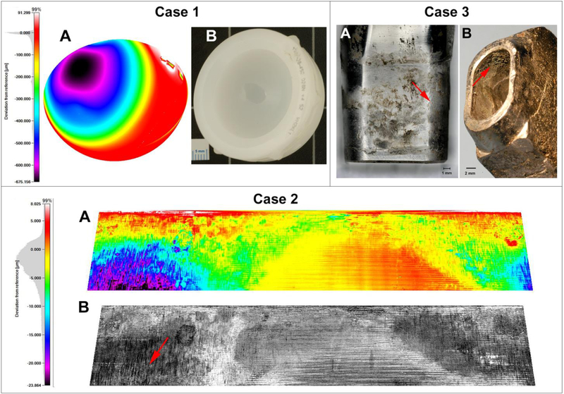
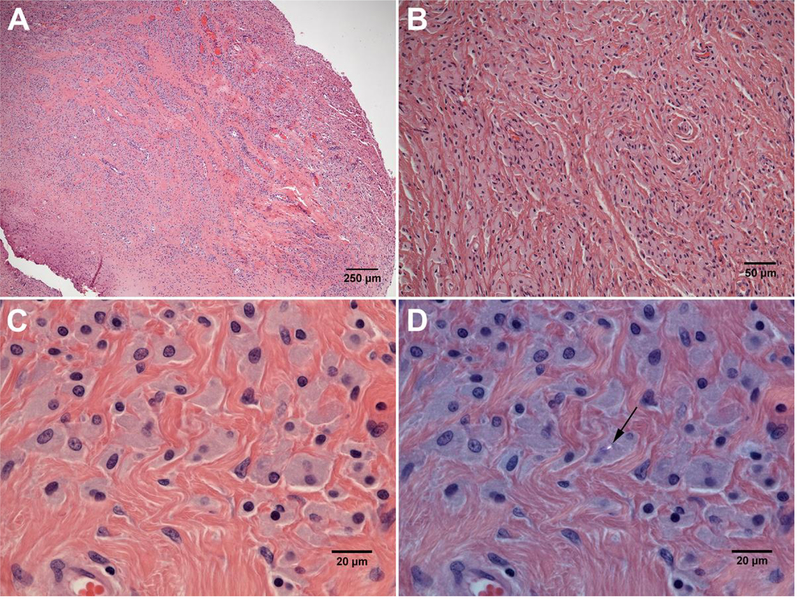
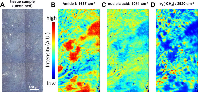
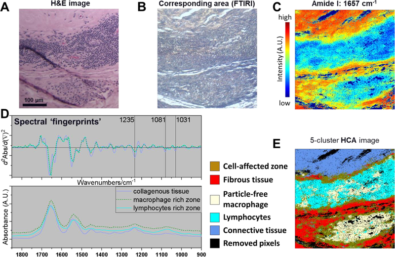
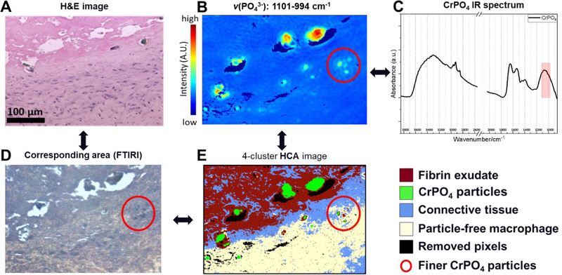
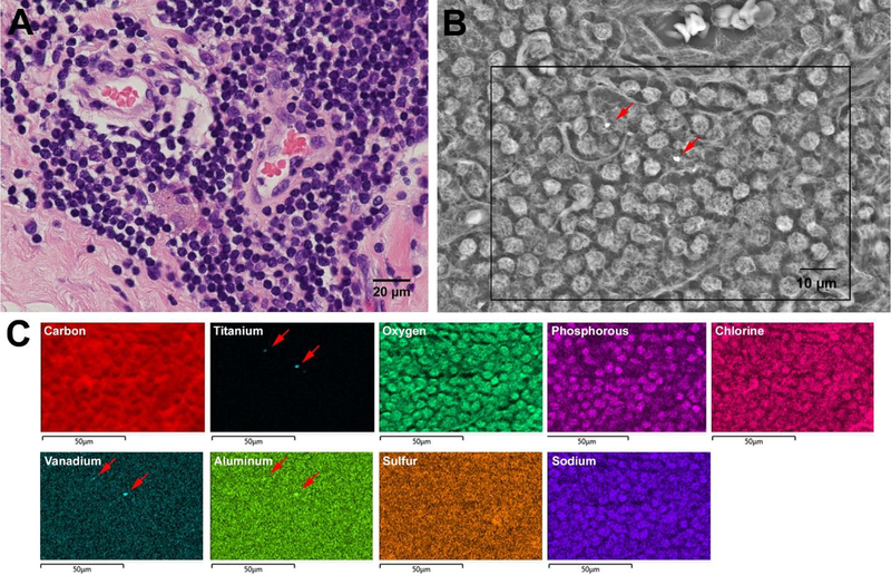
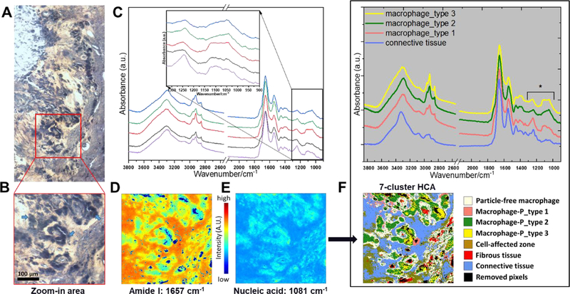
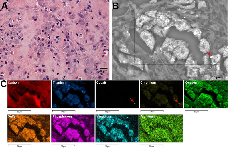
Biotribology is one of the key branches in the field of artificial joint development. Wear and corrosion are among fundamental processes which cause material loss in a joint biotribological system; the characteristics of wear and corrosion debris are central to determining the in vivo bioreactivity. Much effort has been made elucidating the debris-induced tissue responses. However, due to the complexity of the biological environment of the artificial joint, as well as a lack of effective imaging tools, there is still very little understanding of the size, composition, and concentration of the particles needed to trigger adverse local tissue reactions, including periprosthetic osteolysis. Fourier transform infrared spectroscopic imaging (FTIR-I) provides fast biochemical composition analysis in the direct context of underlying physiological conditions with micron-level spatial resolution, and minimal additional sample preparation in conjunction with the standard histopathological analysis workflow. In this study, we have demonstrated that FTIR-I can be utilized to accurately identify fine polyethylene debris accumulation in macrophages that is not achievable using conventional or polarized light microscope with histological staining. Further, a major tribocorrosion product, chromium phosphate, can be characterized within its histological milieu, while simultaneously identifying the involved immune cell such as macrophages and lymphocytes. In addition, we have shown the different spectral features of particle-laden macrophages through image clustering analysis. The presence of particle composition variance inside macrophages could shed light on debris evolution after detachment from the implant surface. The success of applying FTIR-I in the characterization of prosthetic debris within their biological context may very well open a new avenue of research in the orthopedics community.

Keywords: Adverse tissue response; FTIR imaging; Implant wear and corrosion.


Figures

Figure 1.
Figure 1.
Implant damage characterization for three retrieved cases. Case 1: The surface of the polyethylene liner of Case 1 was measured with an optical CMM. A sphere was fitted to unworn regions to visualize and quantify polyethylene wear. A) Heat maps of the articulating surface in the side view illustrates a prominent wear scar with a maximum penetration of −675μm and that yielded a total material loss of 313 mm3. B) The light microscope image of the retrieved PE liner. Case 2: The head taper surface of the CoCrMo femoral head of Case 2 was reconstructed using CMM measurements of a high precision replica. A) The heat map illustrates areas with severe fretting and corrosion damage. The areas of largest material loss (purple and black) were characterized by severe column damage, which is best seen in the light intensity map (B, red arrow). Case 3: Photos of the severely worn and corroded A) male taper of the modular neck component and B) the female taper of the Ti6Al4V alloy stem component. In A) several areas of corrosion damage, deposits, and fretting marks (red arrow) can be seen. The female taper surface in B) mostly exhibited a damage pattern indicating surface fatigue caused by fretting and thick deposits (red arrow).
Figure 2.
Figure 2.
A&B: Low and high magnification of the joint pseudocapsule from Case 1 demonstrating the marked macrophage response dominating the tissue (H&E; A: ×40; B: × 200). C&D: Slate-blue colored particle-laden macrophages containing fine PE particles (×600, C: H&E; D: polarized light). It is worth mentioning that only one PE particle (black arrow) was visibly detected in this region under polarized light.
Figure 3.
Figure 3.
Detection of fine polyethylene wear particles accumulated within macrophages of Case 1 using the univariate FTIR-I. A) Light micrograph of the unstained pseudo-capsule tissue sample adjacent to that shown in Fig. 2, B) The heat map of the sample imaged at 1657 cm−1 (i.e., Amide I group) presented a distribution of collagenous tissue, and C) The cell populated area can be highlighted using IR signal of phosphodiester linkage in nucleic acid at 1081 cm−1 wavenumber. D) Near 2920 cm−1 wavenumber, which νδ(-CH2) presents a strong stretching mode, fine polyethylene debris can be observed as an accumulation inside particle-laden macrophages. Colocalization of cell populated areas, as indicated by the presence of nucleic acid (Fig. 3C), and polyethylene debris, as shown by the strong polyethylene absorbance signal (Fig. 3D), is evident.
Figure 4.
Figure 4.
A) H&E and B) light microscopic image showing strong presence of lymphocytes and macrophages in Case 2. C) an integrated intensity near 1657 cm−1 illustrates the distribution of amide I. D) The subtle difference in infrared spectra were better presented using their corresponding 2nd derivative plots as spectral fingerprints. Here, macrophages, lymphocytes, and collagenous tissue could be distinguished based on the infrared response using corresponding 2nd derivative spectrum. E) 5-color coded HCA image provides clusters representing different cell types or differences in the chemical structure of tissue, which can be validated through H&E image.
Figure 5.
Figure 5.
Capsule tissue from Case 2 with severe taper corrosion. Multiple large, glassy, green particles are observed within the fibrin exudate and at the tissue surface (A: H&E image, and D: light microscopic image on corresponding sample area). B) Univariate FTIR-I showed a strong absorbance in the phosphate region (ν(PO43−): 1101–994 cm−1), and C) extracted spectrum confirmed the chemical structure as CrPO4. E) HCA reconstructed image with classified composition was able to visualize large CrPO4 particles, embedded within fibrin exudate-rich areas adjacent to collagenous tissue without inflammatory cells, and a nearby area with a strong macrophage presence. Some finer CrPO4 particles (red-circle) have also been detected due to the high spatial resolution (~1.1 micron) using the high-magnification scanning mode.
Figure 6.
Figure 6.
A) Accumulation of lymphocytes and macrophages observed within the pseudocapsule tissue of Case 2 (H&E). B) Two particles are highlighted (red arrows) within this backscatter electron SEM image of an adjacent section from A). C) EDS mapping of a subsection of B) shows that that those two particles (red arrows) consisted of TiAlV alloy. However, overall, there were no other implant alloy metals found confirming a very low particle burden, despite severe corrosion of the modular junction.
Figure 7.
Figure 7.
A) Light microscope image of the unstained tissue retrieved from Case 3. A marked macrophage presence (dark areas) is indicative of the foreign body reaction to wear debris. B) The zoom-in light microscope image from area A) was selected for FTIR-I analysis (arrows indicate macrophages). C) Using single point scan to probe randomly selected individual macrophages yielded five spectra with subtle spectral variance in in the 1300–985 cm−1 spectral range (inset). D) and E) Univariate images can provide a heat map for collagenous tissue and cell populated areas. F) The clustering results from HCA showed 3 different clusters of particle-laden macrophages. The averaged spectrum of individual clusters is plotted. The highlighted spectral features (asterisk) could potentially depend on the composition of foreign body or mixtures of foreign bodies that were internalized by the cells.
Figure 8.
Figure 8.
A) Accumulation of particle-laden macrophages within pseudocapsule tissue of Case 3. B) Numerous metal particles can be seen within the macrophages, due to bright contrast in the backscatter electron SEM image of a tissue section adjacent to A). C) EDS mapping reveals that macrophages are packed with titanium and chromium, as well as several TiAlV alloy particles, and one CoCr alloy particle (red arrow). Two possible explanations for the strong presence of chromium, and overall low presence of cobalt could be either that CoCrMo particle are digested during phagocytosis and only chromium oxides remain, or that particles generated at the articulation or during fretting of the modular junction are already primarily consisting of chromium oxide.

Similar articles

Cited by

References

    1. Dowson D Review Paper 2: Whither Tribology? Proc. Inst. Mech. Eng. Conf. Proc (1969). doi:10.1243/PIME_CONF_1969_184_384_02 - DOI
    1. Dowson D Priorities for research on internal prosthesis. Biomaterials 1, 123–124 (1980).
    1. Dumbleton JH Tribology of Natural and Artificial Joints. (Elsevier, 1981).
    1. Fisher J & Dowson D Tribology of Total Artificial Joints. Proc. Inst. Mech. Eng. [H] 205, 73–79 (1991). - PubMed
    1. Shanbhag A, Jacobs J, Glant T, Gilbert J, Black J & Galante J Composition and morphology of wear debris in failed uncemented total hip replacement. J. Bone Joint Surg. Br 76-B, 60–67 (1994). - PubMed

LinkOut - more resources