. 2019 Dec;5(4):373-386.
doi: 10.1007/s40883-019-00094-6. Epub 2019 Feb 27.

Biocompatibility and Viscoelastic Properties of Injectable Resilin-Like Polypeptide and Hyaluronan Hybrid Hydrogels in Rabbit Vocal Folds

Affiliations

Biocompatibility and Viscoelastic Properties of Injectable Resilin-Like Polypeptide and Hyaluronan Hybrid Hydrogels in Rabbit Vocal Folds

Renee E King et al. Regen Eng Transl Med. 2019 Dec.

Abstract

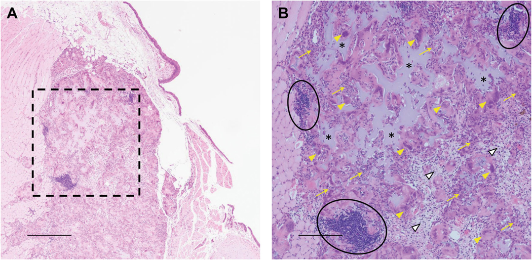
Vocal fold scar, characterized by alterations in the lamina propria extracellular matrix, disrupts normal voice quality and function. Due to a lack of satisfactory clinical treatments, there is a need for tissue engineering strategies to restore voice. Candidate biomaterials for vocal fold tissue engineering must match the unique biomechanical and viscoelastic properties of native tissue without provoking inflammation. We sought to introduce elastomeric properties to hyaluronic acid (HA)-based biomaterials by incorporating resilin-like polypeptide (RLP) into hybrid hydrogels. Physically crosslinked RLP/HA and chemically crosslinked RLP-acrylamide/thiolated HA (RLP-AM/HA-SH) hydrogels were fabricated using cytocompatible chemistries. Mechanical properties of hydrogels were assessed in vitro using oscillatory rheology. Hybrid hydrogels were injected into rabbit vocal folds and tissues were assessed using rheology and histology. A small number of animals underwent acute vocal fold injury followed by injection of RLP-AM/HA-SH hydrogel alone or as a carrier for human bone marrow mesenchymal stem cells (BM-MSCs). Rheological testing confirmed that mechanical properties of materials in vitro resembled native vocal fold tissue and that viscoelasticity of vocal fold mucosa was preserved days 5 and 21 after injection. Histological analysis revealed that hybrid hydrogels provoked only mild inflammation in vocal fold lamina propria with demonstrated safety in the airway for up to 3 weeks, confirming acute biocompatibility of crosslinking chemistries. After acute injury, RLP-AM/HA-SH gel with and without BM-MSCs did not result in adverse effects or increased inflammation. Collectively, results indicate that RLP and HA-based hybrid hydrogels are highly promising for engineering the vocal fold lamina propria.

Keywords: Biocompatibility; Injectable hydrogels; Resilin; Viscoelastic properties; Vocal folds.


Conflict of interest statement

Conflict of Interest The authors declare that they have no conflict of interest.

Figures

Fig. 1
Fig. 1
Schematic of hydrogel formation. Physically crosslinked RLP/HA gel and chemically crosslinked RLP-AM/HA-SH gel crosslinking reactions
Fig. 2
Fig. 2
Oscillatory rheology of physical and chemically crosslinked hydrogels. a Time sweep data for RLP/HA physically crosslinked hydrogels, which formed immediately at 37 °C. b Frequency sweep measurements of RLP/HA physical shows the formation of a physical gel at 37 °C. c Time sweep data for RLP-AM/HA-SH chemically crosslinked hydrogels showing a gel point of approximately 40 min. d Frequency sweep data for RLP-AM/HA-SH gel indicating that stable hydrogels with solid-like properties are formed
Fig. 3
Fig. 3
Cytocompatibility of RLP-AM/HA-SH hydrogels. Confocal z-stack maximum intensity projections images for 3D cultures of encapsulated human BM-MSCs in RLP-AM/HA-SH hydrogels at a day 1, b day 3, c day 7, and d day 14. Colors indicate live cells (calcein, green) and dead cells (ethidium homodimer, red)
Fig. 4
Fig. 4
Viscoelasticity of rabbit vocal folds days 5 and 21 after injection with RLP hydrogels. Data are shown in log–log plots with mean elastic (G′) or viscous modulus (G″) as a function of frequency at day 5 and 21 postinjection of RLP/HA gel (n = 4–5 vocal folds per timepoint), RLP-AM/HA-SH gel (n = 5 per timepoint) or RLP solution (n = 3 per timepoint). Naïve rabbit vocal folds were used as controls (n = 9–10 per timepoint). Due to the logarithmic scale, group variances are not included
Fig. 5
Fig. 5
Rabbit vocal folds days 5 and 21 after injection with RLP hydrogels. Hematoxylin and eosin stain. All demonstrate intact epithelium without inflammation and normal muscularis. a RLP/HA gel at day 5. The lamina propria is essentially unremarkable, with no significant edema or inflammation. b RLP/HA gel at day 21. The lamina propria is more edematous compared to day 5 and has relatively more small spindled fibroblasts and scattered neutrophils and lymphocytes. c RLP-AM/HA-SH gel at day 5. The lamina propria is essentially unremarkable, with no significant edema or inflammation. Lamina propria appears thin due to posterior location and angle of tissue during sectioning. d RLP-AM/HA-SH gel at day 21. The lamina propria is more edematous compared to day 5 and has relatively more small spindled fibroblasts and scattered neutrophils and lymphocytes. e RLP solution at day 5. The lamina propria is essentially unremarkable, with no significant edema or inflammation. f RLP solution at day 21. The lamina propria is essentially unremarkable, with no significant edema or inflammation. g Control naïve vocal fold. Arrows: lymphocytes. Black arrowheads: neutrophils. White arrowheads: fibroblasts. All × 20 magnification; scale bars 100 μm
Fig. 6
Fig. 6
Rabbit vocal fold 21 days after deep RLP-AM/HA-SH gel injection. Hematoxylin and eosin stain. a Low magnification shows intact epithelium and a mild degree of inflammation and edema in the lamina propria. Most evident is a well-demarcated inflammatory mass in the muscularis that contains extracellular amorphous amphophilic material; × 4. Scale bar 500 μm. b High magnification of box in a shows the inflammatory mass comprises amphophilic amorphous extracellular material (asterisks) surrounded by epithelioid histiocytes (yellow arrows), giant cells (yellow arrowheads), plump fibroblasts (white arrowheads), and clusters of lymphocytes (circles) consistent with a foreign body giant cell response; × 10. Scale bar 200 μm
Fig. 7
Fig. 7
Rabbit vocal folds 5 days after injury and immediate injection of RLP-AM/HA-SH gel with and without human BM-MSCs. Hematoxylin and eosin stain. a–c RLP-AM/HA-SH gel. Low magnification shows a nodular mass expanding the lamina propria, covered by focally ulcerated epithelium. The expanded area of the lamina propria comprises cellular granulation tissue with a mixed inflammatory infiltrate, plump fibroblasts, scattered histiocytes, rare giant cells, and increased vascularity. The granulation tissue pushes up against the muscularis. d–f RLP-AM/HA-SH gel + cell, rabbit 3588. Low magnification shows a mass expanding the lamina propria forming a small polyp that is covered by a markedly ulcerated epithelium. The ulcer is demarcated by denuded epithelium with fibrin forming the ulcer base. Numerous neutrophils are entrapped within the fibrin. Deep to the ulcer is a dense inflammatory infiltrate comprising predominantly lymphocytes and neutrophils, with associated fibroblasts and scattered histiocytes, including a few giant cells. This mixed inflammatory infiltrate extends through the entire muscularis layer down to cartilage. g–i RLP-AM/HA-SH gel + cell, rabbit 3589. Low magnification shows a mass expanding the lamina propria forming an ulcerated polyp. The ulcer base is largely comprised of fibroblasts and is beginning to be re-epithelialized. Deep to the ulcer is more mature granulation tissue compared to 3588. The granulation tissue predominantly comprises fibroblasts with relatively fewer inflammatory cells. A few giant cells are present. The muscularis is relatively intact and free from involvement. a, d, g × 4. Scale bar 500 μm. b, e, h × 10 magnification of dashed boxes in a, d, g. Scale bar 200 μm. e Extends slightly superior to box in d. c, f, i × 20 magnification of solid boxes in a, d, g. Scale bar 100 μm. White arrowheads: fibroblasts. Black arrows: lymphocytes. Black arrowheads: neutrophils. Yellow arrows: histiocytes. Yellow arrowheads: giant cells

Similar articles

Cited by

References

    1. Hirano M Morphological structure of the vocal cord as a vibrator and its variations. Folia Phoniatr. 1974;26:89–94. - PubMed
    1. Hirano M, Kurita S, Nakashima T. The structure of the vocal folds. In: Stevens KN, Hirano M, editors. Vocal fold physiology. Tokyo: University of Tokyo Press; 1981. p. 33–43.
    1. Gray SD. Cellular physiology of the vocal folds. Otolaryngol Clin N Am. 2000;33:679–98. - PubMed
    1. Gray SD, Alipour F, Titze IR, Hammond TH. Biomechanical and histologic observations of vocal fold fibrous proteins. Ann Otol Rhinol Laryngol. 2000;109:77–85. - PubMed
    1. Woo P, Casper J, Colton R, Brewer D. Diagnosis and treatment of persistent dysphonia after laryngeal surgery: a retrospective analysis of 62 patients. Laryngoscope. 1994;104:1084–91. - PubMed

LinkOut - more resources