. 2021:267:51-81.
doi: 10.1007/164_2021_454.

High-Resolution Structures of K+ Channels

Affiliations

High-Resolution Structures of K+ Channels

Qiu-Xing Jiang. Handb Exp Pharmacol. 2021.

Abstract

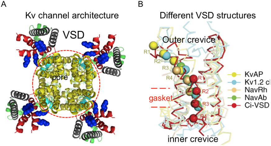
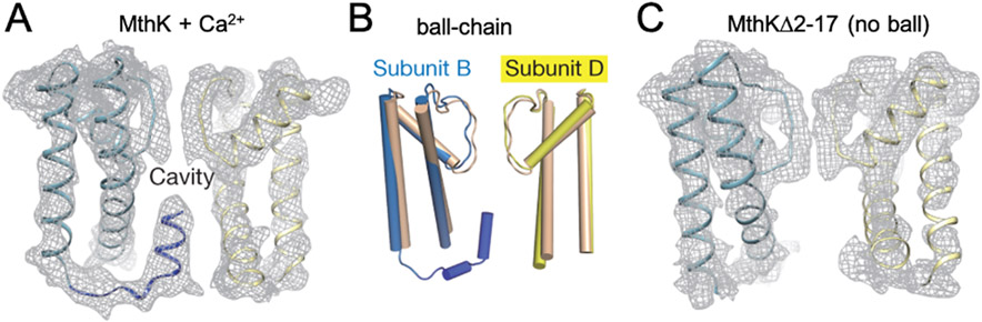
Potassium channels are present in every living cell and essential to setting up a stable, non-zero transmembrane electrostatic potential which manifests the off-equilibrium livelihood of the cell. They are involved in other cellular activities and regulation, such as the controlled release of hormones, the activation of T-cells for immune response, the firing of action potential in muscle cells and neurons, etc. Pharmacological reagents targeting potassium channels are important for treating various human diseases linked to dysfunction of the channels. High-resolution structures of these channels are very useful tools for delineating the detailed chemical basis underlying channel functions and for structure-based design and optimization of their pharmacological and pharmaceutical agents. Structural studies of potassium channels have revolutionized biophysical understandings of key concepts in the field - ion selectivity, conduction, channel gating, and modulation, making them multi-modality targets of pharmacological regulation. In this chapter, I will select a few high-resolution structures to illustrate key structural insights, proposed allostery behind channel functions, disagreements still open to debate, and channel-lipid interactions and co-evolution. The known structural consensus allows the inference of conserved molecular mechanisms shared among subfamilies of K+ channels and makes it possible to develop channel-specific pharmaceutical agents.

Keywords: Activation, deactivation, and inactivation; Co-evolution of channels and lipids; Energetics and allostery; Ligand-gated K+ channels; Lipid-dependent gating; Pharmacological regulators and small molecule compounds; Structure-based drug design; Voltage-gated K+ channels (Kv).


Conflict of interest statement

Declaration: The author claims no conflict of interest.

Figures

Fig. 1
Fig. 1. Transmembrane topology and a simple gating scheme for K+ channels.
A). Each subunit may have only the pore domain which is made of two transmembrane helices and a pore loop made of a short pore helix and a selectivity loop afterwards (I). This is sometimes referred as 1P topology, whose duplication leads to the 2P topology in III. A typical Kv channel has a voltage-sensing domain (VSD) attached to N-terminal side of a 1P domain (II). The Ca2+-activated K channels have an extra transmembrane helix (TM0 or S0) before its voltage-sensor domain (IV). Other structural or functional domains may be attached to the N- and/or C-terminus or the loops between transmembrane segments in some channels. B). A channel can be switched among closed states before reaching the open state (O), and both closed states (Co … Cx) and the open state (O) can switch into the inactivated states (I1 and I2). Some channels may have multiple open states and multiple routes to switch from closed states to the open ones. The structural studies presumably catch channels in some of these gating states, which may leave uncertainty in the interpretation of structural data.
Fig. 2
Fig. 2. Structural basis for ion selectivity and fast permeation in a K+ channel.
A). Structural model of the KcsA channel (PDB ID 1K4C), showing two transmembrane helices and the pore helix of each subunit as blue ribbons and the selectivity loops in yellow. One subunit in the front is taken away for presentation. K+ ions in the selectivity filter (SF) and the cavity are presented as green balls. B). Electronegative carbonyl oxygens (red) of the selectivity loops coordinate and stabilize the K+ ions in four binding sites (K1- K4; here not using the usual S1-S4 labels). C). Throughput cycle of K+ ions switching between two different configurations in SF ([K2, K4] and [K1, K3]) for outward flux. Always two K+ and two water molecules, denoted as green and small red balls respectively, are in the SF. This is named “soft knock-on” because of the separation of two neighboring K+ ions by a water molecule. D). Throughput cycle by a “hard knock-on” model through three distinct configurations in SF for outward flux. The three depicted states all have one pair of K+ ions in two neighboring binding sites without an intervening water. The knock-on of a K+ from the bottom of the SF (cavity site) into the two-ion state (left) would knock off the water molecule in the S4 site such that no water flow would accompany the K+ flux. Figure panels adapted from references (Morais-Cabral et al. 2001; Zhou et al. 2001; Kopfer et al. 2014; Kopec et al. 2018) with permissions in accord with the PMC open access policy.
Fig. 3
Fig. 3. Architecture of a Kv channel and structural variations among VSD structures.
A). A KvAP structure modeled after the Kv1.2/2.1 chimera structure (PDB ID 2R9R) shows a central pore domain (red circle) and four flanking VSDs. The S1/S2 are in grey and the S3/S4 in red. Four Arg residues in the S4 are showed in blue balls. B). Five different VSD structures aligned with S4 Arg residues across the gating pores. Outer (upper) and inner (lower) crevices separated by a hydrophobic gasket (red dashed lines). In a resting (down) state, all four Arg residues (R1-R4) are expected to be in the inner crevice and drive the pore domain into the closed state. Panels modified from references (Long et al. 2005; Li et al. 2014a ) with permissions following the PMC open access policy.
Fig. 4
Fig. 4. Structural evidence for the N-type ball-and-chain inactivation.
A). A cryo-EM structure of the transmembrane part of the Ca2+-activated MthK channel in the open state with an N-terminal ball peptide inside the cavity. Only two subunits are shown with the structural models as blue and yellow ribbons in the map (grey). B). Schematic cartoon shows the ball-and-chain relative to the two subunits of the transmembrane pore region. The model from cryo-EM structure is in blue (subunit B) and yellow (subunit D), and the overlaying model in beige is from the crystal structure (PDB ID 3LDC). C). The cryo-EM structure co-responding to two subunits of the transmembrane domain of the deletion mutant lacking the N-terminal ball peptide. Figure panels were reproduced from (Fan et al. 2020) with permissions following PMC open access policy.
Fig. 5
Fig. 5. Possible structural basis for C-type inactivation.
A). Gating cycle of KcsA. The PDB codes (1K4C, 1K4D, 5VK6, and 5VKE) represent structures of the wild-type channel in high (1K4C) and low (1K4D) K+ solutions and its two mutants --- (E71A) and Y82A. The latter two were assigned to two different functional states. The C/O, O/O, O/I and C/I pairs denote the state of the inner gate (C or O in the first position) and that of the SF gate (O or I in the second position), respectively. The collapse of the SF, affecting the K2 and K3 ion-binding sites in KcsA, correlates with SF inactivation. B). G77dA KcsA pore does not collapse in high or low K+, but it still inactivates as the wild-type channel, suggesting that the SF collapse is not essential for inactivation. C). SFs of the hERG, hERG S631A, EAG1 and Kv1.2/2.1 chimera (KvChim) compared at a level corresponding to the KcsA Y78. hERG C-type inactivation might result from a subtle movement of F627 in the SF, which causes a slight reorientation of the carbonyl oxygens in the SF and may affect the K1 site. D). KvChim mutant V406W has low occupancy of K1 and inactivates quicker than the wild-type, suggesting that small changes that affect the energetics of K+ ions in the site are sufficient to affect inactivation. Panels were adapted from references (Cuello et al. 2010a; Matulef et al. 2016; Pau et al. 2017; Wang and MacKinnon 2017a) with permissions following PMC public access policy.
Fig. 6
Fig. 6. Structures of Ca2+-activated K channels and hydrophobic gating at the inner gate.
A). Structure of acBK channel in high Ca2+/Mg2+. B). Minor structural rearrangements of acBK from the metal-bound (blue) to metal-free (red) state, showing that the inner gate is wide-open, and the RCK domains experience some subtle changes that lead to small rearrangements at the interface between the RCK domains (gating ring) and the transmembrane part, which are not enough to move the inner gate. C). Structure of the Ca2+-free MthK in nanodisc, showing a closed state at the inner gate due to hydrophobic residues and the physical motion of the inner gate. D). The proposed hydrophobic gating of BK due to the switching of side chains of apolar residues (yellow arrow) lining the cavity to make it less favorable for hydrated K+ ions to pass. Panels were adapted from references (Hite et al. 2017; Tao et al. 2017; Jia et al. 2018; Fan et al. 2020) with permissions following the PMC open access policy.
Fig. 7
Fig. 7. Structure of the KATP channel.
A). CryoEM map of the Kir/SUR1 complex at 3.3. Å showing the arrangement of density and binding sites for ATPγS and RPG relative to the membrane. B). Subunit arrangement viewed from the intracellular side. RPG binds inside the SUR1. ATPγS binds to the NBD1 of the SUR1 as well as at the channel/SUR1 interface. C). The TMD0-L0 region of the SUR interfaces with the Kir subunit through hydrophobic interactions. D). The molecular model of the whole octameric complex with one SUR1 subunit close to the reader removed for clarity. The white box locates the ATP-binding site at the Kir/SUR interface and is expanded in (E) to show the key residues important for the ATP binding sites. Panels adapted from original publications (Li et al. 2017b; Ding et al. 2019; Martin et al. 2019) with permissions in accord with the PMC public access policy.
Fig. 8
Fig. 8. Structural basis for GIRK gating.
A). The structure of GIRK2 in a closed state (left; Kir3.2; PDB ID 3SYO). The transmembrane domain forms the pore structure, resembling KcsA, which would have both a SF gate and an inner helix gate. The beta-rich CTD domains form a tetramer containing an ion-conducting tunnel in the middle that is pinched closed at the top of CTD with the G-loop gate right under the presumed inner helix gate. Right: The two lower gates presumably may open by splaying laterally the inner helices (top; PDB ID 3SYA) or the G-loops (bottom; PDB ID 3SYQ). B). Opening of the two lower gates by PIP2 and Gβγ, respectively (PDB ID 4KFM). The PIP2-binding drags the interfacial and inner helices laterally to pen the inner helix gate. The beta-wheel of the Gβγ attached to the CTD laterally through both hydrophobic and electrostatic interactions, causing the opening of the G-loop gate in a stochastic fashion and thus a higher Po than in the absence of Gβγ. Panels adapted from original publications (Whorton and MacKinnon 2011; Whorton and MacKinnon 2013) with permissions under the PMC public access.
Fig. 9
Fig. 9. Structural features of the K2P channels.
A). The crystal structure of a K2P4 channel shows the helical bundle in the cap region right above the SF (the top K0 ion-binding site), leading to two lateral flux pathways marked by red arrows (PDB IDs: 4I9W, 4RUE, 4RUF). The SF is the same as in Fig. 2A. The inner helices at the inner gate region are far away from each other such that the inner gate might not be used to close the pore. There is domain-swapping between the two subunits in the cap region. At the dimer interfaces there are two lateral portals that allow hydrophobic molecules to access the cavity and block the pore. Upward movement of the inner helix 2 is likely to block the lateral portals and relieves the channel from membrane-born blockers. B). A view of the inner gate region and the cavity from the intracellular side along the central axis. K+ ions are shown as purple balls. Panels adapted from original publications (Brohawn et al. 2014a) with permissions under the PMC public access policy.

Similar articles

Cited by

References

    1. Abi-Char J, Maguy A, Coulombe A, et al. (2007) Membrane cholesterol modulates Kv1.5 potassium channel distribution and function in rat cardiomyocytes. J Physiol 582:1205–1217 doi: 10.1113/jphysiol.2007.134809 - DOI - PMC - PubMed
    1. Aggarwal SK, MacKinnon R (1996) Contribution of the S4 segment to gating charge in the Shaker K+ channel. Neuron 16:1169–1177 - PubMed
    1. Ahern CA, Horn R (2005) Focused electric field across the voltage sensor of potassium channels. Neuron 48:25–29 - PubMed
    1. Alam A, Jiang Y (2009a) High-resolution structure of the open NaK channel. Nat Struct Mol Biol 16:30–34 doi: 10.1038/nsmb.1531 - DOI - PMC - PubMed
    1. Alam A, Jiang Y (2009b) Structural analysis of ion selectivity in the NaK channel. Nat Struct Mol Biol 16:35–41 doi: 10.1038/nsmb.1537 - DOI - PMC - PubMed

Substances

LinkOut - more resources