. 2021 Jan-Dec:17:1744806921997206.
doi: 10.1177/1744806921997206.

Systemic administration of a β2-adrenergic receptor agonist reduces mechanical allodynia and suppresses the immune response to surgery in a rat model of persistent post-incisional hypersensitivity

Affiliations

Systemic administration of a β2-adrenergic receptor agonist reduces mechanical allodynia and suppresses the immune response to surgery in a rat model of persistent post-incisional hypersensitivity

Vipin Arora et al. Mol Pain. 2021 Jan-Dec.

Abstract

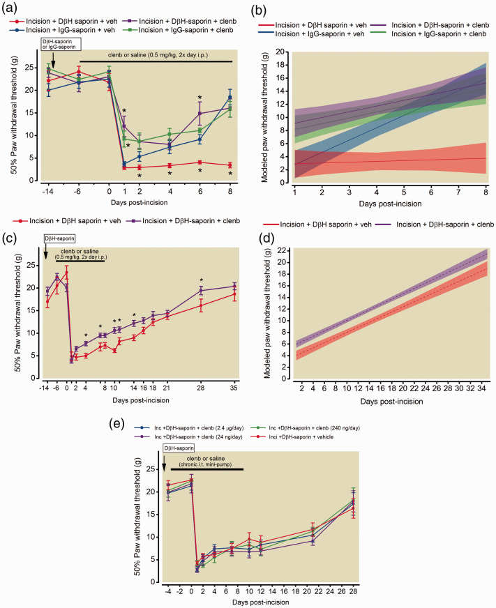
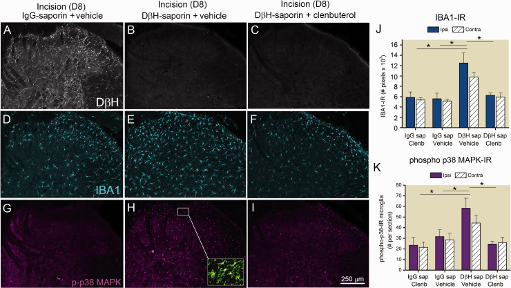
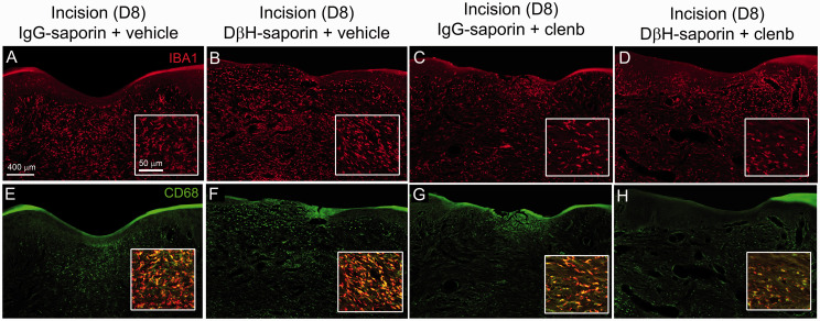
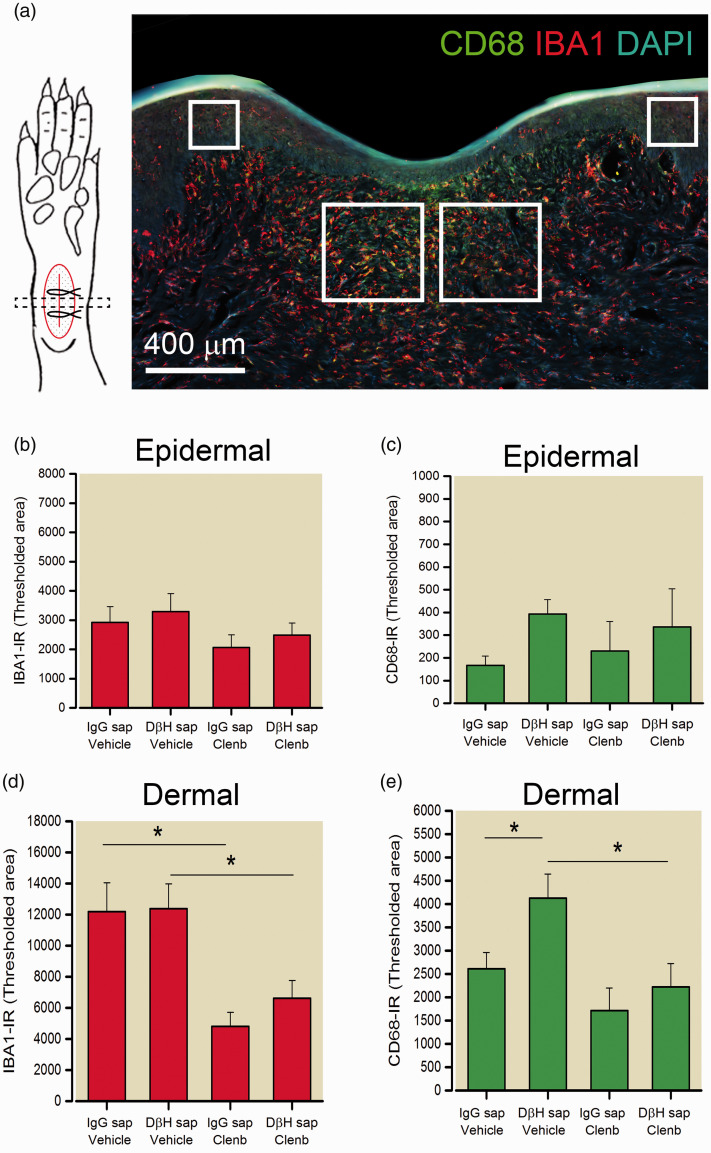
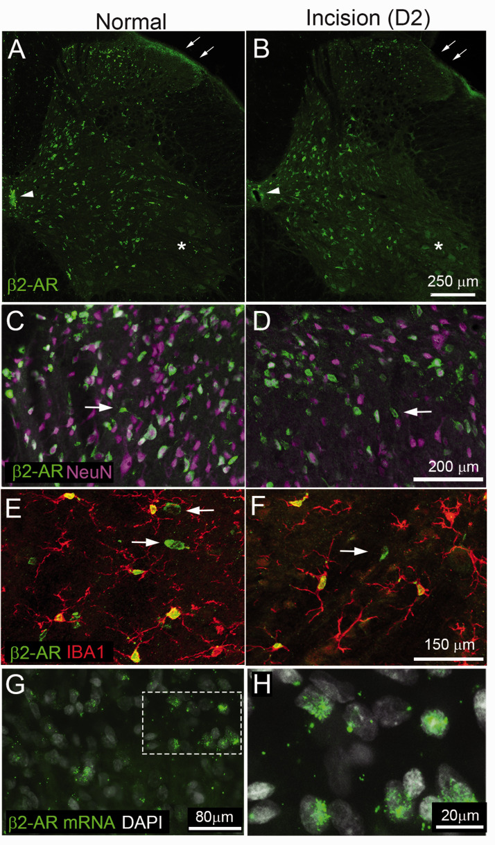
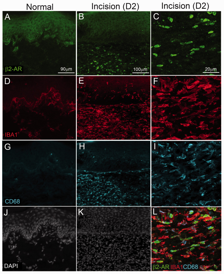
Beta 2 adrenergic receptor (β2 AR) activation in the central and peripheral nervous system has been implicated in nociceptive processing in acute and chronic pain settings with anti-inflammatory and anti-allodynic effects of β2-AR mimetics reported in several pain states. In the current study, we examined the therapeutic efficacy of the β2-AR agonist clenbuterol in a rat model of persistent postsurgical hypersensitivity induced by disruption of descending noradrenergic signaling in rats with plantar incision. We used growth curve modeling of ipsilateral mechanical paw withdrawal thresholds following incision to examine effects of treatment on postoperative trajectories. Depletion of spinal noradrenergic neurons delayed recovery of hypersensitivity following incision evident as a flattened slope compared to non-depleted rats (-1.8 g/day with 95% CI -2.4 to -1.085, p < 0.0001). Chronic administration of clenbuterol reduced mechanical hypersensitivity evident as a greater initial intercept in noradrenergic depleted (6.2 g with 95% CI 1.6 to 10.8, p = 0.013) and non-depleted rats (5.4 g with 95% CI 1.2 to 9.6, p = 0.018) with plantar incision compared to vehicle treated rats. Despite a persistent reduction in mechanical hypersensitivity, clenbuterol did not alter the slope of recovery when modeled over several days (p = 0.053) or five weeks in depleted rats (p = 0.64). Systemic clenbuterol suppressed the enhanced microglial activation in depleted rats and reduced the density of macrophage at the site of incision. Direct spinal infusion of clenbuterol failed to reduce mechanical hypersensitivity in depleted rats with incision suggesting that beneficial effects of β2-AR stimulation in this model are largely peripherally mediated. Lastly, we examined β2-AR distribution in the spinal cord and skin using in-situ hybridization and IHC. These data add to our understanding of the role of β2-ARs in the nervous system on hypersensitivity after surgical incision and extend previously observed anti-inflammatory actions of β2-AR agonists to models of surgical injury.

Keywords: Postoperative pain; acute to chronic pain transition; glial plasticity; growth curve modeling; surgery.


Conflict of interest statement

Authors' Note: Carlos Eduardo Morado-Urbina is now affiliated with Department of Physiology and Pharmacology and Center for Molecular Medicine, Karolinska Institutet, Stockholm, Sweden.

Declaration of conflicting interests: The author(s) declared the following potential conflicts of interest with respect to the research, authorship, and/or publication of this article: In the past 36 months JCE has consulted to Adynxx (San Francisco, CA, USA) and TEVA Pharmaceutical Industries (North Wales, PA, USA) regarding preclinical and clinical analgesic development of pharmaceuticals not related to the current publication. The remaining authors declare no conflicts of interest.

Figures

Figure 1.
Figure 1.
Effects of clenbuterol on resolution of mechanical hypersensitivity in a rat model of persistent postsurgical pain. Fourteen days prior to plantar incision surgery, rats received spinal DβH-saporin (5 µg/10 µl, i.t.) to deplete spinal noradrenergic fibers or IgG-saporin (5 µg/10 µl, i.t.) as a control. Male Sprague Dawley rats were administered clenbuterol (0.5 mg/kg, 2×/day, i.p.) or saline beginning six days prior to surgery and mechanical paw withdrawal thresholds were assessed longitudinally in the ipsilateral paw for eight days postoperatively (a). Data are expressed as mean ± SEM (n = 6 per group, Two way RM-ANOVA with Bonferroni comparisons *P < 0.05 within time point versus IgG-saporin incision + vehicle. Longitudinal behavioral data beginning at day 1 following incisions was also analyzed using mixed effects growth curve modeling to examine differences in acute postoperative pain trajectories (b). Data expressed as group averaged mean trajectories with 95% confidence intervals. Modeled acute postoperative pain trajectories indicated significant differences in initial mechanical hypersensitivity [(Group) p = 0.003] and slope [(Group x Time) p < 0.0001] comparing DβH-saporin vehicle treated rats to IgG-saporin vehicle treated rats. Clenbuterol treated DβH saporin rats had significantly greater intercept [(Group) p = 0.01] but similar slope or rate of recovery [(Group x Time) p = 0.053] compared to DβH-saporin incision vehicle treated rats. In a separate cohort of DβH-saporin incision rats, long term effects of clenbuterol treatment were assessed until 35 days postoperatively (c). Data are expressed as mean ± SEM (Two-way RM-ANOVA with Bonferroni comparisons *P < 0.05 within time point versus DβH-saporin incision + vehicle; n = 8 vehicle threated group; n = 16 clenbuterol treated group). Modeled postoperative pain trajectories indicated significant differences in degree of mechanical hypersensitivity [(Group) p = 0.0014] but similar slopes or rate of recovery over 35 days postoperatively [(Group x Time) p = 0.63] compared to vehicle treated rats (d). Spinal administration of varying doses of clenbuterol for 14 days by mini-osmotic pump beginning four days prior to surgery in DβH-saporin incision rats (f). Data are expressed as mean ± SEM. Two-way RM ANOVA indicate effect of time: p < 0.001 but not dose/group: p = 0.600 or interaction: p = 0.995.
Figure 2.
Figure 2.
Effects of β2 adrenergic receptor (AR) agonist on enhanced spinal microglia activation in a rat model of persistent postoperative pain. Sections of spinal cord were collected from rats 8 days following plantar incision and following treatment with DβH-saporin to deplete spinal noradrenergic terminals or control IgG-saporin. Rats were chronically administered clenbuterol (0.5 mg/kg, 2×/day, i.p.) or saline vehicle 6 days prior to and for 8 days after plantar incision. Depletion of spinal noradrenergic fibers was verified immunohistochemically with an antibody against dopamine β hydroxylase (DβH, (a)–(c)). Representative confocal images of IBA1-IR (blue, (d)–(f)) and phospho-p38 MAPK-IR (purple, (g)–(i)) in the ipsilateral spinal cord of incision rats. Localization of p38 MAPK in microglia was confirmed by colocalization with an antibody against the cell surface antigen CD11b (green, inset in (h)). Quantification of IBA1-IR in ipsilateral and contralateral spinal cord of rats with incision (j). Data represent mean ± SEM, n = 3 rats per group. Two way ANOVA indicated effect of group: p < 0.001 but not side p = 0.184 or interaction: p = 0.59 with SNK pairwise comparisons *p < 0.001 versus Incision+ DβH-saporin+ vehicle. Quantification of phospho-p38 MAPK microglial in the ipsilateral and contralateral spinal cord of rats with incision (k). Data represent mean ± SEM, n = 3 rats per group. Two way ANOVA indicate effect of group: p = 0.002 but not side p = 0.398 or interaction: p = 0.67 with SNK pairwise comparisons * p < 0.005 versus Incision+ DβH-saporin+ vehicle.
Figure 3.
Figure 3.
Confocal images of IBA1-IR monocytes/macrophage and activated CD68-IR macrophage at the incision site in treated rats. Transverse sections of skin were collected from rats 8 days following plantar incision. Sections were labeled with an antibody against IBA1 (red, (a)–(d)) to label all monocytes/macrophage and an antibody against CD68 (green, (e)–(h)) to label M1 or activated macrophage in DβH-saporin and IgG saporin incision rats treated chronically with clenbuterol (0.5 mg/kg 2× day) or vehicle from 6 days prior to 8 days after surgery. Higher magnification insets were obtained from the dermal layer adjacent to the incision site to more clearly show density and morphology of IBA1-IR macrophage ((a)–(d)) and colocalization of CD68 with IBA1-IR in macrophage ((e)–(h)). Note reduced density of IBA1-IR cellular profiles in the dermal skin layer of clenbuterol treated DβH-saporin and IgG saporin incision rats.CD68-IR was also reduced in the dermal skin layer of clenbuterol treated DβH-saporin and IgG saporin incision rats compared to vehicle controls.
Figure 4.
Figure 4.
Quantification of macrophage density and activation at the incision site in treated rats. (a) Illustration of approach for sampling and analyzing transverse skin sections including representative image of IBA1-IR (red), CD68-IR (green) and DAPI positive cellular nuclei (blue) in a transverse section of skin eight days following plantar incision. Immunodensity of IBA1-IR ((b) and (d)) and CD68-IR ((c) and (e)) was quantified in two regions of the epidermis (200 µm2 × 200 µm2 area) and dermis (400 µm2 × 400 µm2 area) indicated by boxes adjacent to the site of incision. The area of labeling within a defined intensity range or threshold was measured in the respective regions and reported as mean ± SEM. n = 6 rats per group. For IBA1 dermal: Two-way ANOVA with SNK pairwise comparisons indicate main effect of treatment: p < 0.001 but not group p = 0.489 **p < 0.002, * p < 0.05 versus vehicle. For CD68 dermal: Two-way ANOVA with Bonferroni contrasts indicates main effect of treatment: p = 0.007 and group p = 0.043. * p < 0.05 versus vehicle. # p < 0.05 versus DβH-saporin.
Figure 5.
Figure 5.
Beta 2-adrenergic receptor immunoreactivity in the spinal cord of rats under naïve conditions and two days following plantar incision. Transverse section of L4 spinal cord of rat reacted with antibody against β2 adrenergic receptor ((a), β2-AR, green). There is a high density of immunoreactivity in cellular profiles throughout dorsal and ventral horn. There is also dense immunoreactivity in axon terminals within the lateral portion of the superficial laminae (arrow) and ependymal cells in the vicinity of the central canal (Arrowhead). Note lack of staining for β2-AR in motor neurons within the ventral horn (asterisk). Higher magnification confocal images show β2-AR-IR ((c), green) is present in a subpopulation of neurons ((d), NeuN, purple) in the dorsal spinal cord. Most β2-AR-IR cellular profiles colocalized with NeuN with the exception of a few non-neuronal profiles with morphology typical of microglia (arrows, (c)–(f)). β2-AR-IR non-neuronal cellular profiles in the spinal cord colocalized with the microglial marker IBA1 (red, (e) and (f)). Arrows in F indicate IBA1 negative neuronal cellular profiles. Representative images of β2 mRNA and DAPI in the dorsal spinal cord (g) with high power image showing colocalization with a subset of nuclei (h).
Figure 6.
Figure 6.
β2-adrenergic receptor immunoreactivity (β2AR-IR) in hindpaw of rats under naïve conditions and following plantar incision. Skin sections were obtained from the hind paw of naïve rats and incision rats two days following surgery. Sixteen-μm-thick sections were stained with antibodies against β2AR-IR (green, (a)–(c)), IBA1 (red, (d)–(f)) to label all monocytes/and macrophage, CD68 (blue, (g)–(i)) for activated M1 macrophage) and DAPI ((j) and (k)) to label all nuclei. β2-AR IR was present in keratinocytes of both naïve and incision rats. Two days following plantar incision there were increased β2-AR IR cellular profiles in predominantly the dermal layers of the skin. Higher magnification confocal images ((c), (f), (i), and (l)) indicate colocalization of β2-AR in IBA1+ cells and a subset of which express CD68-IR. Note in naïve skin IBA1-IR was primarily present at the epidermal/dermal interface and had reduced dermal cellularity (DAPI+ cells) compared to skin adjacent to the wound in incision rats.

Similar articles

Cited by

References

    1. Kehlet H, Jensen TS, Woolf CJ. Persistent postsurgical pain: risk factors and prevention. Lancet 2006; 367: 1618–1625. - PubMed
    1. De Felice M, Sanoja R, Wang R, Vera-Portocarrero L, Oyarzo J, King T, Ossipov MH, Vanderah TW, Lai J, Dussor GO, Fields HL, Price TJ, Porreca F. Engagement of descending inhibition from the rostral ventromedial medulla protects against chronic neuropathic pain. Pain 2011; 152: 2701–2709. - PMC - PubMed
    1. Peters CM, Hayashida K, Suto T, Houle TT, Aschenbrenner CA, Martin TJ, Eisenach JC. Individual differences in acute pain-induced endogenous analgesia predict time to resolution of postoperative pain in the rat. Anesthesiology 2015; 122: 895–907. - PMC - PubMed
    1. Yarnitsky D, Crispel Y, Eisenberg E, Granovsky Y, Ben-Nun A, Sprecher E, Best LA, Granot M. Prediction of chronic post-operative pain: pre-operative DNIC testing identifies patients at risk. Pain 2008; 138: 22–28. - PubMed
    1. Haight ES, Forman TE, Cordonnier SA, James ML, Tawfik VL. Microglial modulation as a target for chronic pain: from the bench to the bedside and back. Anesth Analg 2019; 128: 737–746. - PMC - PubMed

Publication types

MeSH terms

Substances

LinkOut - more resources