Review
. 2021 May 10;10(5):R160-R174.
doi: 10.1530/EC-21-0083.

New gene discoveries in skeletal diseases with short stature

Affiliations
Review

New gene discoveries in skeletal diseases with short stature

Alice Costantini et al. Endocr Connect. .

Abstract

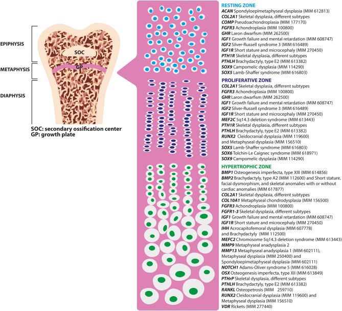
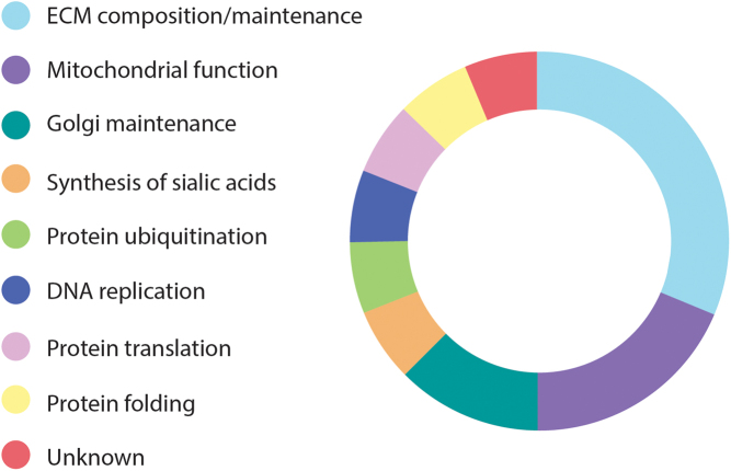
In the last decade, the widespread use of massively parallel sequencing has considerably boosted the number of novel gene discoveries in monogenic skeletal diseases with short stature. Defects in genes playing a role in the maintenance and function of the growth plate, the site of longitudinal bone growth, are a well-known cause of skeletal diseases with short stature. However, several genes involved in extracellular matrix composition or maintenance as well as genes partaking in various biological processes have also been characterized. This review aims to describe the latest genetic findings in spondyloepiphyseal dysplasias, spondyloepimetaphyseal dysplasias, and some monogenic forms of isolated short stature. Some examples of novel genetic mechanisms leading to skeletal conditions with short stature will be described. Strategies on how to successfully characterize novel skeletal phenotypes with short stature and genetic approaches to detect and validate novel gene-disease correlations will be discussed in detail. In summary, we review the latest gene discoveries underlying skeletal diseases with short stature and emphasize the importance of characterizing novel molecular mechanisms for genetic counseling, for an optimal management of the disease, and for therapeutic innovations.


Figures

Figure 1
Figure 1
Structure of a long bone with major focus on the growth plate and the key genes regulating longitudinal bone growth. On the left, structure of a long bone. Pink panel: schematic representation of the chondrocytes within the three different zones of the growth plate. On the right, some key genes regulating longitudinal bone growth and list of monogenic skeletal conditions caused by pathogenic mutations in each of these genes (96, 97, 98). ACAN, aggrecan; BMP1, bone morphogenetic protein 1; COL2A1, collagen type II alpha 1 chain; COL10A1, collagen type X alpha 1 chain; FGFR1-3, fibroblast growth factor receptor 1-3; GHR, growth hormone receptor; IGF1, insulin-like growth factor 1; IGF1R, insulin-like growth factor 1 receptor; IHH, Indian hedgehog signaling molecule; MEF2C, myocyte enhancer factor 2C; MMP9/13, matrix metallopeptidase 9/13; NOTCH1, Notch receptor 1; OSX, Sp7 transcription factor; PTH1R, parathyroid hormone 1 receptor 2; PTHLH, parathyroid hormone-like hormone; RUNX2, Runt-related transcription factor 2; SOX9/5, SRY-Box transcription factor 9/5; MIM, phenotype OMIM number.
Figure 2
Figure 2
Function of the 16 genes recently linked to SMD and SEMD. Most of the genes recently reported as causing SMD and SEMD play a role in the ECM (5/16), in the mitochondria (3/16) and in the Golgi apparatus (2/15). Each of the remaining genes is involved in a different biological mechanism.
Figure 3
Figure 3
Schematic workflow of the genetic approach to identify the disease-causing variant in a patient with a skeletal disease. After an in-depth clinical characterization (step 1), the phenotype of the studied patient might overlap a known condition, several conditions or be a novel/unknown phenotype; this will determine the genetic approach/method to be chosen (step 2). Data analysis could directly lead to a genetic diagnosis, but often further investigations are needed to validate a genetic finding or to pinpoint the genetic defect (step 3). ES, exome sequencing; GS, genome sequencing; SVs, structural variants.

Similar articles

Cited by

References

    1. Krakow D.Skeletal dysplasias. Clinics in Perinatology 2015. 42 301–19. (10.1016/j.clp.2015.03.003) - DOI - PMC - PubMed
    1. Mortier GR, Cohn DH, Cormier-Daire V, Hall C, Krakow D, Mundlos S, Nishimura G, Robertson S, Sangiorgi L, Savarirayan R. et al. Nosology and classification of genetic skeletal disorders: 2019 revision. American Journal of Medical Genetics. Part A 2019. 179 2393–2419. (10.1002/ajmg.a.61366) - DOI - PubMed
    1. Krakow D, Rimoin DL.The skeletal dysplasias. Genetics in Medicine 2010. 12 327–341. (10.1097/GIM.0b013e3181daae9b) - DOI - PubMed
    1. Carpenter TO, Whyte MP, Imel EA, Boot AM, Hogler W, Linglart A, Padidela R, Van't Hoff W, Mao M, Chen CYet al. Burosumab therapy in children with X-linked hypophosphatemia. New England Journal of Medicine 2018. 378 1987–1998. (10.1056/NEJMoa1714641) - DOI - PubMed
    1. Whyte MP, Madson KL, Phillips D, Reeves AL, McAlister WH, Yakimoski A, Mack KE, Hamilton K, Kagan K, Fujita KPet al. Asfotase alfa therapy for children with hypophosphatasia. JCI Insight 2016. 1 e85971. (10.1172/jci.insight.85971) - DOI - PMC - PubMed

LinkOut - more resources