. 2021 Apr 8;6(7):e142205.
doi: 10.1172/jci.insight.142205.

Selective inhibition of JAK3 signaling is sufficient to reverse alopecia areata

Affiliations

Selective inhibition of JAK3 signaling is sufficient to reverse alopecia areata

Zhenpeng Dai et al. JCI Insight. .

Abstract

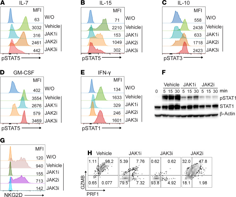
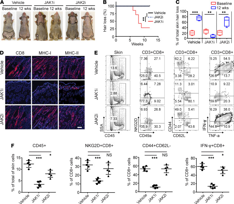
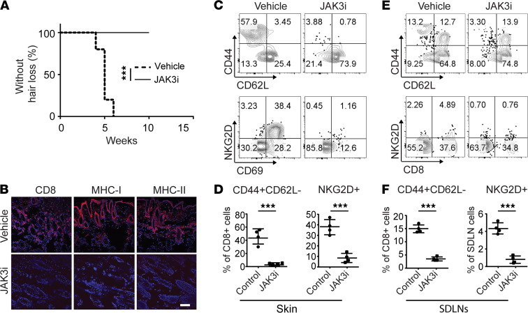
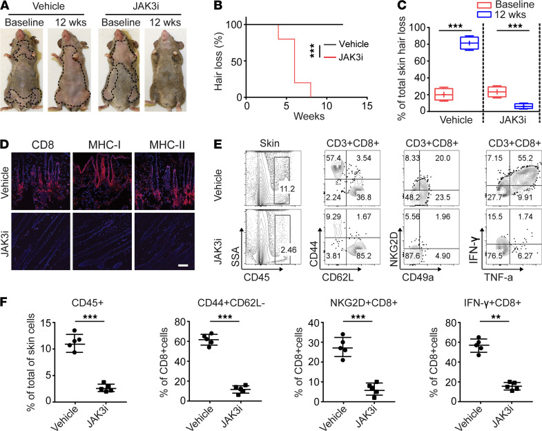
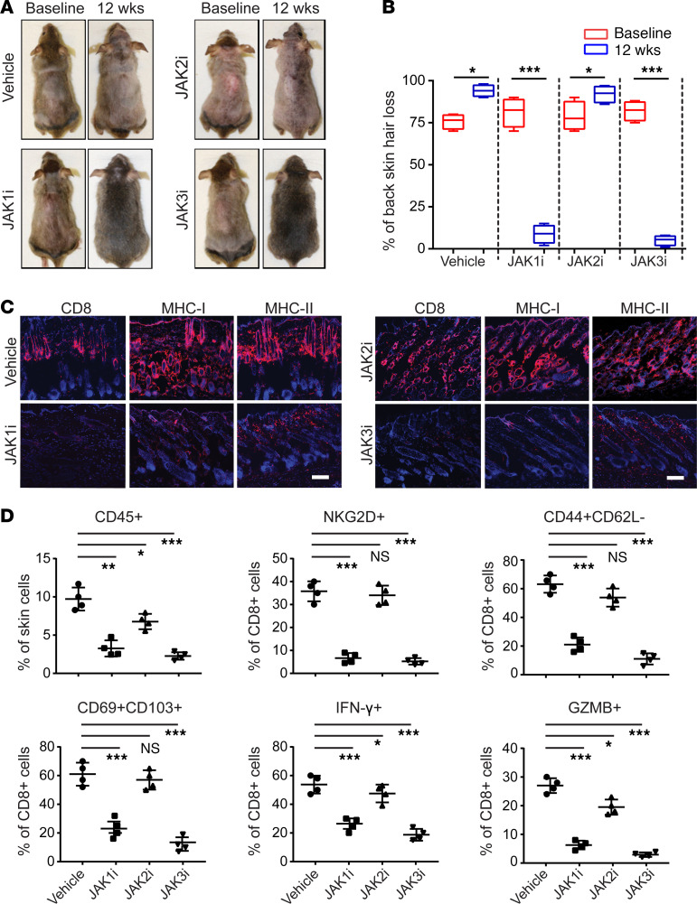
The Janus kinase/signal transducers and activators of transcription (JAK/STAT) are key intracellular mediators in the signal transduction of many cytokines and growth factors. Common γ chain cytokines and interferon-γ that use the JAK/STAT pathway to induce biological responses have been implicated in the pathogenesis of alopecia areata (AA), a T cell-mediated autoimmune disease of the hair follicle. We previously showed that therapeutic targeting of JAK/STAT pathways using the first-generation JAK1/2 inhibitor, ruxolitinib, and the pan-JAK inhibitor, tofacitinib, was highly effective in the treatment of human AA, as well as prevention and reversal of AA in the C3H/HeJ mouse model. To better define the role of individual JAKs in the pathogenesis of AA, in this study, we tested and compared the efficacy of several next-generation JAK-selective inhibitors in the C3H/HeJ mouse model of AA, using both systemic and topical delivery. We found that JAK1-selective inhibitors as well as JAK3-selective inhibitors robustly induced hair regrowth and decreased AA-associated inflammation, whereas several JAK2-selective inhibitors failed to restore hair growth in treated C3H/HeJ mice with AA. Unlike JAK1, which is broadly expressed in many tissues, JAK3 expression is largely restricted to hematopoietic cells. Our study demonstrates inhibiting JAK3 signaling is sufficient to prevent and reverse disease in the preclinical model of AA.

Keywords: Autoimmunity; Inflammation; Skin; T cells.


Conflict of interest statement

Conflict of interest: Columbia University has licensed patents (patent title: Methods for treating hair loss disorders; patent number: US9198911B2) on the use of JAK inhibitors in alopecia areata to Aclaris Therapeutics, Inc. AMC is a consultant to Aclaris Therapeutics, Almirall, and Janssen and a shareholder of Aclaris Therapeutics.

Figures

Figure 1
Figure 1. Inhibition of cytokine-dependent signaling by JAK-selective inhibitors.
(AE) C3H/HeJ mouse splenocytes were pretreated with 1 μM of INCB039110 (JAK1i), CEP-33779 (JAK2i), or PF-06651600 (JAK3i) or vehicle control for 1 hour at 37°C. The treated cells were stimulated with IL-7 (20 ng/mL), IL-15 (40 ng/mL), IL-10 (50 ng/mL), GM-CSF (50 ng/mL), or IFN-γ (50 ng/mL) for 20 minutes at 37°C. Phosphorylated STAT (p-STAT) expression in indicated cell subsets was presented as representative plots and mean fluorescence intensity (MFI). CD3+CD8+ cells (A and B), CD19+ cells (C), CD11b+ cells (D), and CD3+ cells (E) were gated for p-STAT expression. (F) C3H/HeJ mouse HF dermal sheath cells were pretreated with 1 μM of INCB039110, CEP-33779, or vehicle control for 1 hour at 37°C. The treated cells were then stimulated with IFN-γ (50 ng/mL) for indicated time points at 37°C and analyzed by Western blotting for p-STAT1, total STAT1, and the housekeeping protein β-actin. (G and H) C3H/HeJ mouse splenocytes were pretreated with 500 nM of indicated JAKi or vehicle control for 1 hour at 37°C. The treated cells from C3H/HeJ mice were then stimulated with 20 ng/mL IL-15 and 250 nM of indicated JAKi at 37°C for 72 hours. (G) Expression of NKG2D and (H) expression of Granzyme B (GZMB) and Perforin 1 (PRF1) by CD8+ T cells presented as representative plots and MFI. The data shown are from 1 representative experiment out of 2 replicates. W/O, without treatment.
Figure 2
Figure 2. JAK1-selective inhibitor treatment reversed AA.
Five C3H/HeJ AA mice per group were treated systemically with INCB039110 (JAK1i) or CEP-33779 (JAK2i) at a dosage of 50 mg/kg for 12 weeks. (A) Representative images of individual JAK3i or vehicle-treated C3H/HeJ mice before or after 12 weeks’ treatment. (B) Time course of hair regrowth shown as weeks after treatment. **P < 0.01, log-rank test. (C) Percentage of total skin hair loss or regrowth shown before and after treatment. The box plots depict the minimum and maximum values (whiskers), the upper and lower quartiles, and the median. The length of the box represents the interquartile range. **P < 0.01, ***P < 0.001 (unpaired Student’s t test). (D) Representative immunofluorescence images of skin sections from JAKi- or control-treated mice, stained with anti-CD8, anti–MHC class I, or anti–MHC class II mAbs. Scale bar: 200 μm. (E) and (F) Percentages of skin infiltrating CD45+ leukocytes, CD44+CD62LCD8+ T cells, NKG2D+CD8+ T cells, as well as IFN-γ–producing CD8+ T cells within indicated populations within the skin after JAK3i treatment. *P < 0.05, ***P < 0.001 (1-way ANOVA). Two replicate experiments were performed for a total of 10 mice per group.
Figure 3
Figure 3. JAK3-selective inhibitor treatment prevented the onset of AA.
C3H/HeJ grafted mice were given PF-06651600 (JAK3i) at a dosage of 30 mg/kg for 4 weeks. (A) Survival curve analysis depicts the hair loss between JAK3i- and control-treated mice. ***P < 0.01, log-rank test. (B) Representative immunofluorescence images of skin sections from JAK3i- or control-treated mice, stained with anti-CD8, anti–MHC class I, or anti–MHC class II mAbs. Scale bar: 200 μm. (C) Representative FACS plots of the skin single cell in the viable cell gate were acquired for each sample. (D) Summary graphs of the percentages of CD44+CD62LCD8+ T cells as well as NKG2D+CD8+ T cells within the skin after treatment. ***P < 0.001 (unpaired Student’s t test). (E) Representative FACS plots of SDLNs in the viable cell gate were acquired for each sample. (F) Summary graphs of the percentages of CD44+CD62LCD8+ T cells as well as NKG2D+CD8+ T cells within the SDLNs after treatment. ***P < 0.001 (unpaired Student’s t test). Two replicate experiments were performed for 10 mice per group.
Figure 4
Figure 4. JAK3-selective inhibitor treatment reversed AA.
Five C3H/HeJ AA mice per group were treated systemically with PF-06651600 (JAK3i) at a dosage of 30 mg/kg for 12 weeks. (A) Representative images of individual JAK3i- or control-treated C3H/HeJ mice before or after 12 weeks’ treatment. (B) Time course of hair regrowth shown as weeks after treatment. ***P < 0.001, log-rank test. (C) Percentage of total skin hair loss or regrowth shown before and after treatment. The box plots depict the minimum and maximum values (whiskers), the upper and lower quartiles, and the median. The length of the box represents the interquartile range. ***P < 0.001 (unpaired Student’s t test). (D) Representative immunofluorescence images of skin sections from JAK3i- or vehicle-treated mice, stained with anti-CD8, anti–MHC class I, or anti–MHC class II mAbs. Scale bar: 200 μm. (E) and (F) Percentages of skin infiltrating CD45+ leukocytes, CD44+CD62LCD8+ T cells, NKG2D+CD8+ T cells, as well as IFN-γ–producing CD8+ T cells within the skin after treatment. **P < 0.01, ***P < 0.001 (unpaired Student’s t test). Two replicate experiments were performed for a total of 10 mice per group.
Figure 5
Figure 5. Reversal of AA with topical JAK1- or JAK3-selective inhibitor treatment.
C3H/HeJ mice with long standing AA were treated topically with INCB039110 (JAK1i), CEP-33779 (JAK2i), or PF-06651600 (JAK3i) or control daily for 12 weeks, in cohorts of 4 mice per group. (A) Representative images of individual JAK inhibitor– or vehicle-treated C3H/HeJ mice before or after 12 weeks’ treatment. (B) Percentage of dorsal hair loss or regrowth is shown before and after treatment. The box plots depict the minimum and maximum values (whiskers), the upper and lower quartiles, and the median. The length of the box represents the interquartile range. *P < 0.05, ***P < 0.001 (unpaired Student’s t test). (C) Representative immunofluorescence images of skin sections from JAKi- or vehicle-treated mice, stained with anti-CD8, anti–MHC class I, or anti–MHC class II mAbs. Scale bar: 100 μm. (D) Percentages of skin infiltrating CD45+ leukocytes, NKG2D+CD8+ T cells, CD44+CD62LCD8+ T cells, CD103+CD69+CD8+ T cells, IFN-γ–producing CD8+ T cells, as well as GZMB- or PRF1-producing CD8+ T cells within indicated populations within the skin after JAK inhibitor treatment. *P < 0.05, **P < 0.01, ***P < 0.001 (1-way ANOVA). Two replicate experiments were performed for a total of 8 mice per group.

Similar articles

Cited by

References

    1. Papadopoulos AJ, et al. Alopecia areata. Pathogenesis, diagnosis, and therapy. Am J Clin Dermatol. 2000;1(2):101–105. doi: 10.2165/00128071-200001020-00004. - DOI - PubMed
    1. Gupta MA, Gupta AK. Depression and suicidal ideation in dermatology patients with acne, alopecia areata, atopic dermatitis and psoriasis. Br J Dermatol. 1998;139(5):846–850. doi: 10.1046/j.1365-2133.1998.02511.x. - DOI - PubMed
    1. Pratt CH, et al. Alopecia areata. Nat Rev Dis Primers. 2017;3:17011. - PMC - PubMed
    1. Xing L, et al. Alopecia areata is driven by cytotoxic T lymphocytes and is reversed by JAK inhibition. Nat Med. 2014;20(9):1043–1049. doi: 10.1038/nm.3645. - DOI - PMC - PubMed
    1. Dai Z, et al. CXCR3 blockade inhibits T cell migration into the skin and prevents development of alopecia areata. J Immunol. 2016;197(4):1089–1099. doi: 10.4049/jimmunol.1501798. - DOI - PMC - PubMed

Publication types

MeSH terms

Substances