Review
. 2021 Jul;142(1):57-71.
doi: 10.1007/s00401-021-02301-7. Epub 2021 Apr 8.

Tau strains shape disease

Affiliations
Review

Tau strains shape disease

Jaime Vaquer-Alicea et al. Acta Neuropathol. 2021 Jul.

Abstract

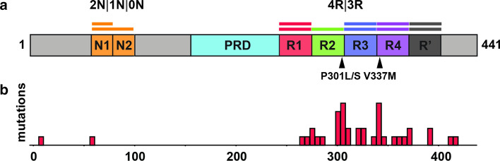
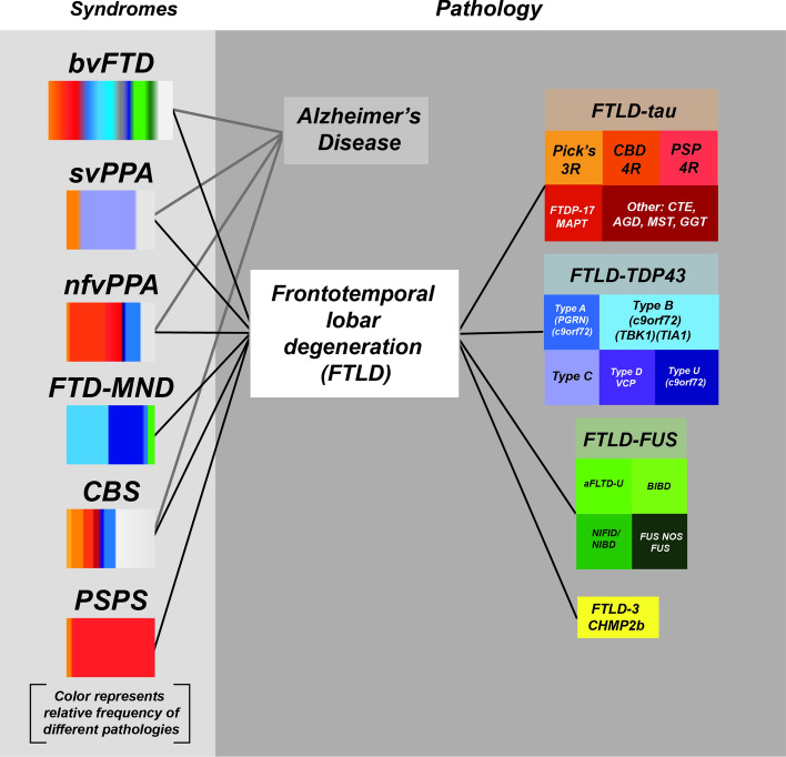
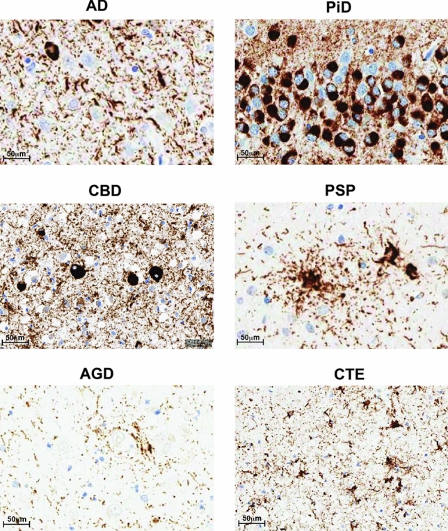
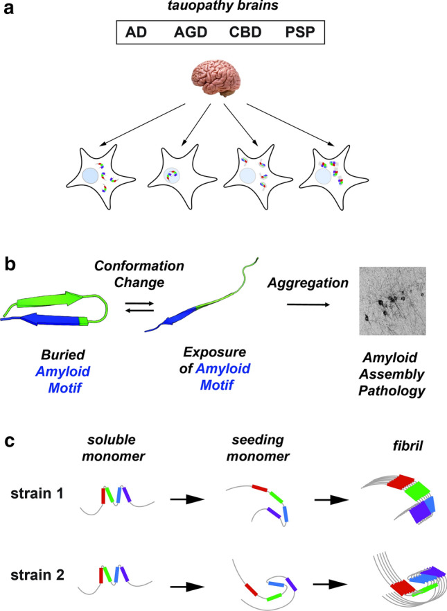
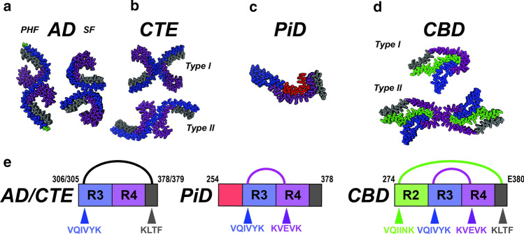
Tauopathies consist of over 25 different neurodegenerative diseases that include argyrophilic grain disease (AGD), progressive supranuclear palsy (PSP), corticobasal degeneration (CBD), and Pick's disease (PiD). Tauopathies are defined by brain accumulation of microtubule-associated protein tau in fibrillar aggregates, whose prevalence strongly correlates with dementia. Dominant mutations in tau cause neurodegenerative diseases, and most increase its aggregation propensity. Pathogenesis of tauopathies may involve pathological tau conformers that serve as templates to recruit native protein into growing assemblies and also move between brain cells to cause disease progression, similar to prions. Prions adopt pathological conformations, termed "strains," that stably propagate in living systems, and create unique patterns of neuropathology. Data from multiple laboratories now suggest that tau acts as a prion. It propagates unique strains indefinitely in cultured cells, and when these are inoculated into mouse models, they create defined neuropathological patterns, which establish a direct link between conformation and disease. In humans, distinct fibril structures are associated with different diseases, but causality has not been established as in mice. Cryo-EM structures of tau fibrils isolated from tauopathy brains reveal distinct fibril cores across disease. Interestingly, the conformation of the tau monomer unit within different fibril subtypes from the same patient appears relatively preserved. This is consistent with data that the tau monomer samples an ensemble of conformations that act as distinct pathologic templates in the formation of restricted numbers of strains. The propensity of a tau monomer to adopt distinct conformations appears to be linked to defined local motifs that expose different patterns of amyloidogenic amino acid sequences. The prion hypothesis, which predicts that protein structure dictates resultant disease, has proved particularly useful to understand the diversity of human tauopathies. The challenge now is to develop methods to rapidly classify patients according to the structure of the underlying pathological protein assemblies to achieve more accurate diagnosis and effective therapy.

Keywords: Aggregation; Amyloid; Diagnosis; Folding; Polymorph; Prion; Propagation; Self-assembly; Strains; Tau; Tauopathy; Therapeutics.


Conflict of interest statement

One of the authors (MID) is a co-inventor of an anti-tau therapeutic antibody (ABBV-8E12) that is in clinical trials.

Figures

Fig. 1
Fig. 1
Tau protein. a Domain organization of tau brain isoforms. Schematic of the 441 residue 2N4R tau isoform highlighting the domains (N1, N2 and R2) which define the isoforms. The tau repeats are colored red (R1; residues 244–274), green (R2; residues 275–305), blue (R3; residues 306–336), purple (R4; residues 337–368) and dark grey (R’; residues 369–400). The proline-rich domain (PRD) is colored in light blue and the N1 and N2 domains are colored in orange. Two key disease-associated mutations are highlighted by arrows: Proline301 to serine or leucine mutations and valine337 to methionine. b Disease-associated mutation frequencies found in human tauopathies. Most mutations are found within the repeat domain
Fig. 2
Fig. 2
Relationship between clinical syndromes and neuropathology. Illustration of the association of different clinical syndromes with the deposition of specific inclusions of proteins including tau as determined by neuropathology. Each disease, defined by neuropathology of specific proteins, is colored differently. The fractional percentage of protein deposition in each clinical syndrome is estimated from the literature. (Figure is adapted from a slide shared by Dr. William Seeley, UCSF)
Fig. 3
Fig. 3
Neuropathology of tauopathies. Representative IHC staining using AT8 on brain sections from different human tauopathy patients AD, CBD, PSP, AGD, PiD and CTE
Fig. 4
Fig. 4
Propagation of tau strains. a Schematic illustrating biosensor-based detection of tau seeds derived from different tauopathies (AD, AGD, CBD and lead to cellular aggregates with different morphologies. b Model of tau domain structural rearrangement and subsequent aggregation. Inert tau monomer (left) has a propensity to form a relatively collapsed conformation, which buries aggregation-prone elements. In the presence of disease-associated mutations, proline isomerization events, or certain splice isoforms, the equilibrium is shifted to disfavor local compact structure. This exposes the aggregation-prone elements and enhances aggregation propensity, leading to subsequent tau pathology. Structural models are shown in cartoon representation and are colored according to repeat domain as in Fig. 1. The aggregation-prone element is colored in blue. c Schematic of tau aggregation pathway for the formation of different strains. Soluble inert tau is shown as a cartoon highlighting local structures surrounding repeat domains, seed-competent monomer highlights structural rearrangements surrounding the aggregation-prone elements and fibrils are shown as an array of ordered monomers. Tau domains are colored as in Fig. 1
Fig. 5
Fig. 5
Unifying themes for diverse tauopathy fibrils. ad Cryo-EM structures of tau fibrils isolated from AD-PHF, AD-SF, CTE (Type I and II), PiD and CBD (Type I and II). The structures are shown in spacefill representation, colored according to the repeat domains as in Fig. 1 and viewed down the fibril axis. e Schematic illustrating key contacts involving aggregation-prone elements observed in the different structures. Amino acids of each fibril are shown as a schematic and colored as in Fig. 1. Amino acids (including aggregation-prone elements) are colored according to the repeat domain and location indicated by an arrow. The linkage between contacts observed in AD/CTE, PiD and CBD are indicated by semi-circles and are colored black, magenta and green. The residues that comprise the amyloid structures are shown in the cartoon schematic

References

    1. Agerschou ED, Schützmann MP, Reppert N, Wördehoff MM, Shaykhalishahi H, Buell AK, Hoyer W. β-Turn exchanges in the α-synuclein segment 44-TKEG-47 reveal high sequence fidelity requirements of amyloid fibril elongation. Biophys Chem. 2020;269:106519. doi: 10.1016/j.bpc.2020.106519. - DOI - PubMed
    1. Akoury E, Pickhardt M, Gajda M, Biernat J, Mandelkow E, Zweckstetter M. Mechanistic basis of phenothiazine-driven inhibition of tau aggregation. Angewandte Chemie Int Ed. 2013;52:3511–3515. doi: 10.1002/anie.201208290. - DOI - PubMed
    1. Al-Hilaly YK, Pollack SJ, Rickard JE, Simpson M, Raulin A-C, Baddeley T, Schellenberger P, Storey JMD, Harrington CR, Wischik CM, Serpell LC. Cysteine-independent inhibition of Alzheimer’s disease-like paired helical filament assembly by leuco-methylthioninium (LMT) J Mol Biol. 2018;430:4119–4131. doi: 10.1016/j.jmb.2018.08.010. - DOI - PubMed
    1. Allen B, Ingram E, Takao M, Smith MJ, Jakes R, Virdee K, Yoshida H, Holzer M, Craxton M, Emson PC, Atzori C, Migheli A, Crowther RA, Ghetti B, Spillantini MG, Goedert M. Abundant tau filaments and nonapoptotic neurodegeneration in transgenic mice expressing human P301S tau protein. J Neurosci. 2002;22:9340–9351. doi: 10.1523/jneurosci.22-21-09340.2002. - DOI - PMC - PubMed
    1. Ambadipudi S, Reddy JG, Biernat J, Mandelkow E, Zweckstetter M. Residue-specific identification of phase separation hot spots of Alzheimer’s-related protein tau. Chem Sci. 2019;10:6503–6507. doi: 10.1039/c9sc00531e. - DOI - PMC - PubMed

Publication types

LinkOut - more resources