

Sirtuin 1 alleviates neuroinflammation-induced apoptosis after traumatic brain injury
- PMID: 33830639
- PMCID: PMC8093975
- DOI: 10.1111/jcmm.16534
Sirtuin 1 alleviates neuroinflammation-induced apoptosis after traumatic brain injury
Erratum in
-
Corrigendum.J Cell Mol Med. 2022 Oct;26(19):5100-5101. doi: 10.1111/jcmm.17548. J Cell Mol Med. 2022. PMID: 36214283 Free PMC article. No abstract available.
Abstract
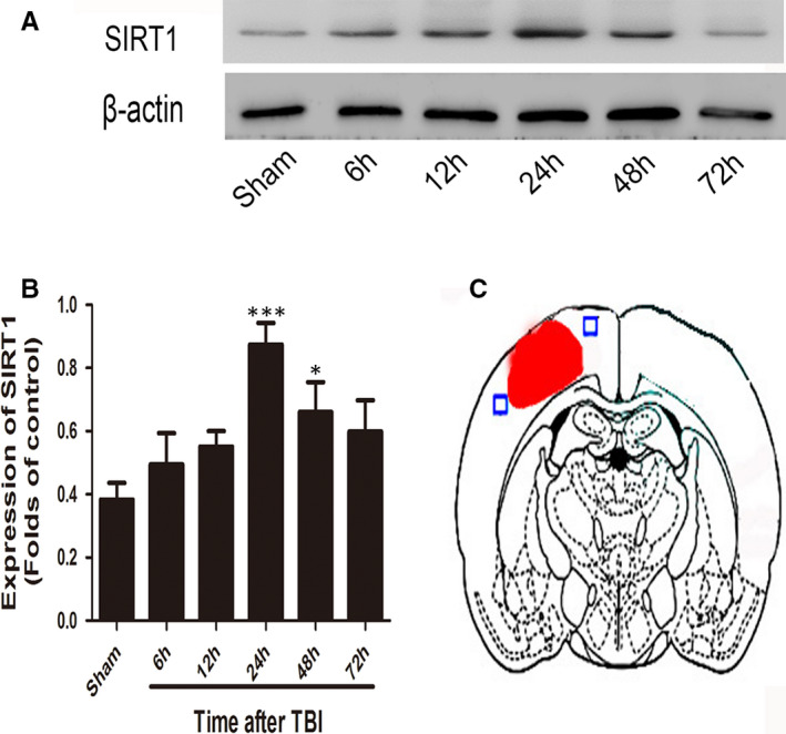
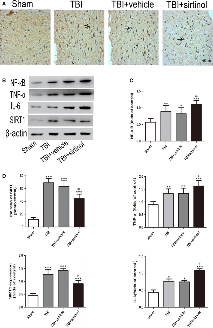
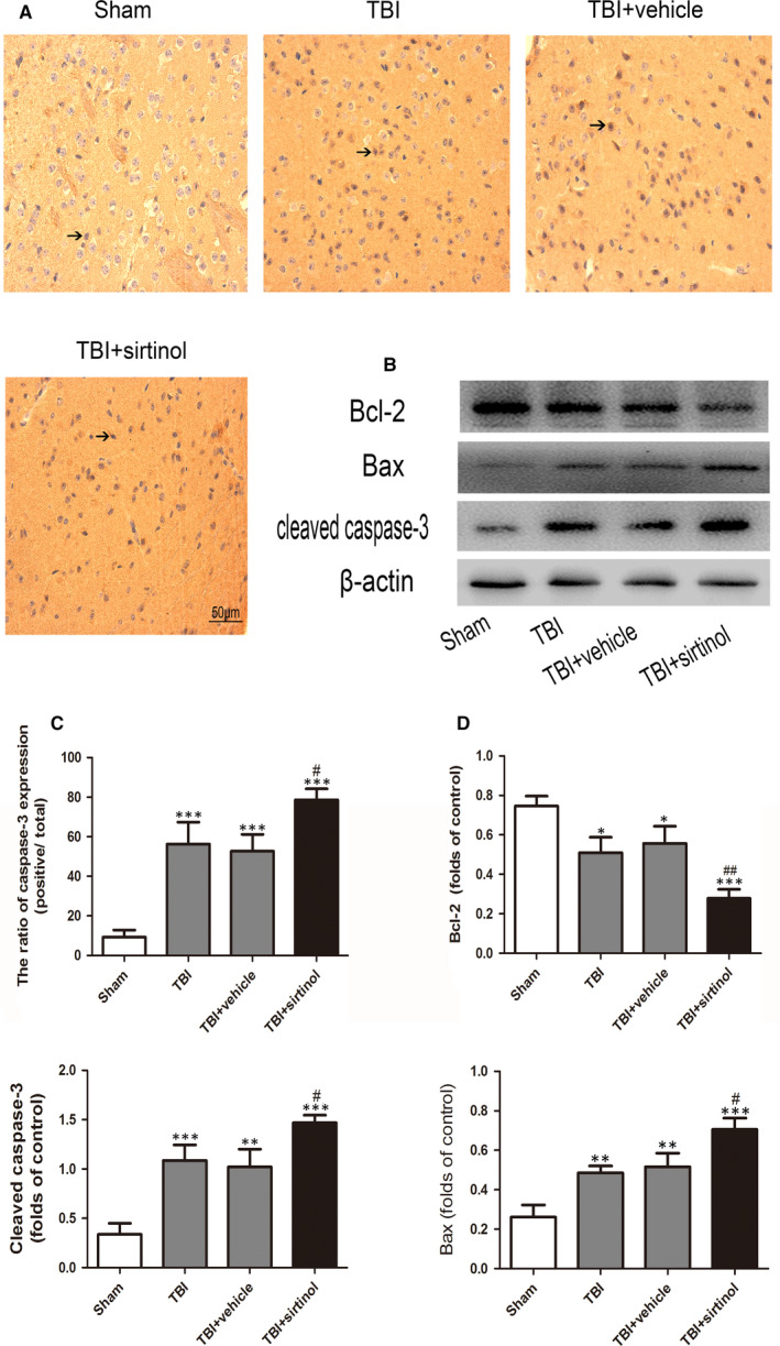
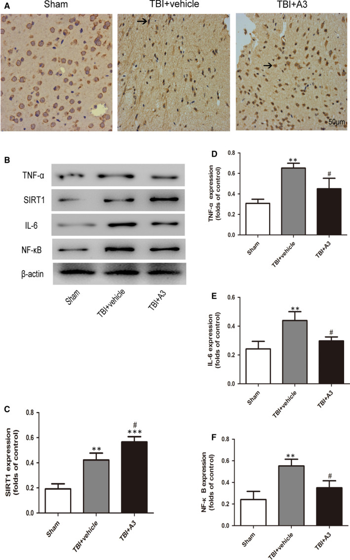
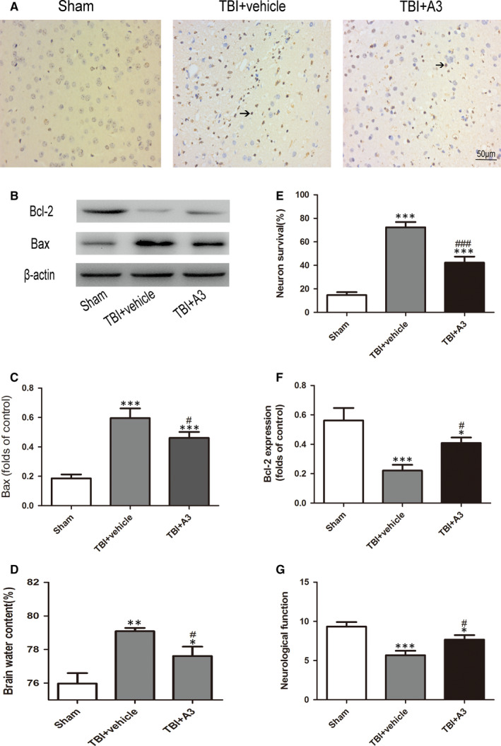
Sirtuin 1 (SIRT1) plays a very important role in a wide range of biological responses, such as metabolism, inflammation and cell apoptosis. Changes in the levels of SIRT1 have been detected in the brain after traumatic brain injury (TBI). Further, SIRT1 has shown a neuroprotective effect in some models of neuronal death; however, its role and working mechanisms are not well understood in the model of TBI. This study aimed to address this issue. SIRT1-specific inhibitor (sirtinol) and activator (A3) were introduced to explore the role of SIRT1 in cell apoptosis. Results of the study suggest that SIRT1 plays an important role in neuronal apoptosis after TBI by inhibiting NF-κB, IL-6 and TNF-α deacetylation and the apoptotic pathway sequentially, possibly by alleviating neuroinflammation.
Keywords: NF-κB; neuroinflammation; neuronal apoptosis; sirtuin 1; traumatic brain injury.
© 2021 The Authors. Journal of Cellular and Molecular Medicine published by Foundation for Cellular and Molecular Medicine and John Wiley & Sons Ltd.
Conflict of interest statement
All of the authors declare that there were no competing interests.
Figures

Similar articles
-
Omega-3 polyunsaturated fatty acid attenuates the inflammatory response by modulating microglia polarization through SIRT1-mediated deacetylation of the HMGB1/NF-κB pathway following experimental traumatic brain injury.J Neuroinflammation. 2018 Apr 20;15(1):116. doi: 10.1186/s12974-018-1151-3. J Neuroinflammation. 2018. PMID: 29678169 Free PMC article.
-
SIRT1 plays a neuroprotective role in traumatic brain injury in rats via inhibiting the p38 MAPK pathway.Acta Pharmacol Sin. 2017 Feb;38(2):168-181. doi: 10.1038/aps.2016.130. Epub 2016 Dec 26. Acta Pharmacol Sin. 2017. PMID: 28017962 Free PMC article.
-
Omega-3 polyunsaturated fatty acid attenuates traumatic brain injury-induced neuronal apoptosis by inducing autophagy through the upregulation of SIRT1-mediated deacetylation of Beclin-1.J Neuroinflammation. 2018 Nov 8;15(1):310. doi: 10.1186/s12974-018-1345-8. J Neuroinflammation. 2018. PMID: 30409173 Free PMC article.
-
The Beneficial Roles of SIRT1 in Neuroinflammation-Related Diseases.Oxid Med Cell Longev. 2020 Sep 14;2020:6782872. doi: 10.1155/2020/6782872. eCollection 2020. Oxid Med Cell Longev. 2020. PMID: 33014276 Free PMC article. Review.
-
Antagonistic crosstalk between NF-κB and SIRT1 in the regulation of inflammation and metabolic disorders.Cell Signal. 2013 Oct;25(10):1939-48. doi: 10.1016/j.cellsig.2013.06.007. Epub 2013 Jun 11. Cell Signal. 2013. PMID: 23770291 Review.
Cited by
-
Fat mass and obesity-mediated N 6 -methyladenosine modification modulates neuroinflammatory responses after traumatic brain injury.Neural Regen Res. 2026 Feb 1;21(2):730-741. doi: 10.4103/NRR.NRR-D-23-01854. Epub 2024 Sep 6. Neural Regen Res. 2026. PMID: 39248160 Free PMC article.
-
Gentamycin Rationally Repositioned to Inhibit miR-34a Ameliorates Oxidative Injury to PC12 Cells.ACS Omega. 2022 Dec 16;8(1):771-781. doi: 10.1021/acsomega.2c06112. eCollection 2023 Jan 10. ACS Omega. 2022. PMID: 36643496 Free PMC article.
-
Fluoride-Induced Mitochondrial Dysfunction and Approaches for Its Intervention.Biol Trace Elem Res. 2024 Mar;202(3):835-849. doi: 10.1007/s12011-023-03720-1. Epub 2023 Jun 10. Biol Trace Elem Res. 2024. PMID: 37300595 Review.
-
Innovative Insights into Traumatic Brain Injuries: Biomarkers and New Pharmacological Targets.Int J Mol Sci. 2024 Feb 17;25(4):2372. doi: 10.3390/ijms25042372. Int J Mol Sci. 2024. PMID: 38397046 Free PMC article. Review.
-
Long-term exposure of sucralose induces neuroinflammation and ferroptosis in human microglia cells via SIRT1/NLRP3/IL-1β/GPx4 signaling pathways.Food Sci Nutr. 2024 Sep 23;12(11):9094-9107. doi: 10.1002/fsn3.4488. eCollection 2024 Nov. Food Sci Nutr. 2024. PMID: 39619997 Free PMC article.
References
-
- Stein DM, Feather CB, Napolitano LM. Traumatic brain injury advances. Crit Care Clin. 2017;33(1):1‐13. - PubMed
-
- Hackenberg K, Unterberg A. Traumatic brain injury. Nervenarzt. 2016;87(2):203‐214; quiz 215‐216. - PubMed
-
- Leikin JB. Traumatic brain injury. Dis Mon. 2019;65(10):100857. - PubMed
-
- Chen X, Chen C, Fan S, et al. Omega‐3 polyunsaturated fatty acid attenuates the inflammatory response by modulating microglia polarization through SIRT1‐mediated deacetylation of the HMGB1/NF‐κB pathway following experimental traumatic brain injury. J Neuroinflammation. 2018;15(1):116. - PMC - PubMed
Publication types
MeSH terms
Substances
Grants and funding
LinkOut - more resources
Full Text Sources
Other Literature Sources
Medical
