

Secondary denervation is a chronic pathophysiologic sequela of volumetric muscle loss
- PMID: 33830817
- PMCID: PMC8354822
- DOI: 10.1152/japplphysiol.00049.2021
Secondary denervation is a chronic pathophysiologic sequela of volumetric muscle loss
Abstract
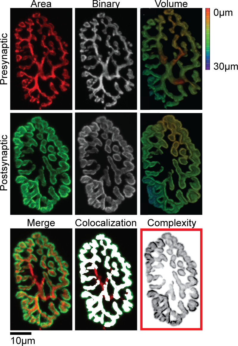
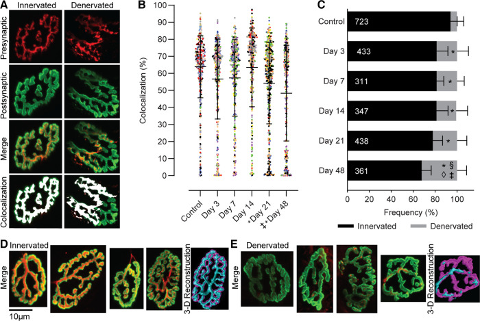
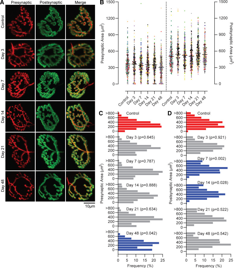
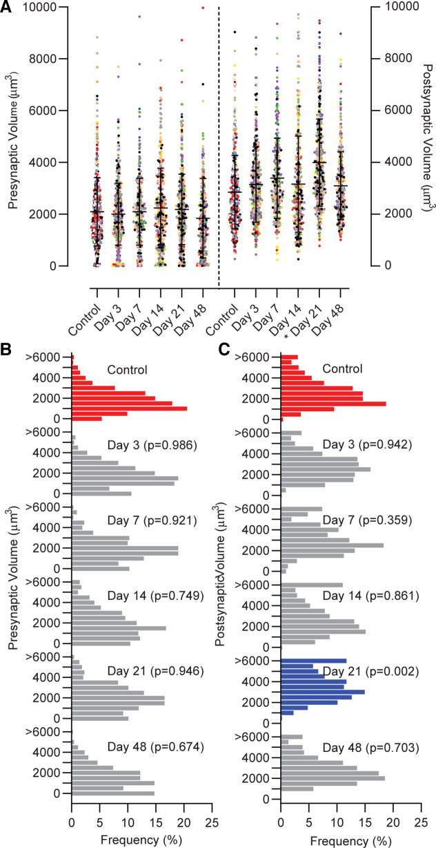
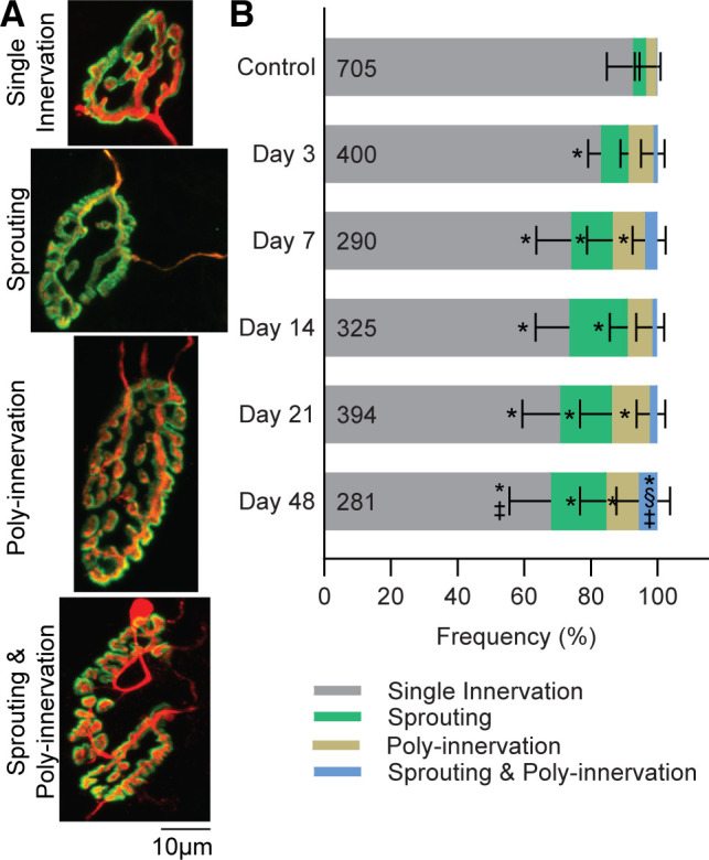
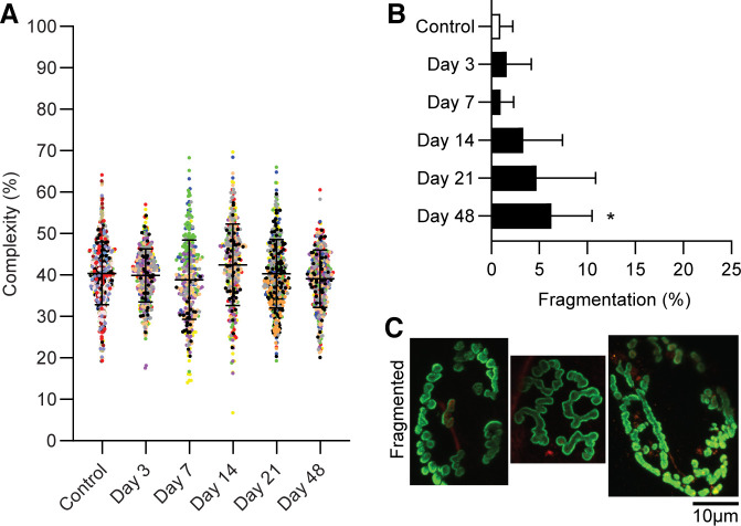
Volumetric muscle loss (VML) is the traumatic loss of muscle tissue that results in long-term functional impairments. Despite the loss of myofibers, there remains an unexplained significant decline in muscle function. VML injury likely extends beyond the defect area, causing negative secondary outcomes to the neuromuscular system, including the neuromuscular junctions (NMJs), yet the extent to which VML induces denervation is unclear. This study systematically examined NMJs surrounding the VML injury, hypothesizing that the sequela of VML includes denervation. The VML injury removed ∼20% of the tibialis anterior (TA) muscle in adult male inbred Lewis rats (n = 43), the noninjured leg served as an intra-animal control. Muscles were harvested up to 48 days post-VML. Synaptic terminals were identified immunohistochemically, and quantitative confocal microscopy evaluated 2,613 individual NMJ. Significant denervation was apparent by 21 and 48 days post-VML. Initially, denervation increased ∼10% within 3 days of injury; with time, denervation further increased to ∼22% and 32% by 21 and 48 days post-VML, respectively, suggesting significant secondary denervation. The appearance of terminal axon sprouting and polyinnervation were observed as early as 7 days post-VML, increasing in number and complexity throughout 48 days. There was no evidence of VML-induced NMJ size alteration, which may be beneficial for interventions aimed at restoring muscle function. This work recognizes VML-induced secondary denervation and poor remodeling of the NMJ as part of the sequela of VML injury; moreover, secondary denervation is a possible contributing factor to the chronic functional impairments and potentially an overlooked treatment target.NEW & NOTEWORTHY This work advances our understanding of the pathophysiologic complexity of volumetric muscle loss injury. Specifically, we identified secondary denervation in the muscle remaining after volumetric muscle loss injuries as a novel aspect of the injury sequela. Denervation increased chronically, in parallel with the appearance of irregular morphological characteristics and destabilization of the neuromuscular junction, which is expected to further confound chronic functional impairments.
Keywords: innervation; neuromuscular junction; polyinnervation; skeletal muscle; sprouting.
Conflict of interest statement
No conflicts of interest, financial or otherwise, are declared by the authors.
Figures
Similar articles
-
Differential evaluation of neuromuscular injuries to understand re-innervation at the neuromuscular junction.Exp Neurol. 2024 Dec;382:114996. doi: 10.1016/j.expneurol.2024.114996. Epub 2024 Oct 10. Exp Neurol. 2024. PMID: 39393669
-
Inhibition of ErbB2 mitigates secondary denervation after traumatic muscle injury.J Physiol. 2025 Mar 3. doi: 10.1113/JP287435. Online ahead of print. J Physiol. 2025. PMID: 40033740
-
Response of terminal Schwann cells following volumetric muscle loss injury.Exp Neurol. 2023 Jul;365:114431. doi: 10.1016/j.expneurol.2023.114431. Epub 2023 May 2. Exp Neurol. 2023. PMID: 37142114 Free PMC article.
-
Pathophysiology of Volumetric Muscle Loss Injury.Cells Tissues Organs. 2016;202(3-4):180-188. doi: 10.1159/000443925. Epub 2016 Nov 9. Cells Tissues Organs. 2016. PMID: 27825160 Review.
-
Vascularized and Innervated Skeletal Muscle Tissue Engineering.Adv Healthc Mater. 2020 Jan;9(1):e1900626. doi: 10.1002/adhm.201900626. Epub 2019 Oct 17. Adv Healthc Mater. 2020. PMID: 31622051 Free PMC article. Review.
Cited by
-
The Neuromuscular Junction: Roles in Aging and Neuromuscular Disease.Int J Mol Sci. 2021 Jul 28;22(15):8058. doi: 10.3390/ijms22158058. Int J Mol Sci. 2021. PMID: 34360831 Free PMC article. Review.
-
Combined regenerative rehabilitation improves recovery following volumetric muscle loss injury in a rat model.J Biomed Mater Res B Appl Biomater. 2024 Jul;112(7):e35438. doi: 10.1002/jbm.b.35438. J Biomed Mater Res B Appl Biomater. 2024. PMID: 38923755 Free PMC article.
-
Human muscle in gene edited pigs for treatment of volumetric muscle loss.Front Genet. 2022 Jul 25;13:948496. doi: 10.3389/fgene.2022.948496. eCollection 2022. Front Genet. 2022. PMID: 35957684 Free PMC article. Review.
-
Traumatic Skeletal Muscle Injury and Recovery.Adv Exp Med Biol. 2025;1478:213-242. doi: 10.1007/978-3-031-88361-3_10. Adv Exp Med Biol. 2025. PMID: 40879942 Review.
-
Differential evaluation of neuromuscular injuries to understand re-innervation at the neuromuscular junction.Exp Neurol. 2024 Dec;382:114996. doi: 10.1016/j.expneurol.2024.114996. Epub 2024 Oct 10. Exp Neurol. 2024. PMID: 39393669
References
-
- Armed Forces Health Surveillance Center (AFHSC). Medical evacuations from Operation Iraqi Freedom/Operation New Dawn, active and reserve components, U.S. Armed Forces, 2003–2011. MSMR 19: 18–21, 2012. - PubMed
Publication types
MeSH terms
LinkOut - more resources
Full Text Sources
Other Literature Sources
Medical