. 2021 Apr 8;17(4):e1009453.
doi: 10.1371/journal.ppat.1009453. eCollection 2021 Apr.

A human coronavirus evolves antigenically to escape antibody immunity

Affiliations

A human coronavirus evolves antigenically to escape antibody immunity

Rachel T Eguia et al. PLoS Pathog. .

Abstract

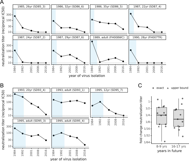
There is intense interest in antibody immunity to coronaviruses. However, it is unknown if coronaviruses evolve to escape such immunity, and if so, how rapidly. Here we address this question by characterizing the historical evolution of human coronavirus 229E. We identify human sera from the 1980s and 1990s that have neutralizing titers against contemporaneous 229E that are comparable to the anti-SARS-CoV-2 titers induced by SARS-CoV-2 infection or vaccination. We test these sera against 229E strains isolated after sera collection, and find that neutralizing titers are lower against these "future" viruses. In some cases, sera that neutralize contemporaneous 229E viral strains with titers >1:100 do not detectably neutralize strains isolated 8-17 years later. The decreased neutralization of "future" viruses is due to antigenic evolution of the viral spike, especially in the receptor-binding domain. If these results extrapolate to other coronaviruses, then it may be advisable to periodically update SARS-CoV-2 vaccines.


Conflict of interest statement

I have read the journal’s policy and the authors of this manuscript have the following competing interests: MJB has consulted for Moderna and Vir Biotechnologies, and received research funding from Regeneron and Vir Biotechnologies. JAE has consulted for Meissa Vaccines and Sanofi Pasteur, and received research funding from Merck, GlaxoSmithKline, Pfizer, and AstraZeneca. The other authors declare no competing interests.

Figures

Fig 1
Fig 1. Spikes used in this study.
(A) Phylogenetic tree of 229E spikes, with tips colored by the country from which the virus was isolated. The spikes used in the experiments are indicated with black text and square shapes. The tree is a maximum-likelihood inference with IQ-TREE [82] with a codon-substitution model and re-scaled with TreeTime [84] to position tips by to date of isolation. S1 Fig shows a tree with branch lengths proportional to divergence rather than time, and validates clock-like evolution. S2 Fig shows recombination does not substantially affect the phylogenetic placements of the spikes used in the experiments. (B) Protein sequence divergence of the spikes used in the experiments, computed over just the receptor-binding domain (RBD) or the full sequence. Divergence is the Levenshtein distance between the amino-acid sequences divided by the number of sites.
Fig 2
Fig 2. The neutralizing activity of human sera is lower against “future” viruses than those that elicited the immunity.
(A) Sera collected between 1985 and 1990 was tested in neutralization assays against spikes from viruses isolated between 1984 and 2016. Each plot facet is a different serum, and black points show its neutralizing titer against viruses from the indicated year. Blue shading indicates the portion of plotted timeframe during which the individual could have been infected prior to serum collection. The dotted gray horizontal line indicates the limit of detection (titer of 1:10). Plot titles give the year of serum collection, the individual’s age when the serum was collected, and the serum ID. (B) Plots like those in (A) but for sera collected between 1993 and 1995. (C) The fold change in neutralization titer against viruses isolated 8–9 or 16–17 years in the “future” relative to the virus isolated just before the serum was collected. Box plots show the median and interquartile range, and each point is the fold change for a single serum. For a few sera (triangles), the fold change is censored (as an upper bound) because the titer against the future virus was below the limit of detection.
Fig 3
Fig 3. Neutralizing titers of sera collected in 2020 are higher against historical viruses that circulated during an individual’s lifetime than viruses isolated before the individual was born.
As in Fig 2A and 2B, each plot facet is a different serum with the title giving the individual’s age and black points indicating the titer against spikes from viruses isolated in the indicated year. Blue shading represents the portion of the plotted timeframe during which the individual was alive: for adults this is the entire timeframe, but for children the left edge of the blue shading indicates the birth year.
Fig 4
Fig 4. Antigenic evolution is primarily due to changes in the spike’s receptor binding domain (RBD).
(A) At top is a schematic of the 229E spike. Within the S1 subunit, the schematic indicates the N-terminal domain (NTD, also known as S1-A) and the RBD (also known as S1-B). The three loops in the RBD that bind the virus’s APN receptor are indicated [53]. Below the schematic is a plot of sequence variability across the alignment of 229E spikes in Fig 1A. Variability is quantified as the effective number of amino acids at a site [86], with a value of one indicating complete conservation and larger values indicating more sequence variability. (B) Site entropy mapped on 229E spike structure (PDB 6U7H, [53]). (C) Neutralizing titers of sera collected between 1985 and 1990 against either the full spike of “future” viruses or chimeras consisting of the 1984 spike containing the RBD from “future” viruses. The plot format and the black circles (full spike) are repeated from Fig 2A with the addition of the orange triangles showing the titers against the chimeric spikes.

Comment in

References

    1. Addetia A, Crawford KHD, Dingens A, Zhu H, Roychoudhury P, Huang M-L, et al. Neutralizing Antibodies Correlate with Protection from SARS-CoV-2 in Humans during a Fishery Vessel Outbreak with a High Attack Rate. J Clin Microbiol. 2020;58. 10.1128/JCM.02107-20 - DOI - PMC - PubMed
    1. Lumley SF, O’Donnell D, Stoesser NE, Matthews PC, Howarth A, Hatch SB, et al. Antibody Status and Incidence of SARS-CoV-2 Infection in Health Care Workers. N Engl J Med. 2021;384: 533–540. 10.1056/NEJMoa2034545 - DOI - PMC - PubMed
    1. Polack FP, Thomas SJ, Kitchin N, Absalon J, Gurtman A, Lockhart S, et al. Safety and Efficacy of the BNT162b2 mRNA Covid-19 Vaccine. N Engl J Med. 2020;0: null. 10.1056/NEJMoa2034577 - DOI - PMC - PubMed
    1. Alsoussi WB, Turner JS, Case JB, Zhao H, Schmitz AJ, Zhou JQ, et al. A Potently Neutralizing Antibody Protects Mice against SARS-CoV-2 Infection. J Immunol. 2020;205: 915–922. 10.4049/jimmunol.2000583 - DOI - PMC - PubMed
    1. McMahan K, Yu J, Mercado NB, Loos C, Tostanoski LH, Chandrashekar A, et al. Correlates of protection against SARS-CoV-2 in rhesus macaques. Nature. 2020; 1–8. 10.1038/s41586-020-03041-6 - DOI - PMC - PubMed

Publication types

MeSH terms