. 2021 Jun:127:193-204.
doi: 10.1016/j.actbio.2021.03.065. Epub 2021 Apr 6.

Tear propagation in vaginal tissue under inflation

Affiliations

Tear propagation in vaginal tissue under inflation

Jeffrey A McGuire et al. Acta Biomater. 2021 Jun.

Abstract

Vaginal tearing at childbirth is extremely common yet understudied despite the long-term serious consequences on women's health. The mechanisms of vaginal tearing remain unknown, and their knowledge could lead to the development of transformative prevention and treatment techniques for maternal injury. In this study, whole rat vaginas with pre-imposed elliptical tears oriented along the axial direction of the organs were pressurized using a custom-built inflation setup, producing large tear propagation. Large deformations of tears through propagation were analyzed, and nonlinear strains around tears were calculated using the digital image correlation technique. Second harmonic generation microscopy was used to examine collagen fiber organization in mechanically untested and tested vaginal specimens. Tears became increasingly circular under pressure, propagating slowly up to the maximum pressure and then more rapidly. Hoop strains were significantly larger than axial strains and displayed a region- and orientation-dependent response with tear propagation. Imaging revealed initially disorganized collagen fibers that aligned along the axial direction with increasing pressure. Fibers in the near-regions of tear tips aligned toward the hoop direction, hampering tear propagation. Changes in tear geometry, regional strains, and fiber orientation revealed the inherent toughening mechanisms of the vaginal tissue. STATEMENT OF SIGNIFICANCE: Women's reproductive health has historically been understudied despite alarming maternal injury and mortality rates in the world. Maternal injury and disability can be reduced by advancing our limited understanding of the large deformations experienced by women's reproductive organs. This manuscript presents, for the first time, the mechanics of tear propagation in vaginal tissue and changes to the underlying collagen microstructure near to and far from the tear. A novel inflation setup capable of maintaining the in vivo tubular geometry of the vagina while propagating a pre-imposed tear was developed. Toughening mechanisms of the vagina to propagation were examined through measurements of tear geometry, strain distributions, and reorientation of collagen fibers. This research draws from current advances in the engineering science and mechanics fields with the goal of improving maternal health care.

Keywords: Digital image correlation; Inflation; Maternal trauma; Second-harmonic generation imaging; Tear propagation; Vaginal tissue.


Conflict of interest statement

Declaration of Competing Interest The authors declare that they have no known competing financial interests or personal relationships that could have appeared to influence the work reported in this paper.

Figures
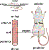
Fig. 1.
Fig. 1.
Schematic of the anatomical regions of the rat vagina.
Fig. 2.
Fig. 2.
(a) Schematic of the custom-made inflation setup for studying the tear propagation in rat vaginal tissue specimens. (b) Transverse cross-section of the latex tube mounted on two opposite coaxial (18-gauge and 16-gauge) needles. (c) Transverse cross-section of the vaginal tissue specimen mounted onto two opposite (6-gauge) needles over the latex tube. (d) Vaginal tissue specimen mounted over the latex tube and speckled for non-contact strain measurements. l: axial length of the latex tube, L: axial length of the vaginal tissue specimen, d: axial distance between the 6-gauge needles.
Fig. 3.
Fig. 3.
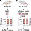
Schematic of the two groups of rats used in this study. (a) Three vaginas from three rats were used only to collect SHG images. (b) Ten vaginas from ten rats were used for inflation testing. Three of the ten vaginas were also used to collect SHG images after inflation testing.
Fig. 4.
Fig. 4.
(a) Pressure and (b) axial load versus normalized tissue volume (NTV) collected up to maximum pressure, Pmax − during inflation tests of rat vaginal tissue specimens (n = 10). Pressure and axial load versus NTV data from the same specimen are represented using the same color.
Fig. 5.
Fig. 5.
Digital image correlation strain data from inflation tests compared by region and normalized tissue volume (NTV). Different colors denote different regions as shown in the insert: orange and red denote small circular regions at tips of the tear closer to the cervix and introitus, respectively, blue and purple denote small circular regions on left and right sides of the mid-region of the tear, respectively, and grey denotes a quadrilateral region enclosing (but not including) the tear. (a) Mean (±std. dev.) Lagrangian hoop strain and (b) mean (±std. dev.) Lagrangian axial strain measured at NTVs of 1, 1.5, 2, and 2.5 across five regions (as shown in the insert) around the tear of rat vaginal tissue specimens (n = 10). For clarity, only statistical differences across regions are reported. (c) Mean (±std. dev.) Lagrangian hoop strain and (d) mean (±std. dev.) Lagrangian axial strain at the initial tear propagation, TPinitial, and the maximum pressure, Pmax, across the same five regions around the tear. Statistical differences across regions and between Pmax and TPinitial are reported. p < 0.05, ∗∗ p < 0.01.
Fig. 6.
Fig. 6.
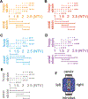
Statistical comparison of hoop strains (dashed lines) and axial strains (continuous lines) between normalized tissue volumes (NTVs) of 1, 1.5, 2, and 2.5 for (a) and (b) small circular regions at tips of the tear closer to the cervix and introitus, respectively, (c) and (d) small circular regions on left and right sides of the mid-region of the tear, respectively, and (e) quadrilateral regions enclosing (but not including) the tear. (f) Regions around the tear, where (hoop and axial) strains were compared between NTVs, are displayed in orange, red, blue, purple, and grey. p < 0.05, ∗∗ p < 0.01, ∗∗∗ p < 0.001.
Fig. 7.
Fig. 7.
Evolution of the pre-imposed, elliptical tear geometry during inflation tests of rat vaginal tissue specimens (n = 10) before tear propagation is observed. (a) Major axis, (b) minor axis, (c) area, and (d) circularity of the pre-imposed, elliptical tears versus normalized tissue volume (NTV). Data from the same specimen are represented using the same color.
Fig. 8.
Fig. 8.
Evolution of tear propagation during inflation tests of rat vaginal tissue specimens (n = 10). (a) Tear propagation toward the cervix (dac), (b) tear propagation toward the introitus (dai), and (c) total tear propagation (dac+dai) versus normalized tissue volume (NTV). (d) Pressure normalized by the maximum pressure achieved, Pmax, versus total tear propagation (dac+dai). Data from the same specimen are represented using the same color.
Fig. 9.
Fig. 9.
Representative SHG images of the collagen fibers within six different regions from (a)–(f) the control (untested) rat vaginal tissue specimens and (g)–(l) the inflation-tested rat vaginal tissue specimens. Data were collected as shown in Fig. 3(a) and (b), respectively.
Fig. 10.
Fig. 10.
Polar histograms of collagen fiber orientations in degrees for the (a)–(f) control (untested) and (g)–(l) inflation-tested rat vaginal tissue specimens in different anatomical regions, with 90° representing the axial direction of the vagina. In each region, radial values represent the frequencies of orientation occurrences (i.e., 0.05 = 5%, 0.075 = 7.5%, 0.1 = 10%, 0.15 = 15% of the total fibers). The average orientation is represented by a solid red line at the center of each polar histogram. Red labels and frame boxes are used for the ventral and dorsal regions and black labels and boxes for the anterior, mid, and posterior regions. Data were collected from 54(= 18 × 3) images from n = 3 rats for the control tissue specimens and from 39(= 13 × 3) images from n = 3 rats for the inflation-tested tissue specimens as shown in Fig. 3(a) and (b), respectively. (A = average orientation and V = circular variance).

References

    1. Samuelsson E, Ladfors L, Lindblom BG, Hagberg H, A prospective observational study on tears during vaginal delivery: occurrences and risk factors, Acta Obstet. Gynecol. Scand. 81 (1) (2002) 44–49. - PubMed
    1. Hopkins LM, Caughey AB, Glidden DV, Laros RK, Racial/ethnic differences in perineal, vaginal and cervical lacerations, Am. J. Obstet. Gynecol 193 (2) (2005) 455–459. - PubMed
    1. Metcalfe A, Tohill S, Williams A, Haldon V, Brown L, Henry L, A pragmatic tool for the measurement of perineal tears, Br. J. Midwifery 10 (7) (2002) 412–417.
    1. Örnö AK, Maršál K, Herbst A, Ultrasonographic anatomy of perineal structures during pregnancy and immediately following obstetric injury, Ultrasound Obstet. Gynecol 32 (4) (2008) 527–534. - PubMed
    1. Fitzpatrick M, O’Herlihy C, Short-term and long-term effects of obstetric anal sphincter injury and their management, Curr. Opin. Obstet. Gynecol 17 (6) (2005) 605–610. - PubMed

Publication types

LinkOut - more resources