. 2021 Jun:127:146-158.
doi: 10.1016/j.actbio.2021.03.060. Epub 2021 Apr 6.

Osteoinductivity and biomechanical assessment of a 3D printed demineralized bone matrix-ceramic composite in a rat spine fusion model

Affiliations

Osteoinductivity and biomechanical assessment of a 3D printed demineralized bone matrix-ceramic composite in a rat spine fusion model

Mark A Plantz et al. Acta Biomater. 2021 Jun.

Abstract

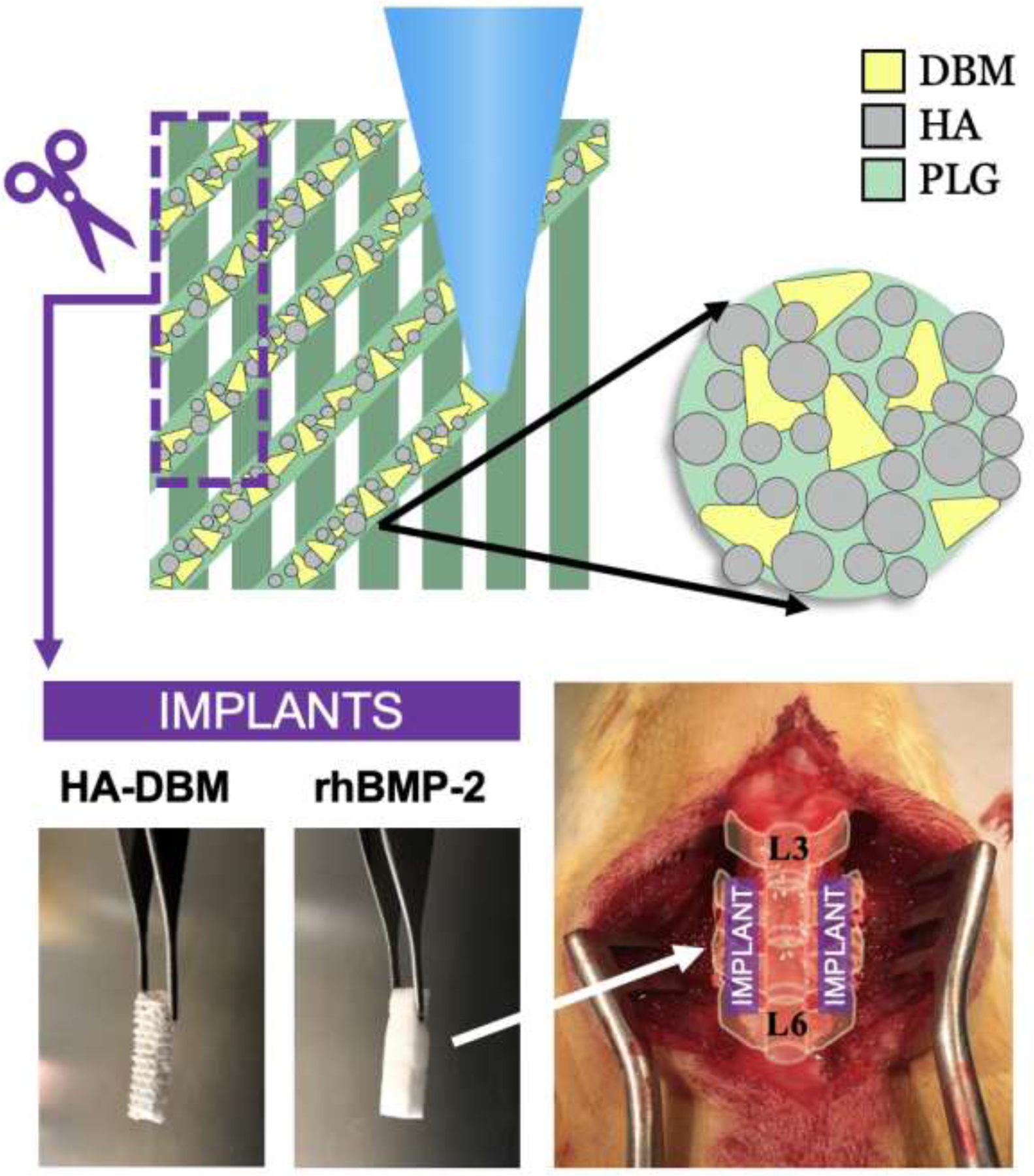
We recently developed a recombinant growth factor-free bone regenerative scaffold composed of stoichiometric hydroxyapatite (HA) ceramic particles and human demineralized bone matrix (DBM) particles (HA-DBM). Here, we performed the first pre-clinical comparative evaluation of HA-DBM relative to the industry standard and established positive control, recombinant human bone morphogenetic protein-2 (rhBMP-2), using a rat posterolateral spinal fusion model (PLF). Female Sprague-Dawley rats underwent bilateral L4-L5 PLF with implantation of the HA-DBM scaffold or rhBMP-2. Fusion was evaluated using radiography and blinded manual palpation, while biomechanical testing quantified the segmental flexion-extension range-of-motion (ROM) and stiffness of the fused segments at 8-weeks postoperatively. For mechanistic studies, pro-osteogenic gene and protein expression at 2-days and 1-, 2-, and 8-weeks postoperatively was assessed with another cohort. Unilateral fusion rates did not differ between the HA-DBM (93%) and rhBMP-2 (100%) groups; however, fusion scores were higher with rhBMP-2 (p = 0.008). Both treatments resulted in significantly reduced segmental ROM (p < 0.001) and greater stiffness (p = 0.009) when compared with non-operated controls; however, the degree of stabilization was significantly higher with rhBMP-2 treatment relative to the HA-DBM scaffold. In the mechanistic studies, PLGA and HA scaffolds were used as negative controls. Both rhBMP-2 and HA-DBM treatments resulted in significant elevations of several osteogenesis-associated genes, including Runx2, Osx, and Alp. The rhBMP-2 treatment led to significantly greater early, mid, and late osteogenic markers, which may be the mechanism in which early clinical complications are seen. The HA-DBM scaffold also induced osteogenic gene expression, but primarily at the 2-week postoperative timepoint. Overall, our findings show promise for this 3D-printed composite as a recombinant growth factor-free bone graft substitute for spinal fusion. STATEMENT OF SIGNIFICANCE: Despite current developments in bone graft technology, there remains a significant void in adequate materials for bone regeneration in clinical applications. Two of the most efficacious bone graft options are the gold-standard iliac crest bone graft and recombinant human-derived bone morphogenetic protein-2 (rhBMP-2), available commercially as Infuse™. Although efficacious, autologous graft is associated with donor-site morbidity, and Infuse™ has known side effects related to its substantial host inflammatory response, possibly associated with a immediate, robust osteoinductive response. Hence, there is a need for a bone graft substitute that provides adequate osteogenesis without associated adverse events. This study represents a significant step in the design of off-the-shelf growth factor-free devices for spine fusion.

Keywords: 3D printing; Bone regeneration; Demineralized bone matrix; Hydroxyapatite; Spine fusion.


Conflict of interest statement

Declaration of Competing Interest M.A.P., S.M., J.G.L., A.G., D.E., M.H., J.Y., S.J., C.Y., K.R.B., R.M.H., M.M., A.G.P., W.K.H., S.R.S., and E.L.H. have nothing to disclose that may create a conflict of interest within this body of work. A.E.J. and R.N.S. are cofounders of the company Dimension Inx, LLC – which aims to create biomaterials, including 3D-printed biologically active materials, that induce tissue regeneration and repair – including some of the materials discussed in this paper. As of October 2020, A.E.J. is the Chief Technology Officer (CTO) and R.N.S. is the Chief Science Officer (CSO) of Dimension Inx, LLC. The trademark for Hyperelastic Bone® is owned by Dimension Inx. The study design, reporting of data, and interpretation of data in this body of work was not influenced by the interests of Dimension Inx LLC.

Figures

Figure 1.
Figure 1.
Materials and experimental model overview. The hydroxyapatite-demineralized bone matrix (HA-DBM) composite is 3D-printed and trimmed to size prior to implantation. The HA-DBM group was compared to recombinant human-derived bone morphogenetic protein-2 (rhBMP-2) delivered on a collagen sponge, known to be highly efficacious in yielding successful fusion. An established L4-L5 posterolateral lumbar fusion (PLF) model in rats was used to compare the efficacy of the HA-DBM to rhBMP-2 in achieving spinal fusion and bone regeneration.
Figure 2.
Figure 2.
Biomechanics Overview. (A) Schematic of the biomechanics testing apparatus from both a posterior and a left lateral view. Samples of explanted L4-L5 vertebrae were loaded with 5 cycles of flexion-extension using a hybrid control algorithm, namely rotation control with a torque limit of 0.01 Nm. (B) Representative load-displacement curve of moment (Nm) versus rotation angle (°). The range-of-motion (ROM) was measured as the difference in rotation angle at the torque limits (−0.01 Nm and 0.01 Nm) of the flexion and extension cycles. The stiffness (K1) was measured as a linear fit to the load-displacement curve from 0 Nm to the first third of the flexion range-of-motion. The red arrows indicate the bounds of the curve to which the linear fit is made. The slope of the red line yields the stiffness (K1). SP = spinous process, TP = transverse process, PMMA = polymethylmethacrylate
Figure 3.
Figure 3.
Comparison of the fusion capacity of rhBMP-2 and HA-DBM groups. (A) Representative radiographs of the lumbar spine at 8 weeks postoperatively for the rhBMP-2 and HA-DBM groups. The fusion area is indicated bilaterally with white arrows. (B) Mean fusion score was significantly greater in the rhBMP-2 group relative to the HA-DBM group. (C) The fusion rate was not significantly different between the rhBMP-2 and HA-DBM groups. * Indicates statistical significance (p<0.05)
Figure 4.
Figure 4.
Histological evaluation of representative spines for each experimental group tested, 2 and 8 weeks post-operatively. Sections are stained with Gill’s hematoxylin and eosin and alcian blue. Acronym legend: transverse process (TP), fusion mass (FM), scaffold struts (ST). Scale bars of large images: 1 mm. Yellow arrows in the 8 weeks rhBMP-2 magnified images indicate trabeculae-like structures. Yellow arrows in the 2 and 8 weeks HA-DBM magnified images indicate DBM particles. The yellow arrow in the 8 weeks HA magnified image indicates remaining HA particles (black). Scale bars of magnified images: 500 µm.
Figure 5.
Figure 5.
Biomechanics assessment of explanted specimens. (A) Representative load-displacement curves of a fused and unfused specimen. The stiffness (K1) – determined by the linear fit of the first third of the flexion cycle for each specimen (indicated by red lines on this plot) – was significantly greater in the fused specimen relative to the unfused specimen. Comparison of the (B) range-of-motion (ROM, °) and (C) stiffness between the non-operated control, rhBMP-2, and HA-DBM groups. The rhBMP-2 group had a significantly lower ROM and greater stiffness than both the HA-DBM and control groups, respectively. Additionally, the HA-DBM had a significantly lower ROM and greater stiffness than the non-operated control group. The first stiffness plot demonstrates the data with an unadjusted y-axis. The second stiffness plot utilizes a y-axis break to illustrate representative differences in stiffness between the three groups. * indicates statistical significance (p<0.05).
Figure 6.
Figure 6.
Gene Expression Studies. A. Expression of select early osteogenic markers in fusion bed tissue 2 days after implantation of 3DP PLGA, 3DP HA, 3DP HA-DBM, or rhBMP-2/ACS scaffolds. B. Heat map representation of the relative expression levels of genes important in osteogenic differentiation in explanted fusion bed tissue taken at 1 and 2 weeks post-operative. Light green signifies significantly elevated expression between 2–5-fold higher than PLGA, and dark green signifies >5 fold-higher expression than the PLGA-treated control group. C. Line graphs depicting the relative expression levels of several key early (Runx2, Osx), early/mid (Alp, Col1a), and late osteogenic markers (Opn, Ocn). Statistical significance (p<0.05) is indicated for HA (#), rhBMP-2 (^) and HA-DBM (*), relative to PLGA control explants.
Figure 7.
Figure 7.
Western Blot Analysis. A. Western blot visualization and B. Quantified protein expression of markers of interest– DLX5, RUNX2, OSX, OPN, and PHEX – for PLGA, HA, HA-DBM, and rhBMP-2 treatment groups at 2-weeks postoperatively. * indicates statistical significance relative to PLGA.

References

    1. Minardi S, Taraballi F, Cabrera F, Van Eps J, Wang X, Gazze S, Fernandez-Mourev JS, Tampieri A, Francis L, Weiner B, Biomimetic hydroxyapatite/collagen composite drives bone niche recapitulation in a rabbit orthotopic model, Materials Today Bio 2 (2019) 100005. - PMC - PubMed
    1. Dimar JR 2nd, Glassman SD, Burkus JK, Pryor PW, Hardacker JW, Carreon LY, Two-year fusion and clinical outcomes in 224 patients treated with a single-level instrumented posterolateral fusion with iliac crest bone graft, Spine J 9(11) (2009) 880–5. - PubMed
    1. Kadam A, Millhouse PW, Kepler CK, Radcliff KE, Fehlings MG, Janssen ME, Sasso RC, Benedict JJ, Vaccaro AR, Bone substitutes and expanders in Spine Surgery: A review of their fusion efficacies, Int J Spine Surg 10 (2016) 33–33. - PMC - PubMed
    1. Tannoury CA, An HS, Complications with the use of bone morphogenetic protein 2 (BMP-2) in spine surgery, The spine journal : official journal of the North American Spine Society 14(3) (2014) 552–9. - PubMed
    1. James AW, LaChaud G, Shen J, Asatrian G, Nguyen V, Zhang X, Ting K, Soo C, A Review of the Clinical Side Effects of Bone Morphogenetic Protein-2, Tissue Eng Part B Rev 22(4) (2016) 284–297. - PMC - PubMed

Publication types

Substances

LinkOut - more resources