. 2021 Apr 8;21(1):186.
doi: 10.1186/s12893-021-01185-4.

Outcomes of surgery for giant hepatic hemangioma

Affiliations

Outcomes of surgery for giant hepatic hemangioma

Qing-Song Xie et al. BMC Surg. .

Abstract

Background: The surgical indications for liver hemangioma remain unclear.

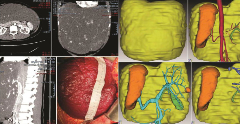
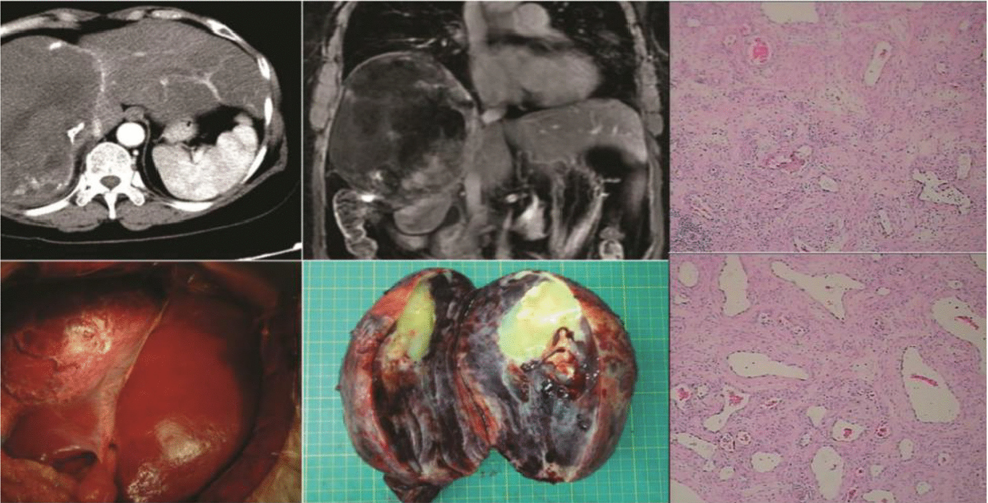
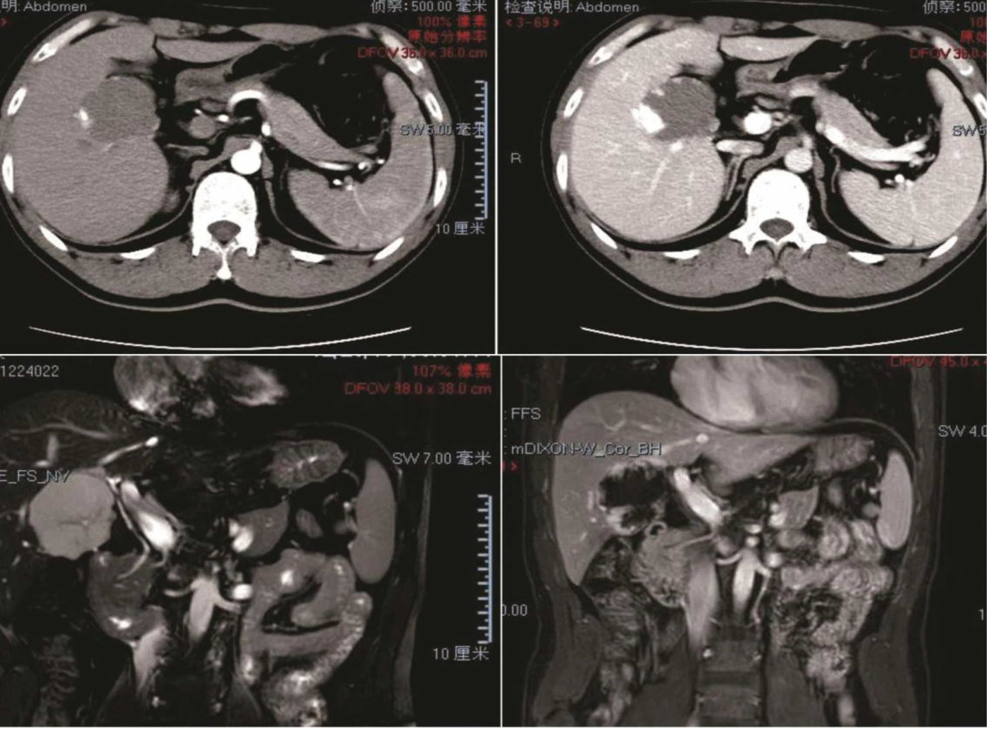
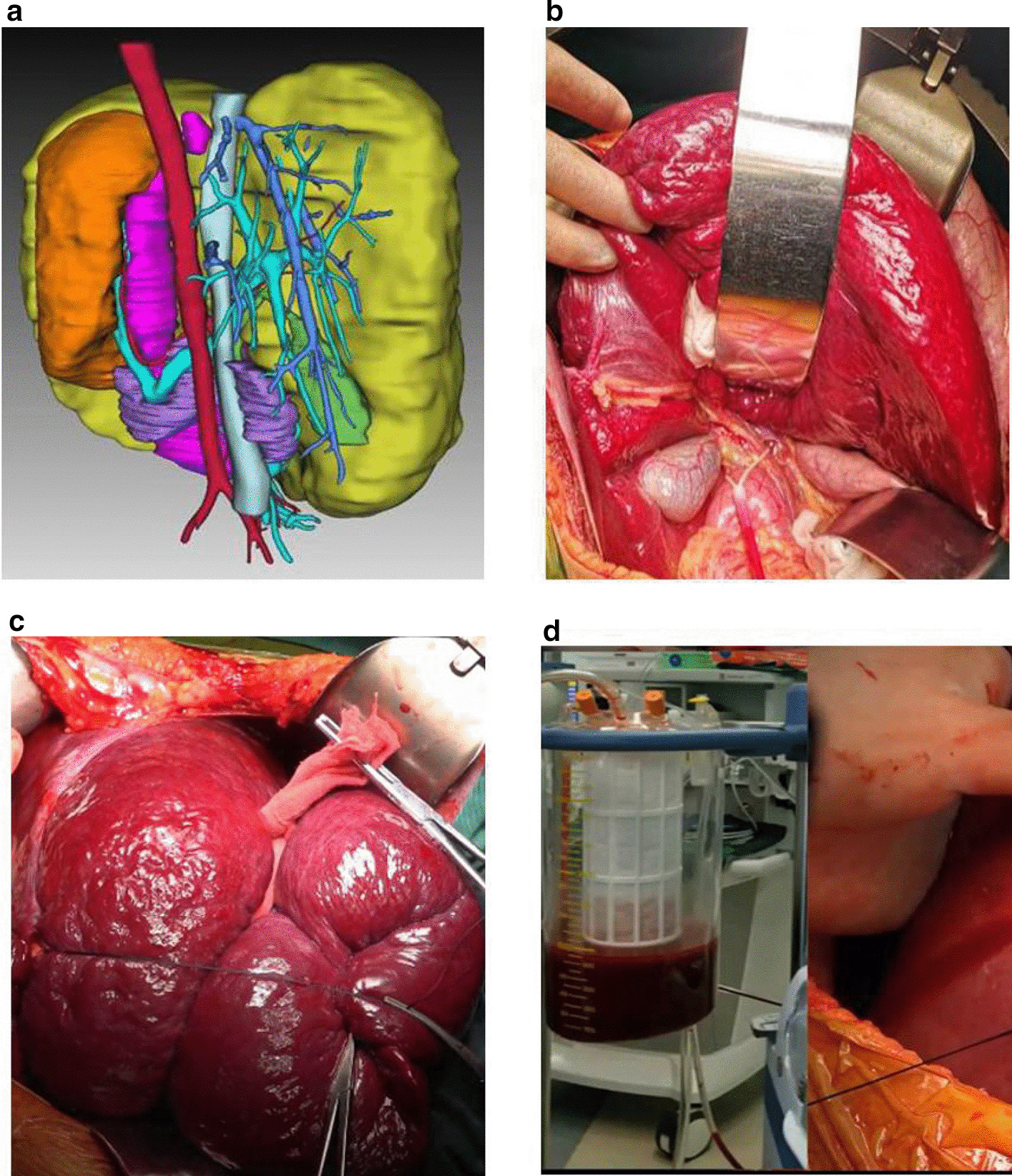
Methods: Data from 152 patients with hepatic hemangioma who underwent hepatectomy between 2004 and 2019 were retrospectively reviewed. We analyzed characteristics including tumor size, surgical parameters, and variables associated with Kasabach-Merritt syndrome and compared the outcomes of laparoscopic and open hepatectomy. Here, we describe surgical techniques for giant hepatic hemangioma and report on two meaningful cases.

Results: Most (63.8%) patients with hepatic hemangioma were asymptomatic. Most (86.4%) tumors from patients with Kasabach-Merritt syndrome were larger than 15 cm. Enucleation (30.9%), sectionectomy (28.9%), hemihepatectomy (25.7%), and the removal of more than half of the liver (14.5%) were performed through open (87.5%) and laparoscopic (12.5%) approaches. Laparoscopic hepatectomy is associated with an operative time, estimated blood loss, and major morbidity and mortality rate similar to those of open hepatectomy, but a shorter length of stay. 3D image reconstruction is an alternative for diagnosis and surgical planning for partial hepatectomy.

Conclusion: The main indication for surgery is giant (> 10 cm) liver hemangioma, with or without symptoms. Laparoscopic hepatectomy was an effective option for hepatic hemangioma treatment. For extremely giant hemangiomas, 3D image reconstruction was indispensable. Hepatectomy should be performed by experienced hepatic surgeons.

Keywords: 3D image reconstruction; Giant; Hepatic hemangioma; Surgical indication; Surgical management; Surgical technique.


Conflict of interest statement

The authors declare no competing interests.

Figures

Fig. 1
Fig. 1
A 24-year-old woman with two children was diagnosed with an extremely giant hepatic hemangioma in the left lobe of her liver after reporting abdominal pain. The tumor, extending below the belly button, had a maximum diameter of 38 cm. The 3D-reconstructed image shows the tumor (yellow), normal liver (orange), and the relationships between the tumor and hepatic vessels. The patient also had Kasabach–Merritt syndrome. The number of platelets increased from 57 × 109/l to 100.5 × 109/l after surgery
Fig. 2
Fig. 2
A 62-year-old female patient with a giant hemangioma in the right lobe of the liver was misdiagnosed with hepatocellular carcinoma. In 2006, the patient underwent physical examination, which revealed a liver mass. The patient was diagnosed with hepatocellular carcinoma and underwent five rounds of transhepatic arterial chemotherapy and embolization and multiple cytokine-induced killer cell treatments at another hospital. In December 2015, the patient was admitted to our hospital and we determined that she was not infected with hepatitis b virus and was α-fetoprotein negative. The CT image shows a large mass in the right lobe of the liver. The patient’s liver reserve function was normal. She underwent extensive right hepatectomy and was given a final pathological diagnosis of hepatic hemangioma
Fig. 3
Fig. 3
A 45-year-old male patient, a seaman, had symptoms of obstructive jaundice and was diagnosed with a hepatic hemangioma about 5 cm in diameter near the first porta hepatis. CT and MRI images show the hepatic hemangioma at the first porta hepatis, compressing the intrahepatic bile duct, which caused obstructive jaundice. We enucleated the tumor and the patient’s bilirubin level returned to normal
Fig. 4
Fig. 4
Methods for the prevention of intraoperative bleeding. a 3D image reconstruction; b Prearrangement of a hepatic portal blocking band; c Bandaging of the tumor; d Autologous blood transfusion

References

    1. Choi BY, Nguyen MH. The diagnosis and management of benign hepatic tumors. J Clin Gastroenterol. 2005;39(5):401–412. doi: 10.1097/01.mcg.0000159226.63037.a2. - DOI - PubMed
    1. Adam YG, Huvos AG, Fortner JG. Giant hemangiomas of the liver. Ann Surg. 1970;172(2):239–245. doi: 10.1097/00000658-197008000-00010. - DOI - PMC - PubMed
    1. Lopez-Arce Angeles G, Barahona-Garrido J, Tellez-Avila FI, et al. A giant hepatic hemangioma treated successfully with hepatic enucleation. Ann Hepatol. 2009;8(4):377–378. doi: 10.1016/S1665-2681(19)31753-3. - DOI - PubMed
    1. Gandolfi L, Leo P, Solmi L, Vitelli E, Verros G, Colecchia A. Natural history of hepatic haemangiomas: clinical and ultrasound study. Gut. 1991;32:677–680. doi: 10.1136/gut.32.6.677. - DOI - PMC - PubMed
    1. Pietrabissa A, Giulianotti P, Campatelli A, et al. Management and follow-up of 78 giant haemangiomas of the liver. Br J Surg. 1996;83:915–918. doi: 10.1002/bjs.1800830710. - DOI - PubMed