Case Reports
. 2021 Apr 8;16(1):29.
doi: 10.1186/s13000-021-01086-3.

Whole-exome sequencing reveals the etiology of the rare primary hepatic mucoepidermoid carcinoma

Affiliations
Case Reports

Whole-exome sequencing reveals the etiology of the rare primary hepatic mucoepidermoid carcinoma

Ping Hou et al. Diagn Pathol. .

Abstract

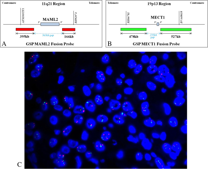
Background: Primary hepatic mucoepidermoid carcinoma (HMEC) is extremely rare and the molecular etiology is still unknown. The CRTC1-MAML2 fusion gene was previously detected in a primary HMEC, which is often associated with MEC of salivary gland in the literature.

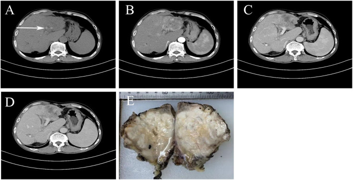
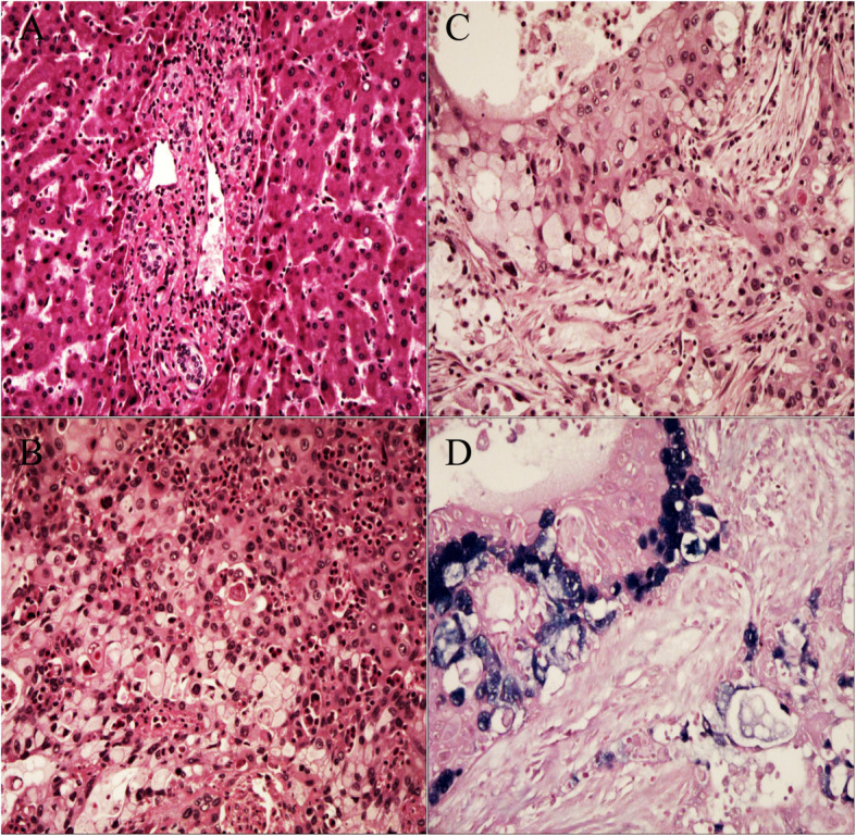
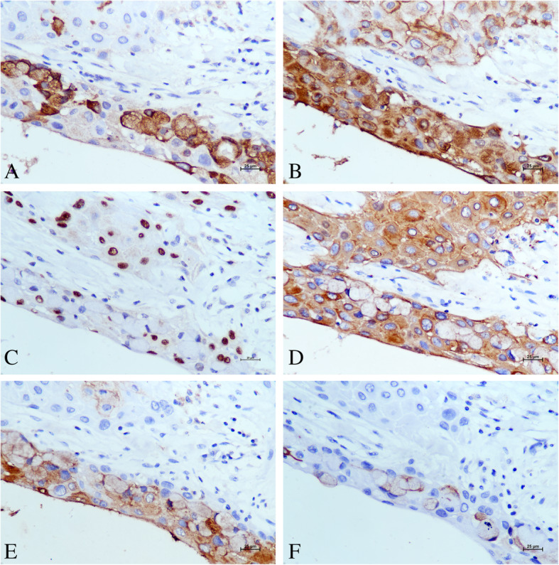
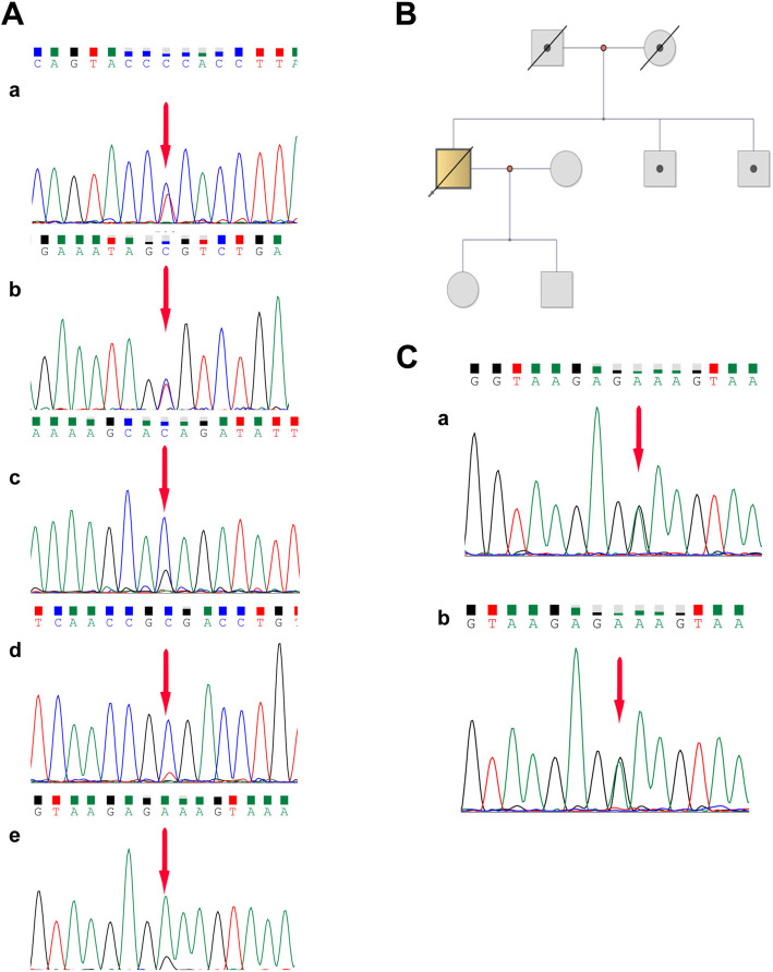
Methods: A 64-year-old male was diagnosed with HMEC based on malignant squamous cells and mucus-secreting cells in immunohistochemical examination. Fluorescence in situ hybridization (FISH) was used to detect the CRTC1-MAML2 fusion gene in HMEC. Whole-exome sequencing and Sanger sequencing were used to reveal the molecular characteristics of HMEC and analysis was performed with public data. Pedigree investigation was performed to identify susceptibility genes.

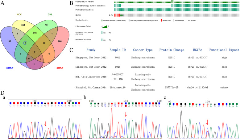
Results: Hematoxylin-eosin staining and immunohistochemistry revealed that the tumor cells were composed of malignant epidermoid malignant cells and mucous cells, indicating a diagnosis of HMEC. The CRTC1-MAML2 fusion gene was not detected in the primary HMEC, and somatic mutations in GNAS, KMT2C and ELF3 genes were identified by sequencing. Analyses of public data revealed somatic GNAS alterations in 2.1% hepatobiliary tumors and relation with parasite infection. Heterozygous germline mutations of FANCA, FANCI, FANCJ/BRIP1 and FAN1 genes were also identified. Pedigree investigation verified that mutation of Fanconi's anemia susceptibility genes were present in the pedigree.

Conclusions: Here we provide the first evidence of the molecular etiology of a rare HMEC associated with germline Fanconi's anemia gene mutations and somatic GNAS R201H mutation.

Keywords: Germline Fanconi’s anemia mutation; Hepatic mucoepidermoid carcinoma (HMEC); Somatic GNAS R201 mutation; Whole exome-sequencing (WES).


Conflict of interest statement

The authors declare no conflict of interests. The authors declare no competing financial interest.

Figures

Fig. 1
Fig. 1
Results of preoperative CT, postoperative gross examination. a Abdominal non-contrast CT scan revealed a low density mass measuring approximate 10 cm in diameter at the left hepatic lobe and intrahepatic bile ducts with multiple stones (white arrow). b Enhanced CT scan (arterial phase) revealed a heterogeneous enhanced mass in the left lobe of the liver. c Enhanced CT scan (portal phase) revealed a persistent inhomogeneous enhanced mass and formation of tumor thrombus in the left branch of portal vein. d Enhanced CT scan (delayed phase) revealed a persistent inhomogeneous enhanced mass. e Gross examination of the resected liver mass indicated an irregular, white solid tumor without a fibrous capsule. The border between the tumor and normal liver tissue was indistinct
Fig. 2
Fig. 2
Pathological results. a Dystrophic arteries and a large number of inflammatory cells, mainly including lymphocyte infiltrations, were observed in the portal area (hematoxylin and eosin (H&E) staining, magnification 100×). b Eosinophil infiltrations were seen in the tumor area (H&E staining, magnification 200×). c H&E staining revealed malignant epidermoid cells and mucus-producing cells with intracytoplasmic mucin. The tumor cells infiltrated into the stroma with occasional keratinization (magnification 200×). d Alcian blue stain-positive material highlights the mucin in mucin-producing cells (magnification 200×)
Fig. 3
Fig. 3
Immunohistochemical results. a Immunopositivity for cytoplasmic MUC5AC was detected in malignant mucinous cells, with negative staining in malignant epidermoid cells (magnification 400×). b Immunopositivity for membrane MUC1 was detected in all malignant epidermoid cells and mucinous cells (magnification 400×). c Immunopositivity for p63 was only detected in the nuclei of malignant epidermoid cells (magnification 400×). d Cytoplasmic CK19 staining was diffusely positive in the malignant epidermoid and mucous component of the tumor (magnification 400×). e Cytoplasmic CK7 staining was focally positive in the malignant epidermoid and mucous component of the tumor (magnification 400×). f Mucous component of the tumor was focally positive for membrane CEA staining (magnification 400×)
Fig. 4
Fig. 4
FISH analysis for the CRTC1/MECT1-MAML2 fusion gene. Probe pattern diagram: GSP CRTC1/MECT1 green fusion probe, GSP MAML2 red fusion probe (top). Non-typical positive signal mode, CRTC1/MECT1-MAML2 fusion gene was negative (bottom)
Fig. 5
Fig. 5
Public data analysis and significant somatic mutation in the proband. (Mutation diagram circles are colored with respect to the corresponding mutation types. In the case of different mutation types at a single position, the color of the circle reflects the most frequent mutation type). A. Venn diagram: six SNVs (in STAT1, TGFBR1, NOTCH1, KMT2C, ELF3 and GNAS genes) in the HMEC overlapped with primary liver tumors (HCC and CHL). Only a SNV in the CHD3 gene in HMEC overlapped with SMEC. B. Somatic GNAS gene mutation occurred in 2.1% (9/445) patients with primary hepatobiliary tumors in public databases. C. Three patients showed GNAS (p.R201H/C) missense mutation (putative driver). Samples (ID W012,T026) were O.pisthorchis viverrini-associated with cholangiocarcinoma (SRP007970)(pentagonal shape). D. Somatic mutations in the proband by Sanger sequencing: a. missense variant in GNAS (chr20.exon8:c.G602A:p.R201H), b. frameshift indel variant in ELF3 (chr1,exon4:c.909dupC:p.F303fs), and c. nonsense variant in KMT2C (chr7,exon4:c.C1519T:p.Q507X) in tumor tissue
Fig. 6
Fig. 6
Germline heterozygous mutations in the proband and pedigree screening. A. a. FANCI (chr15.exon4:c.C257T:p.A86V); b. FANCI (chr15.exon22:c.G2225C:p.C742S); c. FANCJ/BRIP1 (chr17.exon19:c.T2755C:p.S919P), d. FANCA (chr16.exon9:c.A796G:p.T266A); e. FAN1 (chr15.exon2:c.G 698A:p.G233E) (corresponding non-tumor liver tissue). B. Pedigree map: the proband’s parents died from cardiovascular and cerebrovascular diseases; there was no obvious Fanconi’s anemia disease in the proband’s offspring and brothers. C. Siblings A and B, FAN1 (exon2:c.G698A:p.G233E) (blood)

Similar articles

Cited by

References

    1. Nallacheruvu Y, Gaur K, Sakhuja P, Agarwal AK, Srivastava S. Mucoepidermoid carcinoma of the gallbladder: a case-based study of an extremely rare tumor highlighting the role of immunohistochemical profiling. Int J Surg Pathol. 2019;27(4):418–422. doi: 10.1177/1066896918821436. - DOI - PubMed
    1. Tonon G, Modi S, Wu L, Kubo A, Coxon AB, Komiya T, O’Neil K, Stover K, el-Naggar A, Griffin JD, Kirsch IR, Kaye FJ. t(11;19)(q21;p13) translocation in mucoepidermoid carcinoma creates a novel fusion product that disrupts a Notch signaling pathway. Nat Genet. 2003;33(2):208–213. doi: 10.1038/ng1083. - DOI - PubMed
    1. Lennerz JK, Perry A, Mills JC, Huettner PC, Pfeifer JD. Mucoepidermoid carcinoma of the cervix: another tumor with the t(11;19)-associated CRTC1-MAML2 gene fusion. Am J Surg Pathol. 2009;33(6):835–843. doi: 10.1097/PAS.0b013e318190cf5b. - DOI - PubMed
    1. Achcar RDOD, Nikiforova MN, Dacic S, Nicholson AG, Yousem SA. Mammalian mastermind like 2 11q21 gene rearrangement in bronchopulmonary mucoepidermoid carcinoma. Hum Pathol. 2009;40(6):854–860. doi: 10.1016/j.humpath.2008.11.007. - DOI - PubMed
    1. Saeki K, Ohishi Y, Matsuda R, Mochidome N, Miyasaka Y, Yamamoto H, Koga Y, Maehara Y, Nakamura M, Oda Y. “Pancreatic mucoepidermoid carcinoma” is not a pancreatic counterpart of CRTC1/3-MAML2 fusion gene-related mucoepidermoid carcinoma of the salivary gland, and may more appropriately be termed pancreatic adenosquamous carcinoma with mucoepidermoid carcinoma-like features. Am J Surg Pathol. 2018;42(11):1419–1428. doi: 10.1097/PAS.0000000000001135. - DOI - PubMed

Publication types

MeSH terms