

Identification of novel HLA-restricted preferentially expressed antigen in melanoma peptides to facilitate off-the-shelf tumor-associated antigen-specific T-cell therapies
- PMID: 33832817
- PMCID: PMC8316284
- DOI: 10.1016/j.jcyt.2021.03.001
Identification of novel HLA-restricted preferentially expressed antigen in melanoma peptides to facilitate off-the-shelf tumor-associated antigen-specific T-cell therapies
Abstract
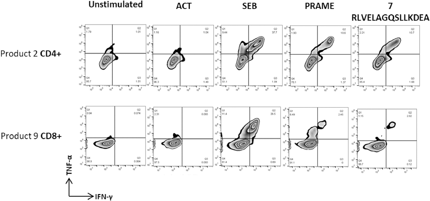
Background aims: Preferentially expressed antigen in melanoma (PRAME) is a cancer/testis antigen that is overexpressed in many human malignancies and poorly expressed or absent in healthy tissues, making it a good target for anti-cancer immunotherapy. Development of an effective off-the-shelf adoptive T-cell therapy for patients with relapsed or refractory solid tumors and hematological malignancies expressing PRAME antigen requires the identification of major histocompatibility complex (MHC) class I and II PRAME antigens recognized by the tumor-associated antigen (TAA) T-cell product. The authors therefore set out to extend the repertoire of HLA-restricted PRAME peptide epitopes beyond the few already characterized.
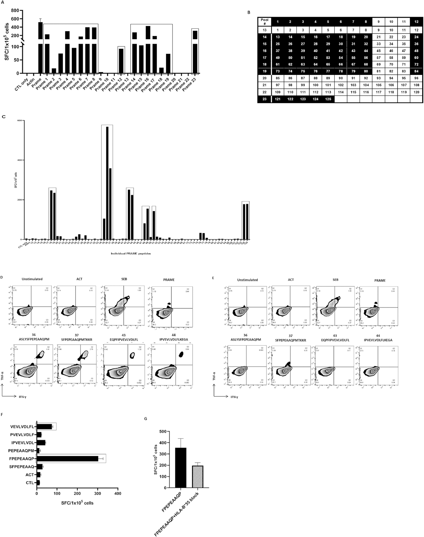
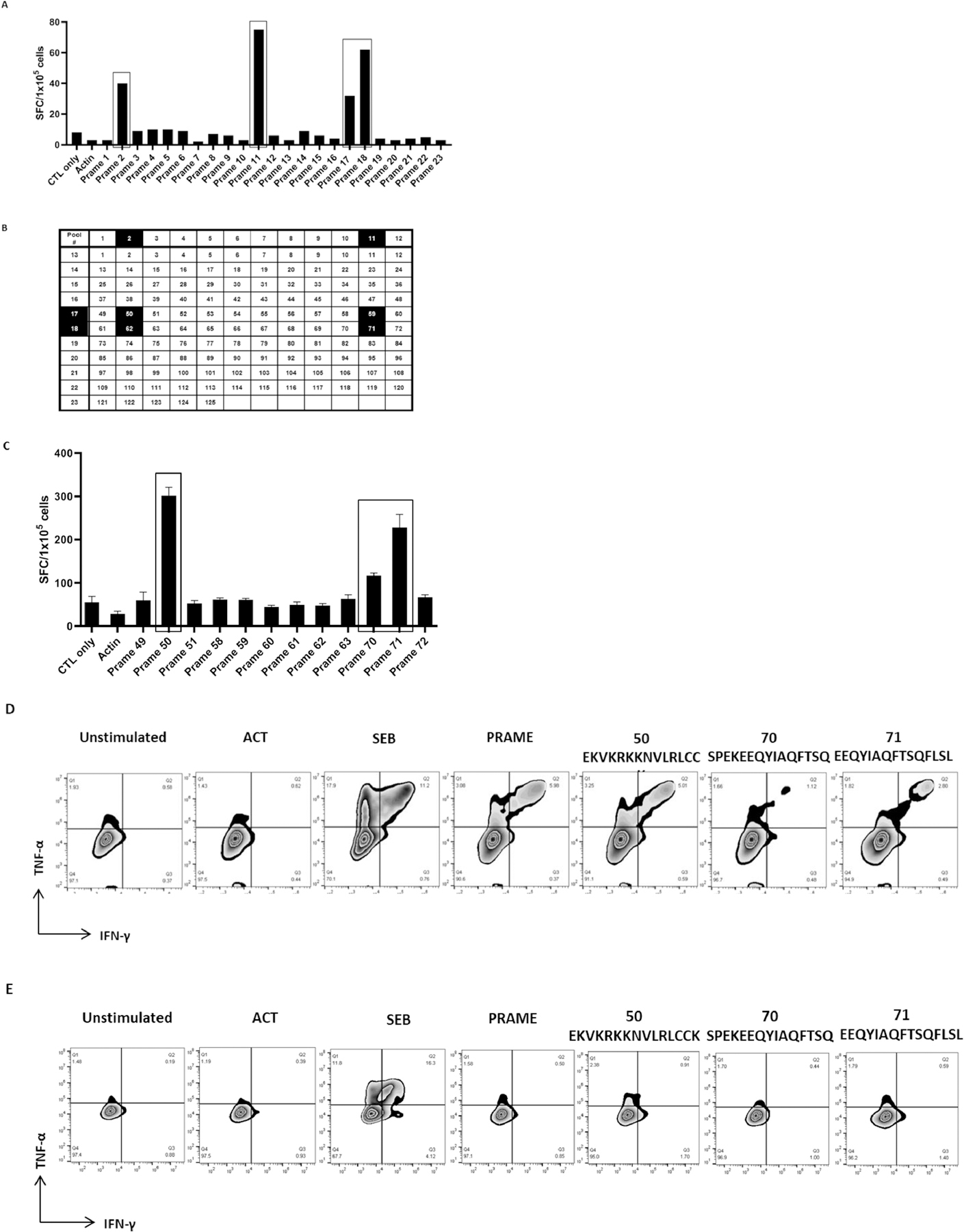
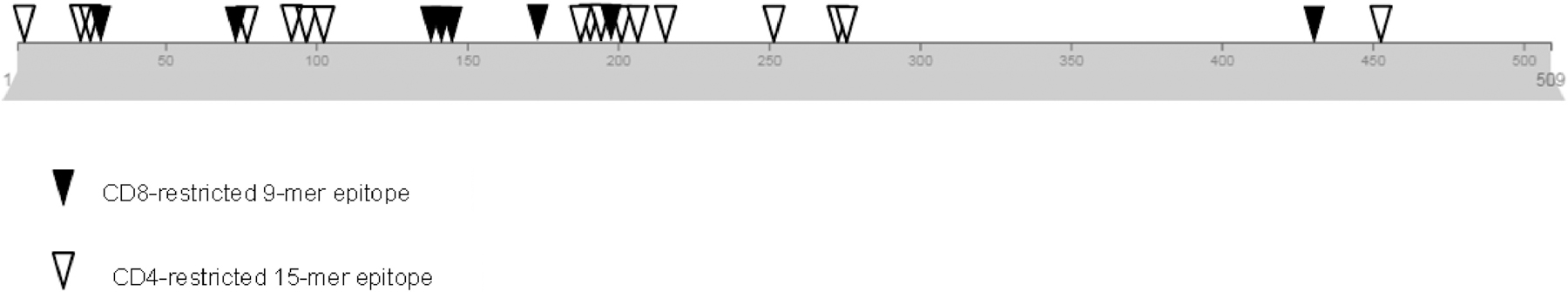
Methods: Peptide libraries of 125 overlapping 15-mer peptides spanning the entire PRAME protein sequence were used to identify HLA class I- and II-restricted epitopes. The authors also determined the HLA restriction of the identified epitopes.
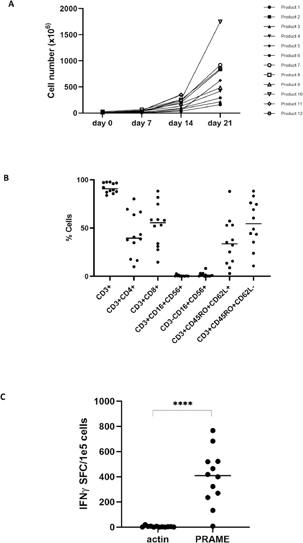
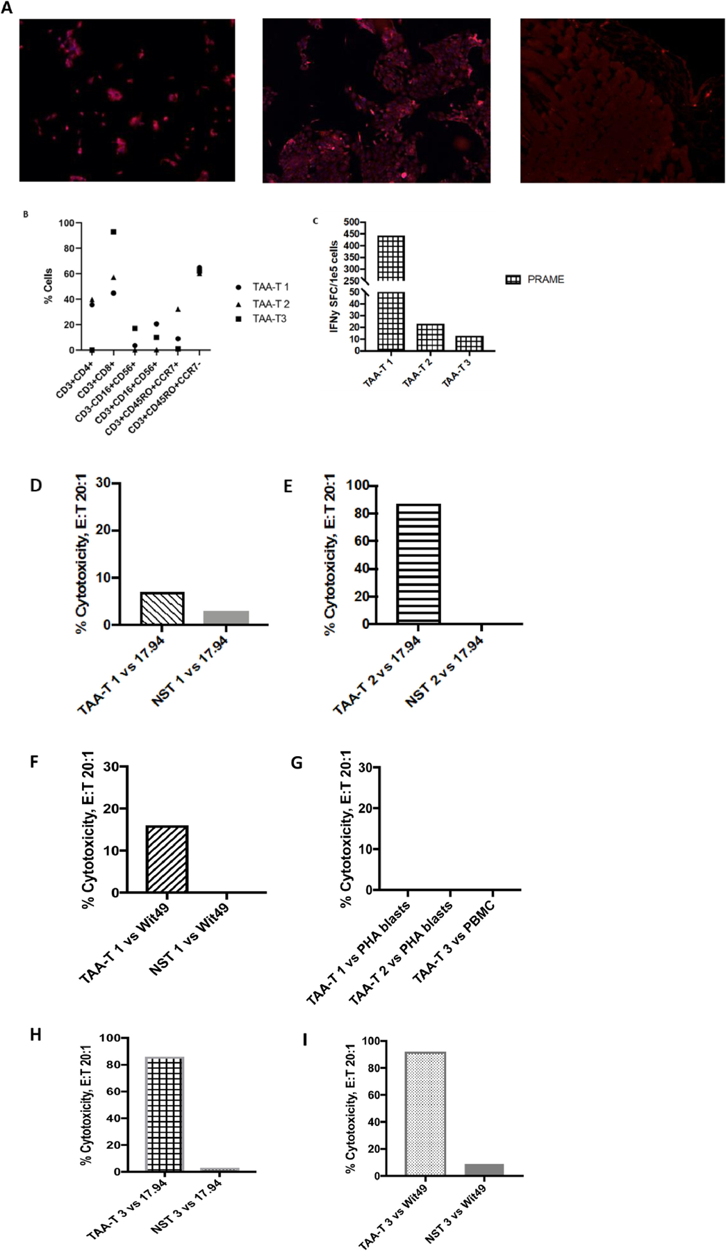
Results: PRAME-specific T-cell products were successfully generated from peripheral blood mononuclear cells of 12 healthy donors. Ex vivo-expanded T cells were polyclonal, consisting of both CD4+ and CD8+ T cells, which elicited anti-tumor activity in vitro. Nine MHC class I-restricted PRAME epitopes were identified (seven novel and two previously described). The authors also characterized 16 individual 15-mer peptide sequences confirmed as CD4-restricted epitopes.
Conclusions: TAA T cells derived from healthy donors recognize a broad range of CD4+ and CD8+ HLA-restricted PRAME epitopes, which could be used to select suitable donors for generating off-the-shelf TAA-specific T cells.
Keywords: PRAME; T-cell epitope; cancer immunotherapy; off-the-shelf T-cell therapy.
Copyright © 2021 International Society for Cell & Gene Therapy. Published by Elsevier Inc. All rights reserved.
Conflict of interest statement
Declaration of Competing Interest CMB is on the advisory board of Cellectis and the scientific advisory boards of Catamaran Bio and Mana Therapeutics, with stock and/or ownership. CMB is also on the board of directors of Cabaletta Bio, with stock options, and has stock in Neximmune and Torque Therapeutics. PJH and CRYC are co-founders of Mana Therapeutics, and PJH is on the board of directors of Mana Therapeutics. PJH is also on the scientific advisory board of Cellevolve. MDK is on the scientific advisory panel of Gilead Sciences. CMB, CRYC, PJH and MS have filed a patent application based on the findings in this article.
Figures
References
-
- Boon K, et al. , Comparison of medulloblastoma and normal neural transcriptomes identifies a restricted set of activated genes. Oncogene, 2003. 22(48): p. 7687–7694. - PubMed
-
- Ikeda H, et al. , Characterization of an antigen that is recognized on a melanoma showing partial HLA loss by CTL expressing an NK inhibitory receptor. Immunity, 1997. 6(2): p. 199–208. - PubMed
-
- Oberthuer A, et al. , The tumor-associated antigen PRAME is universally expressed in high-stage neuroblastoma and associated with poor outcome. Clinical Cancer Research, 2004. 10(13): p. 4307–4313. - PubMed
-
- Steinbach D, et al. , PRAME gene expression in childhood acute lymphoblastic leukemia. Cancer Genetics and Cytogenetics, 2002. 138(1): p. 89–91. - PubMed
Publication types
MeSH terms
Substances
Grants and funding
LinkOut - more resources
Full Text Sources
Other Literature Sources
Medical
Molecular Biology Databases
Research Materials
Miscellaneous
