Review
. 2021 Sep;23(9):757-773.
doi: 10.1016/j.jcyt.2021.02.005. Epub 2021 Apr 6.

Options for imaging cellular therapeutics in vivo: a multi-stakeholder perspective

Affiliations
Review

Options for imaging cellular therapeutics in vivo: a multi-stakeholder perspective

Brooke M Helfer et al. Cytotherapy. 2021 Sep.

Abstract

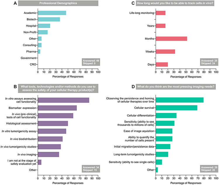
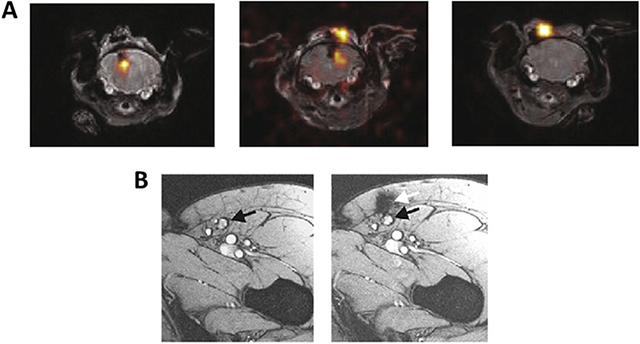
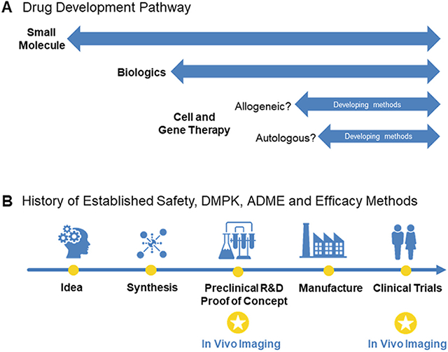
Cell-based therapies have been making great advances toward clinical reality. Despite the increase in trial activity, few therapies have successfully navigated late-phase clinical trials and received market authorization. One possible explanation for this is that additional tools and technologies to enable their development have only recently become available. To support the safety evaluation of cell therapies, the Health and Environmental Sciences Institute Cell Therapy-Tracking, Circulation and Safety Committee, a multisector collaborative committee, polled the attendees of the 2017 International Society for Cell & Gene Therapy conference in London, UK, to understand the gaps and needs that cell therapy developers have encountered regarding safety evaluations in vivo. The goal of the survey was to collect information to inform stakeholders of areas of interest that can help ensure the safe use of cellular therapeutics in the clinic. This review is a response to the cellular imaging interests of those respondents. The authors offer a brief overview of available technologies and then highlight the areas of interest from the survey by describing how imaging technologies can meet those needs. The areas of interest include imaging of cells over time, sensitivity of imaging modalities, ability to quantify cells, imaging cellular survival and differentiation and safety concerns around adding imaging agents to cellular therapy protocols. The Health and Environmental Sciences Institute Cell Therapy-Tracking, Circulation and Safety Committee believes that the ability to understand therapeutic cell fate is vital for determining and understanding cell therapy efficacy and safety and offers this review to aid in those needs. An aim of this article is to share the available imaging technologies with the cell therapy community to demonstrate how these technologies can accomplish unmet needs throughout the translational process and strengthen the understanding of cellular therapeutics.

Keywords: biodistribution; cell therapy; fate; in vivo tracking; non-invasive; safety.


Figures

Figure 1.
Figure 1.
Results of the 2017 CT-TRACS stakeholder survey (“Assessing the needs for cell-based therapies translation”), administered at the 2017 ISCT Annual Meeting. CRO, contract research organization; ISCT, International Society for Cell & Gene Therapy; Pharma, pharmaceutical.
Figure 2.
Figure 2.
PET/CT images of a 65-year-old man with a history of anterior wall infarction. After percutaneous intervention, 18F-FDG-labeled stem cells were injected via an intracoronary catheter. PET/CT images were obtained 2 h after injection. Stem cell accumulation at the myocardium is well visualized (arrow). The total amount of stem cells at the myocardium was 2.1% of the injected dose. Reproduced with permission from [35] © Society of Nuclear Medicine and Molecular Imaging (2006).
Figure 3.
Figure 3.
HSV1-tk was fused in-frame with the hygromycin phosphotransferase gene and expressed as a fusion protein. Through imaging of HSV1-tk reporter gene expression using [18F]FHBG, which is an 18F-radiolabeled analog of the anti-herpes drug penciclovir, [18F]FHBG trapping in the brain tumor could be imaged, which corresponds to CTL accumulation. The [18F]FHBG PET imaging was performed in a patient with a recurrent right frontoparietal glioblastoma (A) before and (B) 1 week after tumor-specific HSV1tk-transduced CAR T-cell infusions. Allogeneic CAR T cells and IL-2 were injected intratumorally (red arrows). Tumor recurrence was monitored by T1W MRI (top panels). The [18F]FHBG PET images were fused with MR images (bottom panels), and 3D volumes of interest were drawn using a 50% [18F]FHBG SUVmax threshold (outlined in yellow). (C) Voxel-wise analysis of[18F]FHBG SUV in pre- and post-CTL infusion scans. Reproduced with permission from [111] © American Association for the Advancement of Science (2017). CTL, cytotoxic T lymphocyte; [18F]FHBG, 9-[4-[18F]fluoro-3-(hydroxymethyl)butyl] guanine; HSV1-tk, herpes simplex virus 1-thymidine kinase; SUV, standard uptake value; 3D, three-dimensional; T1W, T1-weighted.
Figure 4.
Figure 4.
Serial imaging of ferumoxytol-labeled bone marrow cells. (A) T1W, T2W and x-ray images of the right femur show osteonecrosis (arrows) in the femoral epiphysis with typical fat-equivalent center and serpiginous borders. (B) At 24 h after intravenous injection of ferumoxytol, the normal bone marrow in the iliac crest shows hypointense (dark) enhancement (asterisks) on T2-weighted MR images. (C) One week after core decompression and injection of iron-labeled bone marrow cells, a hypointense (dark) signal is noted in the decompression track (arrows), consistent with delivery of iron-labeled cells. (D,E) MRI follow-up at 4 weeks and 24 weeks after core decompression and transplantation of labeled cells shows decline in iron signal over time. The femoral epiphysis did not collapse during this 6-month follow-up period. Reproduced with permission from [63] © American Association for Cancer Research (2018). STIR, short tau inversion recovery; T1W, T1-weighted; T2W, T2-weighted.
Box 1.
Box 1.
Administration and misadministration. (A) 19F-labeled NK cells were injected intracranially to treat medulloblastoma. Images demonstrate successful administration, partial success and delivery failure of labeled NK cells (left to right respectively). Reproduced with permission from [93] © Springer Nature (2019). (B) Imaging the administration of dendritic cells. MRI before dendritic cell vaccination (left), with the inguinal lymph node to be injected, with cells identified by black arrow. MRI after injection (right) showing that dendritic cells were not accurately delivered into the lymph node (black arrow) but into the subcutaneous fat adjacent to the lymph node (white arrow). Reproduced with permission from [52] © Springer Nature (2015). NK, natural killer.
Box 2.
Box 2.
Can imaging add knowledge and experience to the development of cellular therapeutics? (A) In the discovery of small molecules, there are decades of experience and knowledge to draw upon, and these data have shaped the drug discovery pathway, from the idea for a therapeutic target to a licensed product. Many of the in vitro and pre-clinical tests to determine drug safety are also applicable to large molecules and biologics; however, there are fewer established methods for the field of cellular therapies. Arguably, some pre-clinical testing may be relevant, but options are limited for autologous and allogeneic cellular therapies. By and large, the established tools for safety assessment do not transfer well to cellular therapies, but in vivo imaging can play a major part in enabling the gathering of biodistribution data. This will be essential as the field moves to treating solid tumors, where evidence to prove that the cells reach their target will be required, and it is difficult to apply pre-clinical animal data to meet this need for cellular therapeutics. (B) In the development of cellular therapeutics, imaging can help proof-of-concept studies demonstrate mechanism of action. Additionally, imaging stands to play a large role in safety and efficacy in clinical trials. These approaches may help optimize delivery dose and route of administration, demonstrate efficacy of delivery or migration, show how/why some people respond better than others (i.e., responders versus non-responders) and lay the groundwork for co-therapeutics. ADME, absorption, distribution, metabolism and excretion; DMPK, drug metabolism and pharmacokinetics.

References

    1. European Medicines Agency. Advanced therapy medicinal products: overview. https://www.ema.europa.eu/en/human-regulatory/overview/advanced-therapy-...; 2020. [accessed 20 March 2020].
    1. Boyiadzis MM, Dhodapkar MV, Brentjens RJ, Kochenderfer JN, Neelapu SS, Maus MV, et al. Chimeric antigen receptor (CAR) T therapies for the treatment of hematologic malignancies: clinical perspective and significance. J Immunother Cancer 2018;6:137. - PMC - PubMed
    1. June CH, O'Connor RS, Kawalekar OU, Ghassemi S, Milone MC. CAR T cell immunotherapy for human cancer. Science 2018;359:1361–5. - PubMed
    1. Maude SL, Laetsch TW, Buechner J, Rives S, Boyer M, Bittencourt H, et al. Tisagenlecleucel in children and young adults with B-cell lymphoblastic leukemia. N Engl J Med 2018;378:439–48. - PMC - PubMed
    1. Kochenderfer JN, Rosenberg SA. Treating B-cell cancer with T cells expressing anti-CD19 chimeric antigen receptors. Nat Rev Clin Oncol 2013;10:267–76. - PMC - PubMed

Publication types

LinkOut - more resources