. 2021 Apr 13;118(15):e2018505118.
doi: 10.1073/pnas.2018505118.

Establishment of bovine expanded potential stem cells

Affiliations

Establishment of bovine expanded potential stem cells

Lixia Zhao et al. Proc Natl Acad Sci U S A. .

Abstract

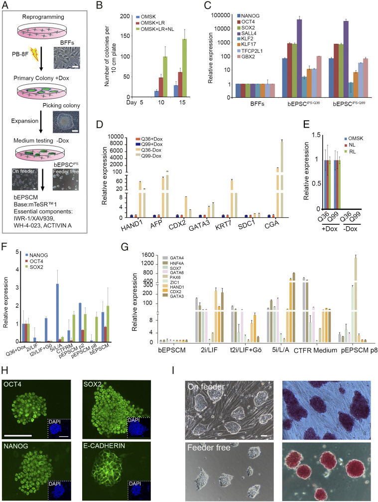
Embryonic stem cells (ESCs) and induced pluripotent stem cells have the potential to differentiate to all cell types of an adult individual and are useful for studying development and for translational research. However, extrapolation of mouse and human ESC knowledge to deriving stable ESC lines of domestic ungulates and large livestock species has been challenging. In contrast to ESCs that are usually established from the blastocyst, mouse expanded potential stem cells (EPSCs) are derived from four-cell and eight-cell embryos. We have recently used the EPSC approach and established stem cells from porcine and human preimplantation embryos. EPSCs are molecularly similar across species and have broader developmental potential to generate embryonic and extraembryonic cell lineages. We further explore the EPSC technology for mammalian species refractory to the standard ESC approaches and report here the successful establishment of bovine EPSCs (bEPSCs) from preimplantation embryos of both wild-type and somatic cell nuclear transfer. bEPSCs express high levels of pluripotency genes, propagate robustly in feeder-free culture, and are genetically stable in long-term culture. bEPSCs have enriched transcriptomic features of early preimplantation embryos and differentiate in vitro to cells of the three somatic germ layers and, in chimeras, contribute to both the embryonic (fetal) and extraembryonic cell lineages. Importantly, precise gene editing is efficiently achieved in bEPSCs, and genetically modified bEPSCs can be used as donors in somatic cell nuclear transfer. bEPSCs therefore hold the potential to substantially advance biotechnology and agriculture.

Keywords: bovine; expanded potential stem cell; nuclear transfer.


Conflict of interest statement

The authors declare no competing interest.

Figures

Fig. 1.
Fig. 1.
Reprogramming BFFs to Dox-inducible iPSCs for testing bovine stem cell culture condition. (A) Schematic illustration of reprogramming BFFs to iPSCs. PB-8F: bOMSK+ pN–hLIN + hRL. bOMSK (bovine OCT4, MYC, SOX2, and KLF4 cDNAs), pN–hLIN (porcine NANOG and human LIN28 cDNAs); hRL (human RARG and LRH1 cDNAs). BFFs: bovine fetal fibroblasts; Dox: doxycycline. (Scale bar, 50 μm.) (B) Coexpression of LIN28 (L), NANOG (N), LRH1, and RARG (LR) along with four Yamanaka factors substantially increased the reprogrammed colony numbers. (C) Relative expression of key endogenous pluripotency genes in two iPSC lines cultured in bEPSCM, bEPSCiPS-Q36, and bEPSCiPS-Q99. Data represent the mean ± SD, n = 3 independent experiments. (D) Expression of lineage genes in RT-qPCR of iPSC lines in the presence or absence of Dox in M15 medium. Q36: biPS-Q36 with Dox, Q99: biPS-Q99 with Dox. Data represent the mean ± SD, n = 3 independent experiments. (E) No detectable leaky expression of the exogenous reprogramming factors in iPSCs in RT-qPCR. (F and G) RT-qPCR analysis of pluripotency (F) and lineage genes (G) in bovine iPSCs under several culture conditions in the absence of Dox. These conditions include 2i/LIF, t2iL+Gӧ, and 5i/L/A on day 8; CTFR medium (passage 4); and pEPSCM (cells of passage 2 and passage 8 for analyzing pluripotency genes, and cells of passage 8 for analyzing lineage genes). Cells cultured in bEPSCM for passage 36 were used in the analysis. pEPSCM: porcine expanded potential stem cells medium, bEPSCM: bovine expanded potential stem cells medium, CTFRM: custom TeSR1 base medium supplemented with FGF2 and IWR1. Data represent the mean ± SD, n = 3 independent experiments. (H) Immunostaining of NANOG, OCT4, SOX2, and E-CADHERIN in bovinebEPSCiPS. (Scale bar, 100 μm.) (I) The morphology and alkaline phosphatase (AP) staining of bEPSCiPS-Q36 on feeder cells (Upper) or feeder free (Lower). (Scale bar, 50 μm.)
Fig. 2.
Fig. 2.
Establishment of bEPSCs from preimplantation embryos. (A) Schematic diagram of establishment of bEPSCES from bovine day 6 in vivo fertilization embryos. (Scale bar, 50 μm.) (B) bEPSCES lines of three breeds: Holstein, Angus, and Montbéliarde. (C) Karyotyping analysis of bEPSCsES-A6 (female, passage 82) and bEPSCsES-A15 (male, passage 76). (D) Immunostaining of NANOG, OCT4, SOX2, and E-CADHERIN in bEPSCs ES-A15 of passage 38. (Scale bar, 100 μm.) (E) Relative expression of core pluripotency genes OCT4, NANOG and SOX2 in two bEPSC lines (bEPSCsES-A15 on passage 32 and bEPSCiPS-Q36 on passage 36) on feeders or feeder-free. The relative expressions above were normalized to control and housekeeping gene. Data represent the mean ± SD, n = 3 independent experiments. (F) Morphology and AP staining of bEPSCES-A15 on feeder cells (Upper, passage 36) or feeder free (Lower, passage 30). (Scale bar, 50 μm.) (G) Teratoma derived from bEPSCES-A15 (passage 40). (H and E) Analysis revealed the presence of glandular epithelium (endoderm, i), muscle (mesoderm, ii), cartilage (mesoderm, iii), and mature neural tissue (glia and neurons, ectoderm, iv). (Scale bar, 50 μm.)
Fig. 3.
Fig. 3.
Transcriptomic and epigenetic features of bEPSCs. (A) DNA methylation levels in bEPSCs by whole-genome bisulfite sequencing analysis. Boxplots of the averaged DNA methylation levels (CpG sites) of 5 kb tiles in bEPSCiPS-Q36, bEPSCES-A6, and BFFs. The bottom and top of the boxes indicate the first and third quartiles, respectively, and the lines inside the boxes indicate the medians of the data. (B) DNA methylation in DMR of bovine-imprinted genes, including DLK1-DIO3 cluster, H19 cluster, IGF2R, MEG3, PEG3, PEG10, PLAGL1, SNRPPN, and ZIM2 in bEPSCs. (C) Immunostaining detection of H3K27me3 foci in female bEPSCES. The male bEPSCES were the negative control. (Scale bar, 50 μm.) (D) PCA of global gene expression (RNA-seq) of bEPSCs, bovine-primed ESCs, and BFFs. (E and F) Expression of pluripotency genes, lineage genes, and DNA methylation genes in bEPSCs, pEPSCs, and BFFs. Bovine-primed ESCs, n = 10; BFFs, n = 2; bEPSCsiPS-Q36, n = 2; bEPSCsES-A6, n = 2; n represents the number of biologically independent samples. (G) PCA of global gene expression (RNA-seq) of EPSCs and bovine preimplantation embryos (GSE59186) (60). Two replicates in each sample were used. (H) Expression levels of all annotated bovine histone genes in bovine-primed ESCs, EPSCs, and BFFs.
Fig. 4.
Fig. 4.
bEPSC’s developmental potential in chimeras. (A) Schematic diagram of chimera experiments using bEPSCs. (B) Contribution of bEPSCs in bovine preimplantation embryo development. The tdTomato+ donor bEPSCsES-A15 (passage 20) were injected into bovine morula embryos, which developed into blastocysts in 48 h in vitro. Panel (i), Injected tdTomato+ bEPSCsES-A15 in the blastocyst; Panel (ii), Several tdTomato+ cells (arrow) expressed trophectoderm factor CDX2. (Scale bar, 100 μm.) TdT, tdTomato. (C) Whole-mount fluorescence and bright field images of 40 d conceptuses from transferred preimplantation embryos. Chimera no. F1506 appeared to have tdTomato+ cells. (Scale bar, 0.5 cm.) (D) Detection of bEPSCsES-A15 tdTomato+ descendants in the chorionic placenta (PL-1+; i) and in smooth muscles (SMA+; ii) in chimera F1506. The control embryos have no tdTomato+ cell injected. DAPI stains nuclei. (Scale bar, 50 μm.) TdT, tdTomato. (E) Genome editing in bEPSCsES-A15. Knock-in of the T2A-H2B-mCherry cassette into the OCT4 locus using the CRISPR/Cas9. The targeting vector with short homology arms from the OCT4 locus flanking the T2A-H2B-mCherry and a Puromycin-resistance cassette was constructed and transfected into bEPSCs. The Puro-resistant transfectant colonies were picked 10 d after transfection. The correctly targeted colonies were identified in genomic DNA PCR. GT primers are for genotyping. (F) Bright field and fluorescence images of the correctly targeted bEPSCs colonies. Eleven out of forty-eight colonies examined were correctly targeted ones. (G and H) tdTomato+ bEPSCES-A15 as the donor in SCNT. Injection of tdTomato+ bEPSCES-A15 (passage 32) donor cells into the perivitelline space of oocytes was shown. (Scale bar, 50 μm.) (I) Derivation of secondary bEPSCs from SCNT (cloned) blastocysts. An outgrowth of day 16 from a SCNT blastocyst with bEPSCs as the donor cell (Upper) was picked for establishing the secondary EPSCs (Lower, Passage 4). (Scale bar, 50 μm.)

References

    1. Evans M. J., Kaufman M. H., Establishment in culture of pluripotential cells from mouse embryos. Nature 292, 154–156 (1981). - PubMed
    1. Martin G. R., Isolation of a pluripotent cell line from early mouse embryos cultured in medium conditioned by teratocarcinoma stem cells. Proc. Natl. Acad. Sci. U.S.A. 78, 7634–7638 (1981). - PMC - PubMed
    1. Buehr M., et al. ., Capture of authentic embryonic stem cells from rat blastocysts. Cell 135, 1287–1298 (2008). - PubMed
    1. Thomson J. A., et al. ., Embryonic stem cell lines derived from human blastocysts. Science 282, 1145–1147 (1998). - PubMed
    1. Thomson J. A., et al. ., Isolation of a primate embryonic stem cell line. Proc. Natl. Acad. Sci. U.S.A. 92, 7844–7848 (1995). - PMC - PubMed

Publication types