. 2021 May 3;32(5):1249-1261.
doi: 10.1681/ASN.2020081165. Epub 2021 Apr 8.

Protocadherin 7-Associated Membranous Nephropathy

Affiliations

Protocadherin 7-Associated Membranous Nephropathy

Sanjeev Sethi et al. J Am Soc Nephrol. .

Abstract

Background: Membranous nephropathy (MN) results from deposition of antigen-antibody complexes along the glomerular basement membrane (GBM). PLA2R, THSD7A, NELL1, and SEMA3B account for 80%-90% of target antigens in MN.

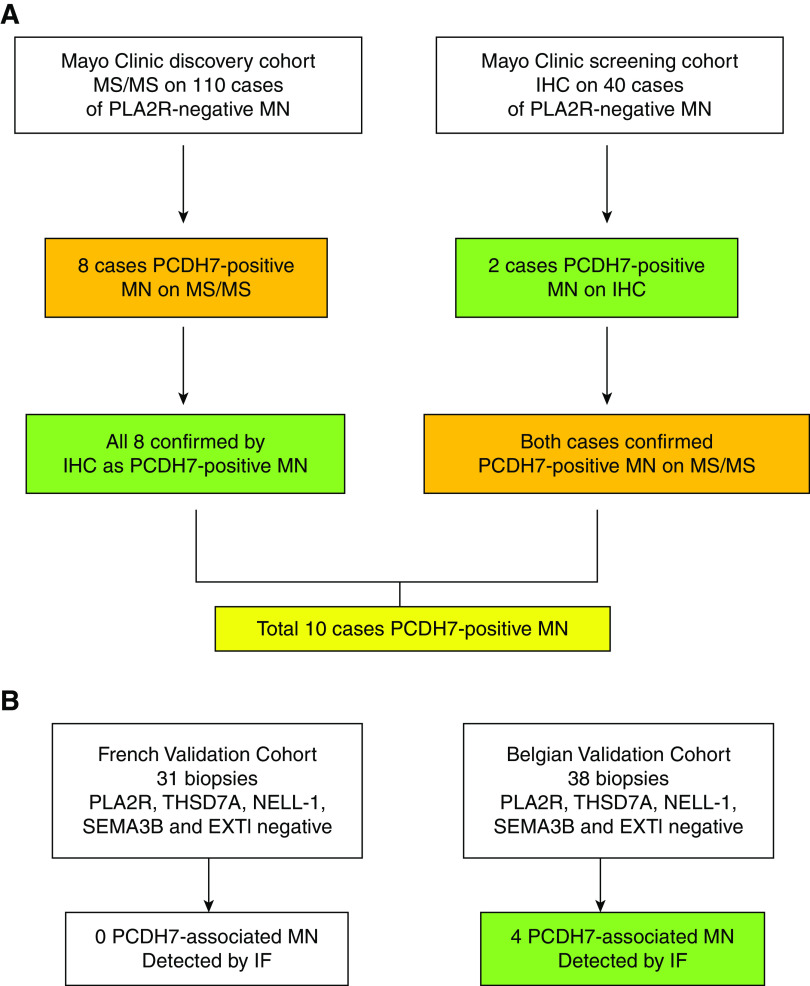
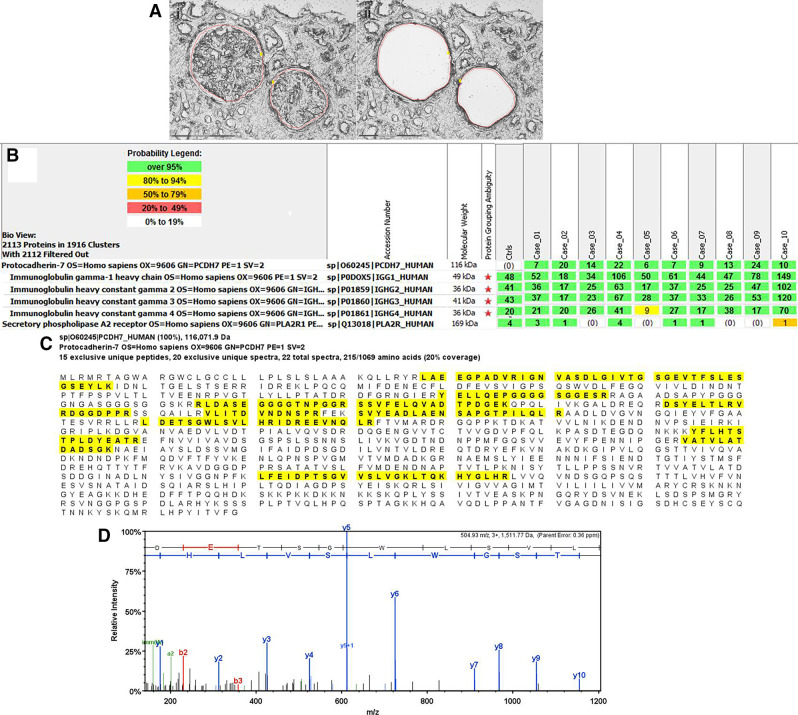
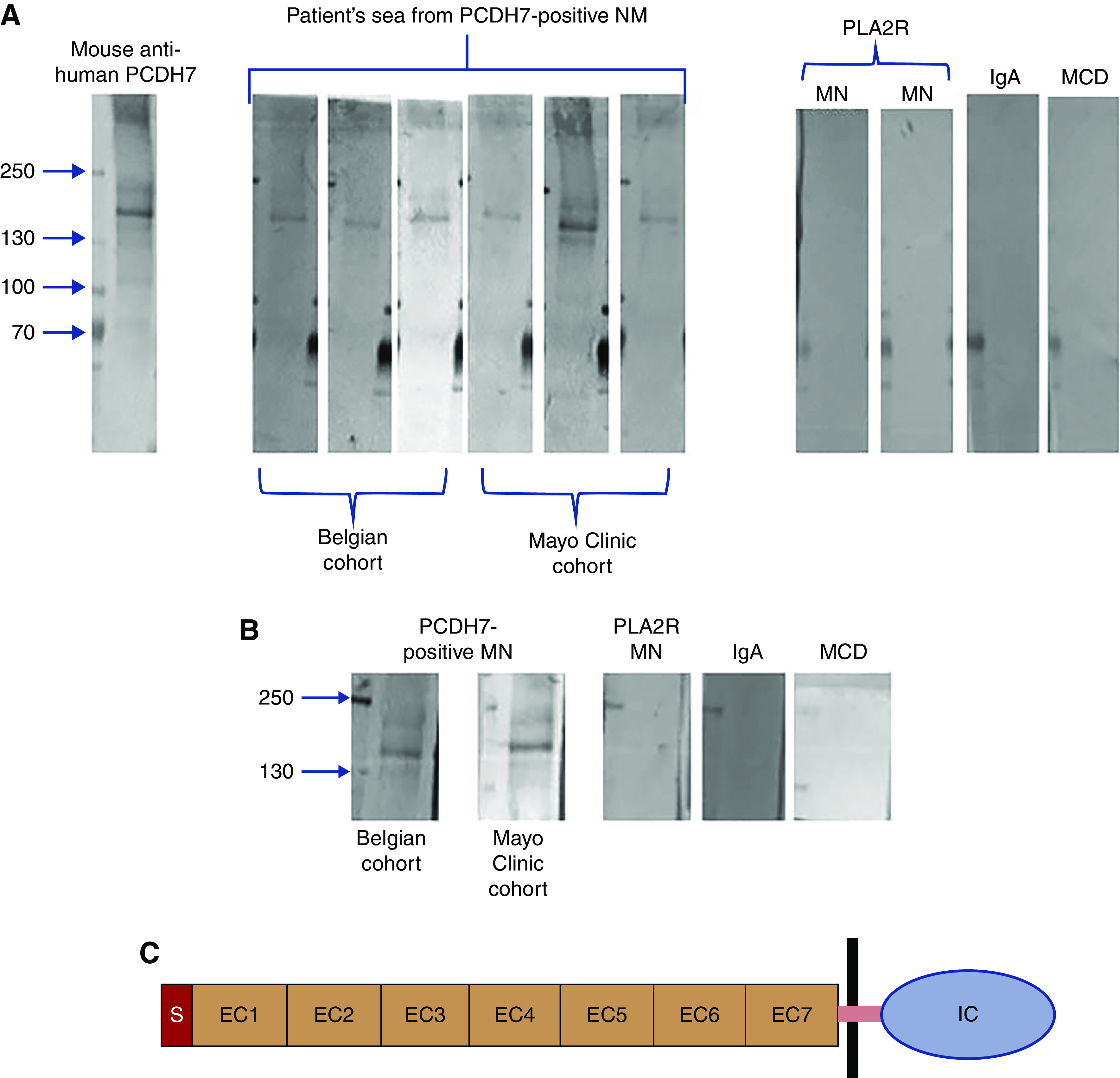
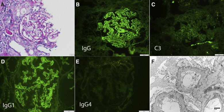
Methods: We performed laser microdissection and mass spectrometry (MS/MS) in kidney biopsies from 135 individuals with PLA2R-negative MN, and used immunohistochemistry/immunofluorescence and confocal microscopy to confirm the MS/MS finding, detect additional cases, and localize the novel protein. We also performed MS/MS and immunohistochemistry on 116 controls and used immunofluorescence microscopy to screen biopsy samples from two validation cohorts. Western blot and elution studies were performed to detect antibodies in serum and biopsy tissue.

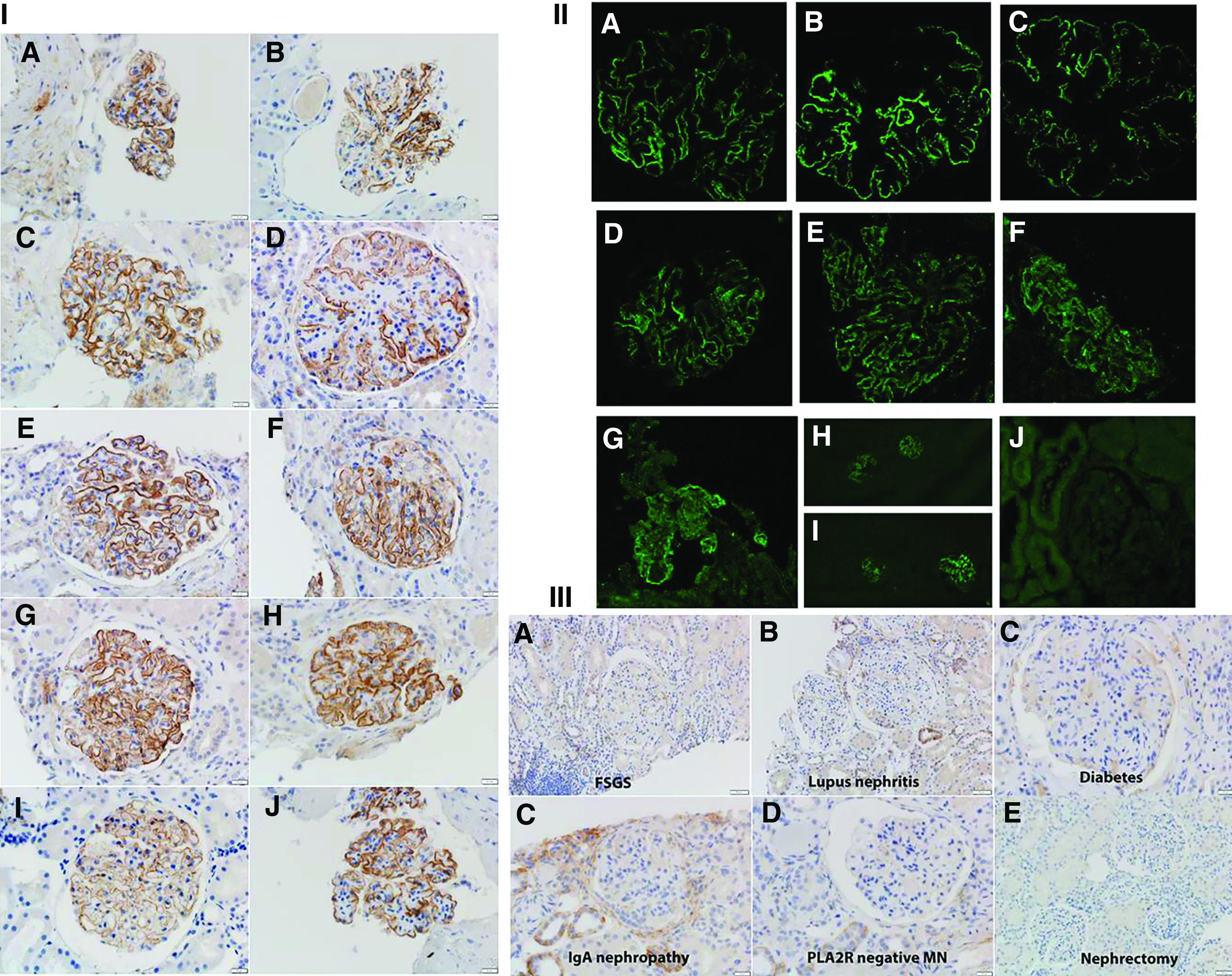
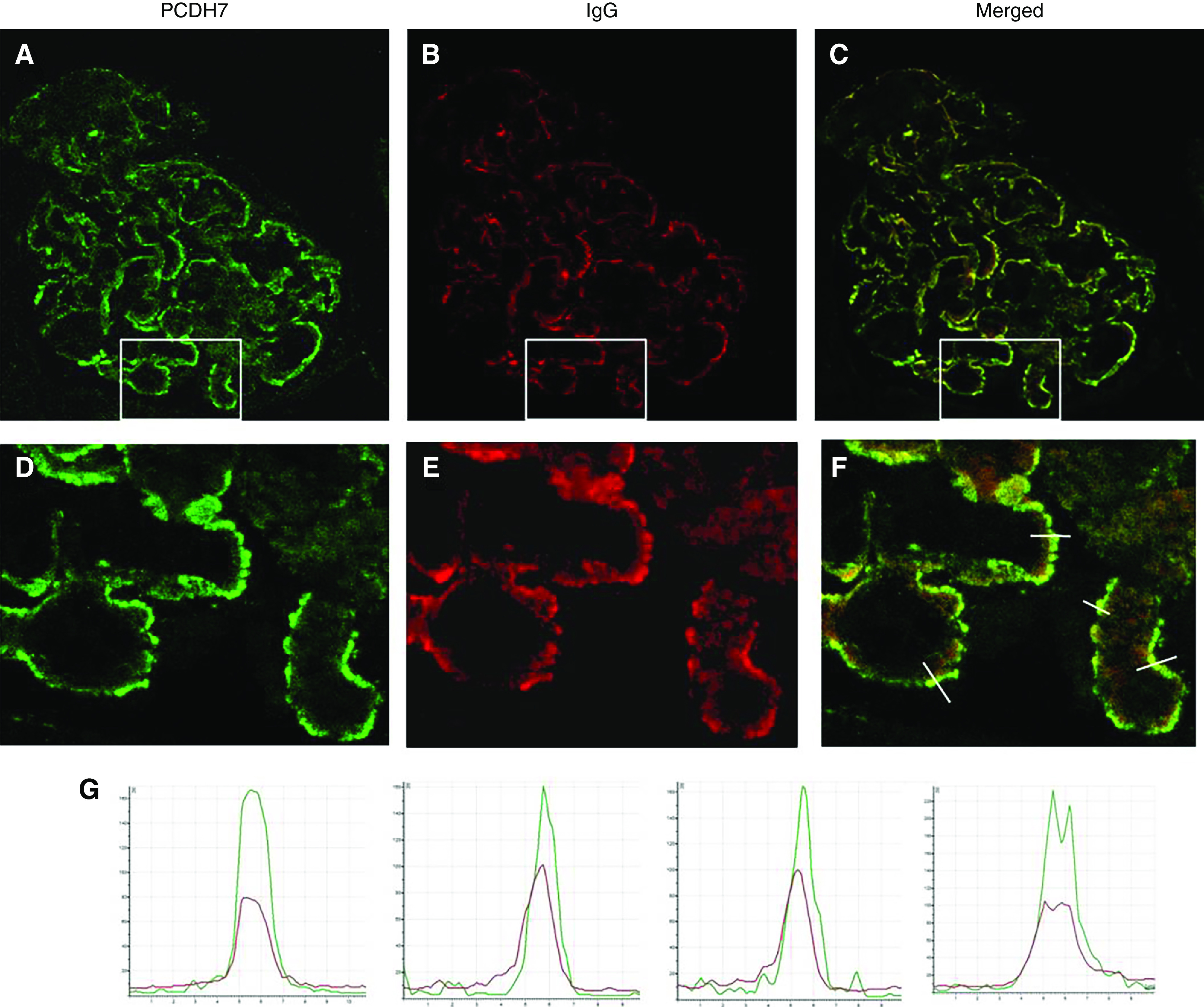
Results: MS/MS studies detected a unique protein, protocadherin 7 (PCDH7), in glomeruli of ten (5.7%) PLA2R-negative MN cases, which also were negative for PLA2R, THSD7A, EXT1/EXT2, NELL1, and SEMA3B. Spectral counts ranged from six to 24 (average 13.2 [SD 6.6]). MS/MS did not detect PCDH7 in controls (which included 28 PLA2R-positive cases). In all ten PCDH7-positive cases, immunohistochemistry showed bright granular staining along the GBM, which was absent in the remaining cases of PLA2R-negative MN and control cases. Four of 69 (5.8%) cases in the validation cohorts (all of which were negative for PLA2R, THSD7A, EXT1, NELL1, and SEMA3B) were PCDH7-positive MN. Kidney biopsy showed minimal complement deposition in 12 of the 14 PCDH7-associated cases. Confocal microscopy showed colocalization of PCDH7 and IgG along the GBM. Western blot analysis using sera from six patients showed antibodies to nonreduced PCDH7. Elution of IgG from frozen tissue of PCDH7-associated MN showed reactivity against PCDH7.

Conclusions: MN associated with the protocadherin PCDH7 appears to be a distinct, previously unidentified type of MN.

Keywords: glomerular disease; immunology and pathology; membranous nephropathy; nephrotic syndrome; renal biopsy; renal pathology.


Figures

Figure 1.
Figure 1.
Discovery, screening, and validation cohorts of PCDH7-positive MN. (A) In the discovery cohort, MS/MS was performed in 135 cases to look for novel proteins in PLA2R-negative MN. Eight cases were positive for a unique protein PCDH7. IHC for PCDH7 was performed on the eight cases and showed granular GBM staining for PCDH7. In the screening cohort, we performed IHC on 40 cases of PLA2R-negative MN and found two cases of PCDH7-positive MN. The findings were confirmed by MS/MS in both cases. (B) In the validation cohorts, IF for PCDH7 was performed in two cohorts. No positive cases were detected in the French cohort of 31 cases, whereas four positive cases were detected in the Belgian cohort of 38 cases.
Figure 2.
Figure 2.
Proteomic identification of PCDH7 in PLA2R-negative MN. Glomeruli were microdissected and analyzed using mass spectrometry as described in Methods. (A) shows (1) two glomeruli marked for dissection and (2) vacant space on slide following microdissection. Scale, 20×. (B) shows moderate spectral counts of PCDH7 in ten cases of PLA2R-negative MN. Numbers in green boxes represent spectral counts of MS/MS matches to a respective protein. All ten cases show moderate total spectral counts for PCDH7 and Igs; baseline spectral counts of PLA2R were detected in six of ten cases. For comparison, the average total spectral counts from six control cases (day 0 protocol transplant biopsies) are also shown. (C) Representative sequence coverage map of PCDH7 from one case. Amino acids highlighted in bold letters over yellow background are the amino acids detected. Note that the majority of the amino acids detected are in the first 570 amino acids toward the N terminus. (D) An example of MS/MS spectra match to a sequence from PCDH7. Example MS/MS spectra of 504.93 m/z 3+ ion matched to the PCDH7 peptide sequence LDETSGWLSVLHR.
Figure 3.
Figure 3.
IHC and IF for PCDH7-associated MN and control cases. (I, A–J) PCDH7-associated MN (Mayo Clinic cohort). Ten cases show bright granular capillary wall staining for PCDH7 along the GBMs. Each panel shows two cases. (A) Patient 1. (B) Patient 2. (C) Patient 3. (D) Patient 4. (E) Patient 5. (F) Patient 6. (G) Patient 7 (low power, ×10; ×40). (H) Patient 8 (×40). (I) Patient 9 (×40). (J) Patient 10 (×40). (II, A–J) PCDH7-associated MN (validation cohort). IF shows (A–C) bright capillary wall staining for PCDH7 of three cases of the Mayo cohort, (D–G) bright capillary wall staining for PCDH7 in four cases of the Belgian validation cohort, and (H and I) bright staining for PCDH7 in (H) one patient from the Mayo cohort and (I) one patient from the Belgian cohort (low-power view, ×20). (J) PCDH7 staining is negative in a case of PLA2R-negative MN. (III, A–F) Control cases. PCDH7 staining is negative in a case of FSGS, lupus nephritis, diabetes, IgA nephropathy, PLA2R-negative MN, and a nephrectomy specimen.
Figure 4.
Figure 4.
Confocal IF analysis: detection of PCDH7 and IgG in glomerular immune deposits in PCDH7-associated MN. Glomeruli double labeled with (A) anti-PCDH7 (green) and (B) anti-human IgG (red). (C) The merged image. (D–F) These images are enlarged images of the boxed areas in A–C, respectively. (G) The graphs show quantitative analyses of the fluorescence recorded across sections of a representative capillary loop (indicated by lines in (F)). Note the superimposition of the two signals, which indicates that subepithelial immune deposits contain PCDH7 (green) and IgG (red).
Figure 5.
Figure 5.
Detection of anti-PCDH7 protein antibodies by western blot analysis. (A) Under nonreducing conditions, PCDH7 was detected using mouse anti-human PCDH7 antibody as a band at 140 kD. The same band was recognized by sera from patients with PCDH7-associated MN but not by sera from patients with other kidney diseases. Each lane shows an individual patient from the discovery cohort (Mayo Clinic) or from the Belgian validation cohort. The three Mayo Clinic cohort patients are patients 1, 9, and 10, and the three Belgian cohort patients are patients 11–13. (B) Reactivity of eluted IgG from biopsy specimens. Each lane represents the eluates from three pooled biopsies from the Belgian cohort and the Mayo Clinic cohort. The three lanes on the right represent eluates from three pooled biopsies from patients with PLA2R MN, IgA nephropathy, or minimal change disease (MCD). Only IgG eluted from the PCDH7-associated MN samples reacted with the recombinant PCDH7 protein at the expected mol wt. (C) Schematic representation of PCDH7. The cadherin family is characterized by repeating motifs of extracellular cadherin (EC) domain. PCDH7 has a signal (S) peptide, seven EC domains, a single pass transmembrane domain (purple bar), and an intracellular cytoplasmic (IC) domain.
Figure 6.
Figure 6.
Biopsy finding of a representative case (patient 5) of PCDH7-associated MN. (A) Light microscopy shows thickened GBMs (periodic acid–Schiff stain ×40). IF shows (B) bright 3+ capillary wall staining for IgG, (C) mild 1+ staining for C3, (D) bright 3+ staining for IgG1 along the capillary walls, and (E) negative staining for IgG4. (F) EM shows subepithelial electron-dense deposits (×7140).

Similar articles

Cited by

References

    1. Beck LH Jr., Salant DJ: Membranous nephropathy: From models to man. J Clin Invest 124: 2307–2314, 2014. - PMC - PubMed
    1. Ronco P, Debiec H: Pathophysiological advances in membranous nephropathy: Time for a shift in patient’s care. Lancet 385: 1983–1992, 2015. - PubMed
    1. Couser WG: Primary membranous nephropathy. Clin J Am Soc Nephrol 12: 983–997, 2017. - PMC - PubMed
    1. Beck LH Jr., Bonegio RGB, Lambeau G, Beck DM, Powell DW, Cummins TD, et al. .: M-type phospholipase A2 receptor as target antigen in idiopathic membranous nephropathy. N Engl J Med 361: 11–21, 2009. - PMC - PubMed
    1. Tomas NM, Beck LH Jr., Meyer-Schwesinger C, Seitz-Polski B, Ma H, Zahner G, et al. .: Thrombospondin type-1 domain-containing 7A in idiopathic membranous nephropathy. N Engl J Med 371: 2277–2287, 2014. - PMC - PubMed

Publication types