. 2021 Apr 8;12(1):2105.
doi: 10.1038/s41467-021-22212-1.

Symbiotic polyamine metabolism regulates epithelial proliferation and macrophage differentiation in the colon

Affiliations

Symbiotic polyamine metabolism regulates epithelial proliferation and macrophage differentiation in the colon

Atsuo Nakamura et al. Nat Commun. .

Abstract

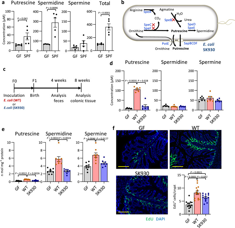
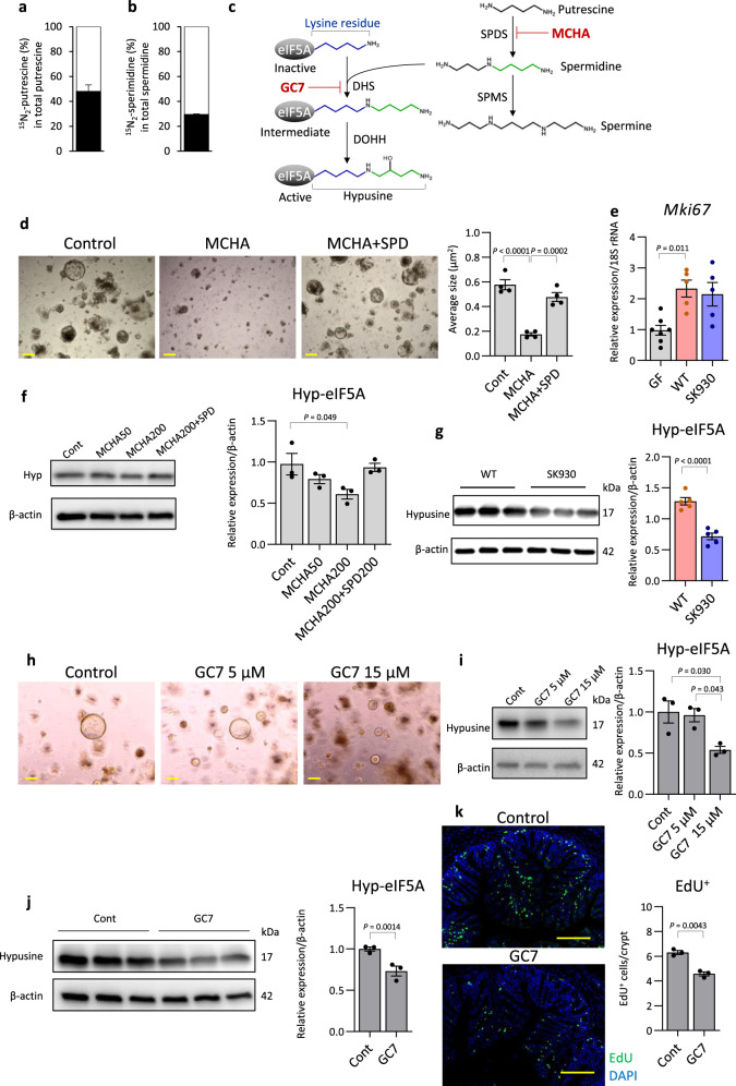
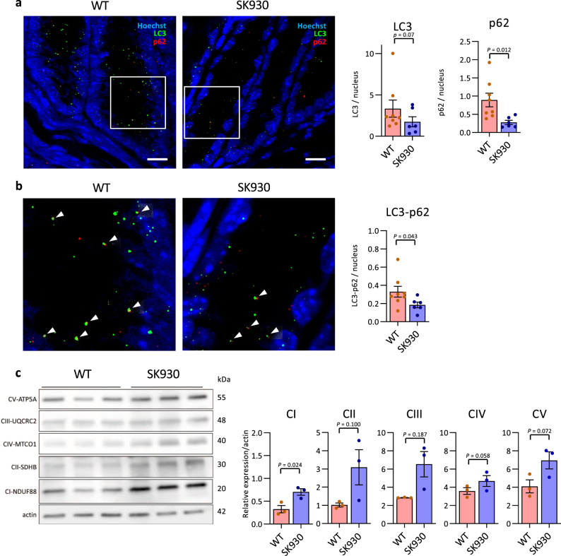
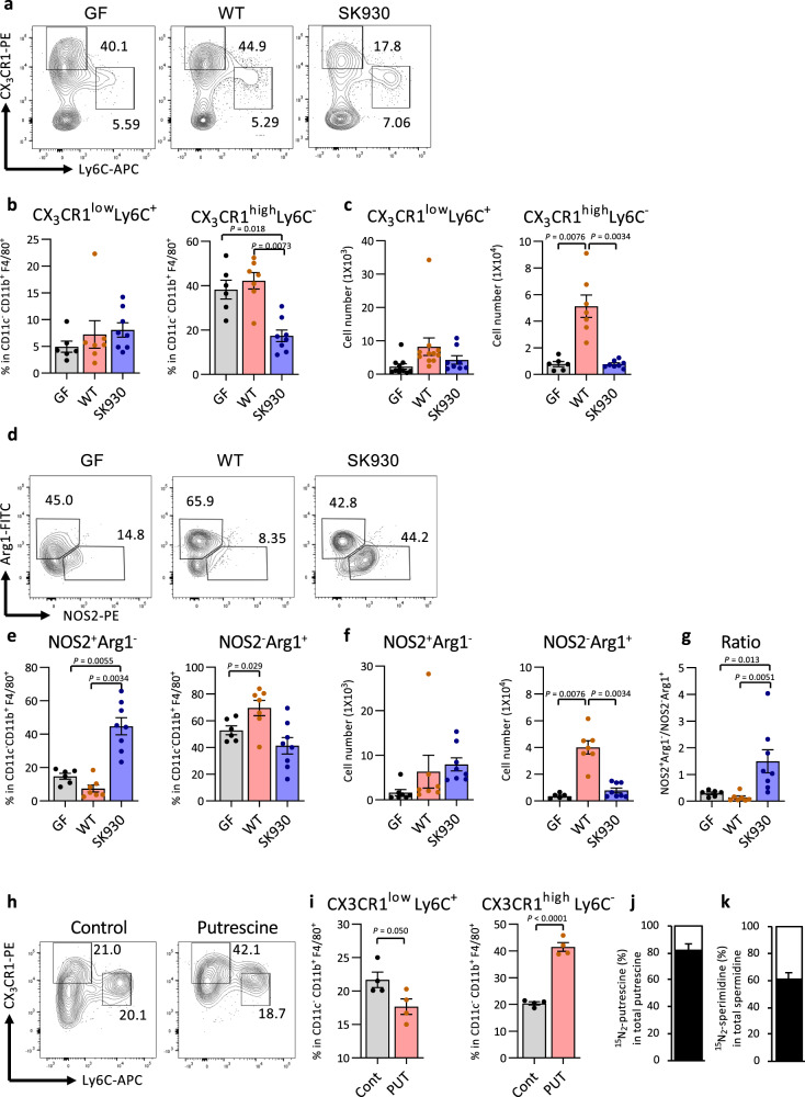
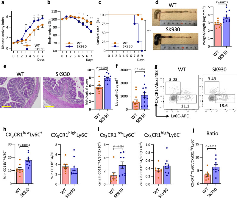
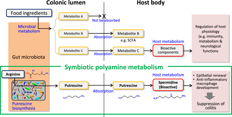
Intestinal microbiota-derived metabolites have biological importance for the host. Polyamines, such as putrescine and spermidine, are produced by the intestinal microbiota and regulate multiple biological processes. Increased colonic luminal polyamines promote longevity in mice. However, no direct evidence has shown that microbial polyamines are incorporated into host cells to regulate cellular responses. Here, we show that microbial polyamines reinforce colonic epithelial proliferation and regulate macrophage differentiation. Colonisation by wild-type, but not polyamine biosynthesis-deficient, Escherichia coli in germ-free mice raises intracellular polyamine levels in colonocytes, accelerating epithelial renewal. Commensal bacterium-derived putrescine increases the abundance of anti-inflammatory macrophages in the colon. The bacterial polyamines ameliorate symptoms of dextran sulfate sodium-induced colitis in mice. These effects mainly result from enhanced hypusination of eukaryotic initiation translation factor. We conclude that bacterial putrescine functions as a substrate for symbiotic metabolism and is further absorbed and metabolised by the host, thus helping maintain mucosal homoeostasis in the intestine.


Conflict of interest statement

The authors declare no competing interests.

Figures

Fig. 1
Fig. 1. Bacterial putrescine facilitates epithelial cell proliferation in the colon.
a Polyamine concentrations in faeces of eight-weeks old GF and SPF mice (n = 5). b Polyamine synthesis pathway in E. coli and the deleted genes (marked with X) in an E. coli strain SK930. Blue letters represent enzymes and transporters: arginine decarboxylase (SpeA), agmatine ureohydrolase (SpeB), ornithine decarboxylase (SpeC and SpeF), adenosylmethionine decarboxylase (SpeD), spermidine synthase (SpeE), putrescine-ornithine antiport transporter (PotE) and putrescine exporters (SapBCDF). c Experimental design for gnotobiotic mice colonised with WT or SK930 strain. d The polyamine concentrations in faeces of F1 mice. e The polyamine concentrations in CECs (n = 8, independent animals). f Representative microscopic images of EdU assay and quantification of EdU-positive cells/crypt in each group. EdU-positive cells (green). Nuclei are counterstained with DAPI (blue) (n = 8, independent animals). Scale bars: 100 µm. Three randomly selected areas were examined per slide (n = 13). All data shown represent the mean ± SEM. Statistical significance was calculated using a the Welch’s t-test (two-sided), d, e Kruskal–Wallis followed by Steel–Dwass post hoc test, f one-way ANOVA followed by Tukey’s post-hoc test.
Fig. 2
Fig. 2. Inhibition of hypusination of eIF5A arrests colonic organoid growth.
a, b Isotope ratio of putrescine and spermidine in colon organoids cultured with isotope-labelled 15N2 putrescine were analysed via GC-MS. The black bar indicates the percent putrescine (PUT) or spermidine (SPD) of 15N2 putrescine or 15N2 spermidine enrichment in total corresponding polyamines, respectively (n = 3, independent cultures). c Polyamine synthesis pathway and eIF5A hypusination. Hypusine is formed by conjugation of the aminobutyl moiety (green) of spermidine to a lysine residue. MCHA and GC7 inhibit SPDS and DHS, respectively. SPMS: spermine synthase, DOHH: deoxyhypusine hydrogenase. d Representative microscopy images of organoid cultured with or without MCHA and spermidine (SPD). Scale bars: 200 µm. The average size of each organoid was quantified using Image J software (n = 4, independent cultures). e Mki67 mRNA expression was analysed by q-PCR (GF: n = 8, WT: n = 6, SK930: n = 8, independent animals). Western blot analysis of hyp-eIF5A in f organoids treated with MCHA (n = 3) and in g CECs of gnotobiotic mice (n = 5). h Representative microscopy images of organoids cultured with or without GC7. Scale bars: 200 µm. Reproducibility of results was confirmed by performing two independent experiments. i Western blot analysis of hyp-eIF5A in organoids treated with MCHA (n = 3, independent cultures). j Western blot analysis of hyp-eIF5A in CECs of SPF mice with GC7 treatment (n = 3, independent animals). k Representative microscopic images of EdU assay and quantification of EdU-positive cells/crypt in control or GC7 treated mice. EdU-positive cells (green). Nuclei are counterstained with DAPI (blue). Scale bars: 100 µm. Three randomly selected areas were examined per slide (n = 3, independent animals). All data shown represent the mean ± SEM. Statistical significance was calculated using d, f the one-way ANOVA followed by Tukey’s post-hoc test, e, i Welch’s ANOVA followed by Dunnett’s T3 post-hoc test and g, j, k Student’s t-test (two-sided).
Fig. 3
Fig. 3. Bacterial putrescine activates autophagy and OXPHOS in CECs.
a, b Representative microscopic images of LC3 and p62 and its enlarged view. Green and red dots indicate LC3 and p62, respectively. Nuclei are stained with Hoechst (blue). The white arrowhead indicates LC3-p62 double-positive dots. Scale bars: 10 µm. Three randomly selected areas were examined per slide (WT: n = 8, SK930: n = 6, independent animals). c Western blot analysis of OXPHOS complex I-V (CI-CV) in CECs of gnotobiotic mice (n = 3, independent animals). All data shown represent the mean ± SEM. Statistical significance was calculated using the Welch’s t-test (a, p62 and c) (two-sided), Mann–Whitney test with (a, LC3 and b, LC3-p62) (two-sided).
Fig. 4
Fig. 4. Bacterial putrescine regulates the development of macrophage subsets.
CX3CR1 and Ly6C expression were analysed in CD45+CD11b+F4/80+ cells via flow cytometry. Representative flow cytometry images (a), frequency (b), number (c) of CX3CR1lowLy6C+ monocyte/macrophage and CX3CR1highLy6C macrophages and representative flow cytometry images (d), frequency (e), number (f) of NOS2+Arg1 and NOS2Arg1+ macrophages and the ratio of the former to the latter subset (g) in cLP of gnotobiotic mice colonised with WT (n = 7) or SK930 (n = 8) strain and GF mice (n = 6). CX3CR1 and Ly6C expression in CD45+CD11cCD11b+F4/80+cells from BMDMs cultured in medium with or without putrescine (n = 4). Representative flow cytometry images (h) and frequency (i). j, k Isotope ratio of putrescine and spermidine in BMDMs cultured with isotope-labelled 15N2-putrescine were analysed via GC-MS. The black bar indicates the percent putrescine (PUT) or spermidine (SPD) of 15N2-putrescine or 15N2-spermidine enrichment in total corresponding polyamines, respectively (n = 3, independent cultures). All data shown represent the mean ± SEM. Kruskal-Wallis followed by Steel-Dwass post hoc test (bg) and Student’s t-test (i) (two-sided).
Fig. 5
Fig. 5. Bacterial putrescine ameliorates DSS-induced colitis.
2% DSS in the drinking water to the F1 gnotobiotic mice colonised WT or SK930 strain to induce experimental colitis for six days, then regular drinking water thereafter. ac: Red and blue indicate WT-strain-associated and SK930-strain-associated mice. a DAI score. b Body weight changes were measured daily and calculated as the percentage change from day 0. c Survival rate in mice after DSS administration. The survival curve was calculated using the Kaplan-Meier method and statistical significance was calculated using the log rank test (WT: n = 10, SK930: n = 8). dj Mice were analysed on day 5 after starting 2% DSS (n = 10). d Representative image of the colons and values of weight/length of the colon. e Haematoxylin and eosin-stained images of representative histopathologic changes in the WT and SK930 strain-associated mice. Scale bar: 100 µm. Mucosal damage was estimated using a histological scoring system. f Faecal lipocalin-2 concentrations in faeces at day 5 after starting 2% DSS. Expression of CX3CR1 and Ly6C were analysed in CD45+CD11b+F4/80+ cells via flow cytometry. Representative flow cytometry images (g), and frequency (h) and number (i) of CX3CR1lowLy6C+ monocyte/macrophages and CX3CR1highLy6C macrophages and the ratio of the former to the latter subset (j) in the cLP of gnotobiotic mice colonised with WT or SK930 strain (WT: n = 8, SK930: n = 9). All data shown represent the mean ± SEM. Statistical significance was calculated using the Welch’s t-test (e, h, i) (two-sided) with Bonferroni adjustments (a), Mann–Whitney test with Bonferroni adjustments (b), Student’s t-test (d, f, j) (two-sided) (*P < 0.05, **P < 0.01, ***P < 0.001).
Fig. 6
Fig. 6. Symbiotic metabolism.
Low-molecular metabolites produced by the intestinal microbiota remain in the lumen (Metabolite A) or are absorbed from the intestinal lumen to systemic circulation (Metabolite B and C). Metabolite B, which are represented by short-chain fatty acids, directory act on host cells to exert physiological activities. Meanwhile, a part of inactive metabolites may be further converted to bioactive substances via host metabolic pathways after absorption (Metabolite C). The entities of such symbiotic metabolism, however, remain obscure. Our study using gnotobiotic mice demonstrated that microbial putrescine is uptaken by the colonic epithelial cells to produce bioactive spermidine that accelerates epithelial renewal and increases the abundance of anti-inflammatory macrophages in the colon. Thus, microbial putrescine serves as a source (precursor) of bioactive substances in the host animals. This observation provides evidence for symbiotic metabolism contributing to the maintenance of intestinal homoeostasis.

Similar articles

Cited by

References

    1. Pegg AE. Functions of polyamines in mammals. J. Biol. Chem. 2016;291:14904–14912. doi: 10.1074/jbc.R116.731661. - DOI - PMC - PubMed
    1. Igarashi K, Kashiwagi K. Polyamines: mysterious modulators of cellular functions. Biochem. Biophys. Res. Commun. 2000;271:559–564. doi: 10.1006/bbrc.2000.2601. - DOI - PubMed
    1. Matsumoto M, Kakizoe K, Benno Y. Comparison of fecal microbiota and polyamine concentration in adult patients with intractable atopic dermatitis and healthy adults. Microbiol. Immunol. 2007;51:37–46. doi: 10.1111/j.1348-0421.2007.tb03888.x. - DOI - PubMed
    1. Matsumoto M, et al. Impact of intestinal microbiota on intestinal luminal metabolome. Sci. Rep. 2012;2:233. doi: 10.1038/srep00233. - DOI - PMC - PubMed
    1. Kibe R, et al. Upregulation of colonic luminal polyamines produced by intestinal microbiota delays senescence in mice. Sci. Rep. 2014;4:4548. doi: 10.1038/srep04548. - DOI - PMC - PubMed

Publication types

MeSH terms

LinkOut - more resources