. 2021 Apr 8;12(1):2099.
doi: 10.1038/s41467-021-22305-x.

Defective dystrophic thymus determines degenerative changes in skeletal muscle

Affiliations

Defective dystrophic thymus determines degenerative changes in skeletal muscle

Andrea Farini et al. Nat Commun. .

Abstract

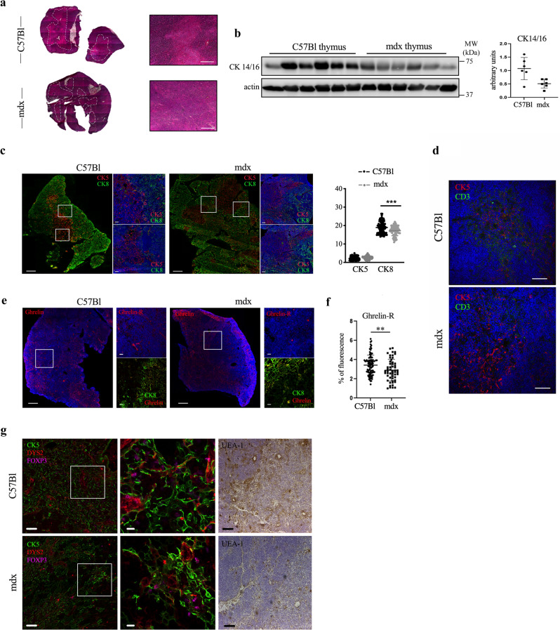
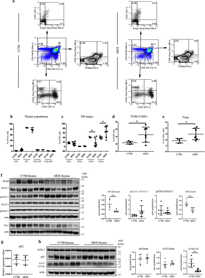
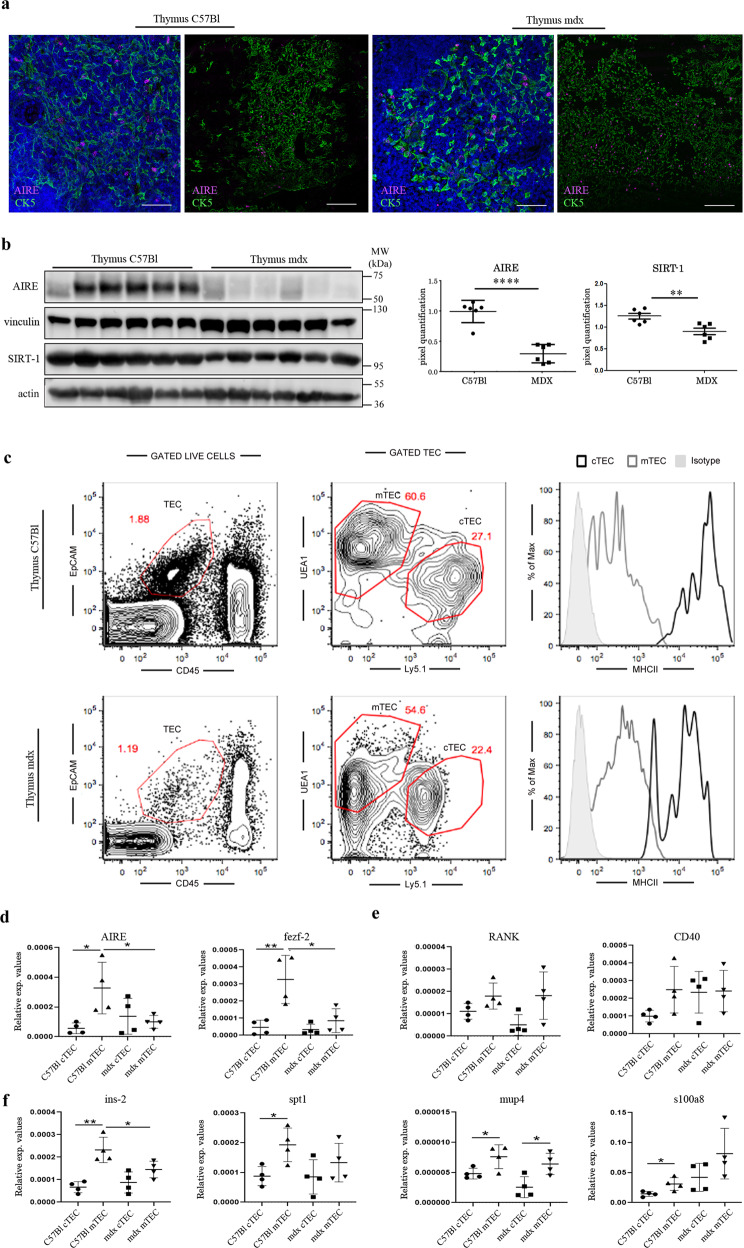
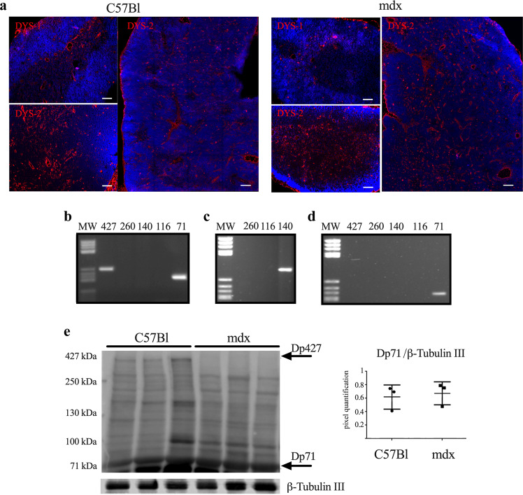
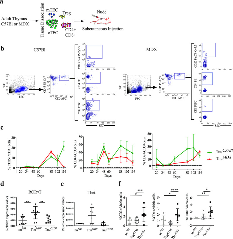
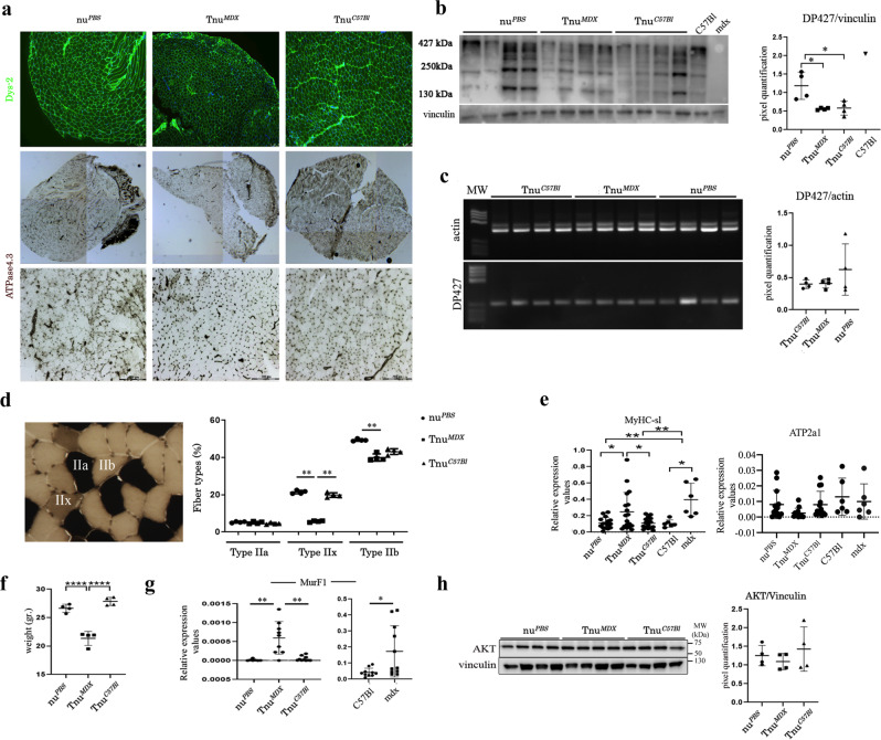
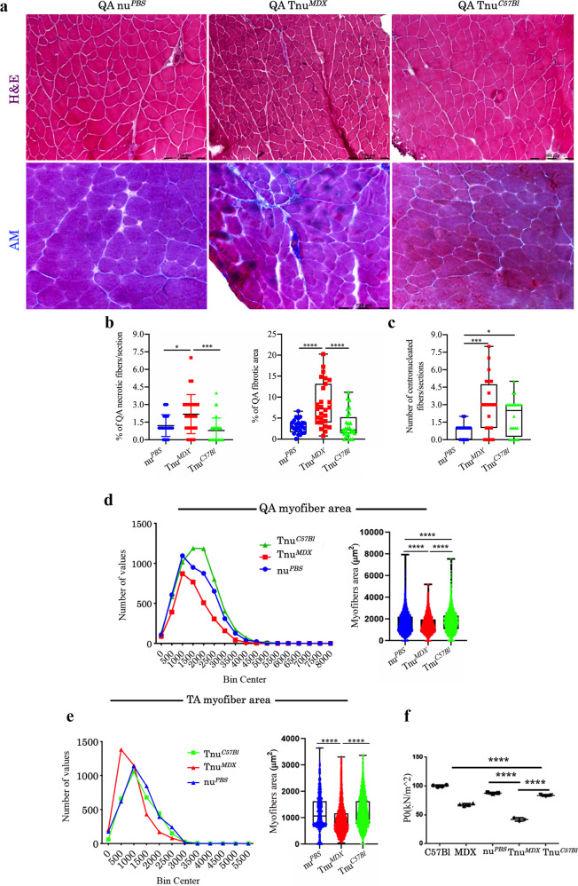
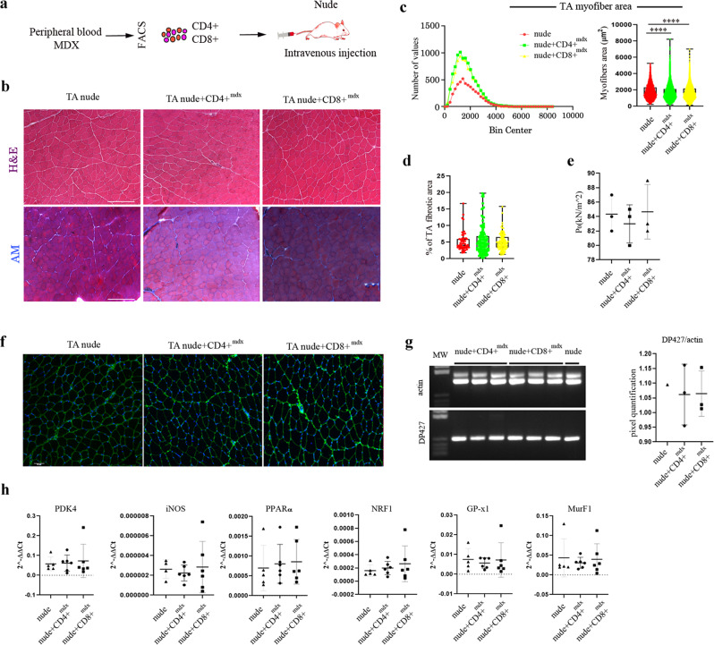
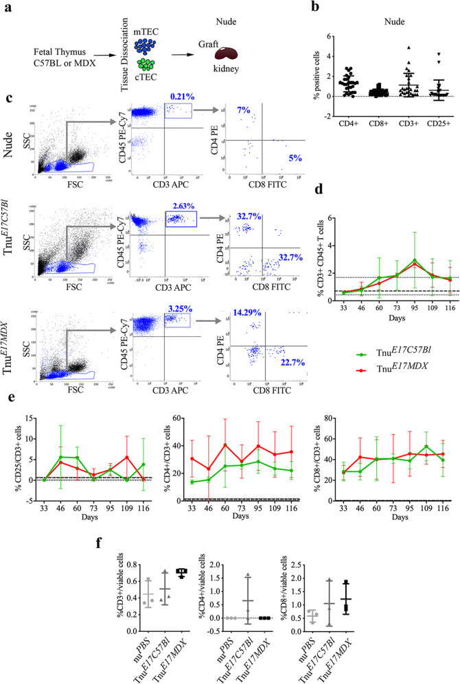
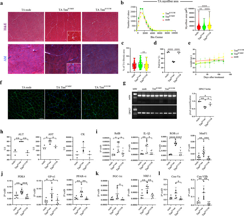
In Duchenne muscular dystrophy (DMD), sarcolemma fragility and myofiber necrosis produce cellular debris that attract inflammatory cells. Macrophages and T-lymphocytes infiltrate muscles in response to damage-associated molecular pattern signalling and the release of TNF-α, TGF-β and interleukins prevent skeletal muscle improvement from the inflammation. This immunological scenario was extended by the discovery of a specific response to muscle antigens and a role for regulatory T cells (Tregs) in muscle regeneration. Normally, autoimmunity is avoided by autoreactive T-lymphocyte deletion within thymus, while in the periphery Tregs monitor effector T-cells escaping from central regulatory control. Here, we report impairment of thymus architecture of mdx mice together with decreased expression of ghrelin, autophagy dysfunction and AIRE down-regulation. Transplantation of dystrophic thymus in recipient nude mice determine the up-regulation of inflammatory/fibrotic markers, marked metabolic breakdown that leads to muscle atrophy and loss of force. These results indicate that involution of dystrophic thymus exacerbates muscular dystrophy by altering central immune tolerance.


Conflict of interest statement

The authors declare no competing interests.

Figures

Fig. 1
Fig. 1. Altered thymic architecture in mdx mice.
Representative images of H&E staining of thymus of 3-month-old C57Bl and mdx mice revealed differences in medullary/cortex boundaries between animals (dashed white line) (a). WB analysis showed the downregulation of cytokeratin 14/16 in mdx thymus related to C57Bl (b). Thymic architecture of C57Bl and mdx mice characterized by immunofluorescence staining for cortical cytokeratin CK8 (green) and medullary cytokeratin CK5 (red) confirmed changes in dystrophic thymic environment. Graph displays fluorescence area % occupied by CK5 and CK8, as calculated by ImageJ software (c). Double immunofluorescence staining for CK5 (red) and CD3 (green) of C57Bl and mdx thymi portrayed a loosen embedding of CD3+ cells within dystrophic medulla. d Staining of ghrelin (GHR) and ghrelin receptor (GHS-R) (red) showed a comparable distribution of GHR between animals, but a prevalent expression of GHS-R in thymus of C57Bl mice. Of note, GHR was preferentially found in proximity of cortical CK8 (green). e Graph displays fluorescence area % occupied by GHS-R, as calculated by ImageJ software, in C57Bl and mdx mice (f). Expression of FoxP3+ cells (magenta) was evaluated by immunofluorescence staining within CK5+ thymic medulla (green). C-terminal containing dystrophin isoforms were detected by DYS-2 antibody (red) to identify a specific protein distribution within thymus. mTEC maturation level was evaluated by immunohistochemistry staining of C57Bl and mdx thyme with UEA-1. For fluorescence microscopy, nuclei were counterstained with DAPI (g). Scale bars: 200 μm (a); 100 μm for confocal tile scan reconstruction (left) and 20 μm for higher magnification confocal microscope images (right) (c, e); 50 μm (d); 50 μm (left) and 10 μm for higher magnification confocal microscope images (right) (g). The comparisons between the averages of the two groups were evaluated using two-sided Student’s t-test. b *p = 0.0495. c ***p = 0.0007. f **p = 0.0046. Data are presented as mean ± SD of three independent experiments with n = 6 mice/group. For Ck5–Ck8 immunofluorescence staining n = 12 images/mice have been quantified, GHS-R staining was quantified in n = 12 and n = 8 images of C57Bl and mdx mice, respectively. Source data are provided as a Source Data file.
Fig. 2
Fig. 2. Cellularity, NF-kB/STATs expression, and autophagy in thymus of C57Bl and mdx mice.
FACS analysis of thymus homogenate from mdx and C57Bl mice at 8 weeks of age demonstrates no significant alteration of T cells (a, b), and few differences in CD4−CD8−DN stages, in particular DN3 (CD44−CD25+) and DN4 (CD44+CD25+) (c). The number of TCRβ+CD69+ cells (d) and of Foxp3+CD25+ cells (e) was significantly increased in thymus of mdx mice. Cropped image of a representative WB and densitometric analysis revealed a downregulation of NF-kB, IKKi, and STAT3 in mdx thymus (f). RT-qPCR of p62 expression is shown in g. Autophagy markers such as Atg7, p62 and LC3 were also assessed by WB analysis. Representative WB image and quantification of LC3-II/LC3-I showed the impairment of the autophagic flux (h). All protein expression was normalized on actin, as a loading control. The comparisons between the averages of the groups were evaluated using two-sided Student’s t-test. c *p = 0.0177 (DN3), *p = 0.0351 (DN4). d *p = 0.043. e *p = 0.0332. f **p = 0.0048 (NF-κB), **p = 0.0018 (IKKi). h **p = 0.0026. Data are presented as mean ± SD of three independent experiments with n = 7 (mdx) and n = 5 (C57Bl) mice (ae); n = 6 mice/group (f, h); n = 3 mice (mdx) and n = 6 mice (C57Bl) (g). Source data are provided as a Source Data file.
Fig. 3
Fig. 3. AIRE dysregulation in thymus of 3-month-old mdx mice.
Representative confocal microscope images (left) and tile scan reconstruction (right) of thymic lobes from 3-month-old C57Bl and mdx mice. Despite a comparable AIRE+ cell pattern distribution embedded within CK5+ thymic medulla of both mice, in mdx thymus immunofluorescence staining for AIRE appeared less evident. Nuclei were counterstained with DAPI (a). Representative images of western blot analysis showing the expression of the AIRE and SIRT-1 proteins in the thymus of C57Bl and mdx mice. Densitometric analyses are shown as the AIRE/vinculin ratio and SIRT-1/actin ratio (b). For the identification and sorting of cTEC and mTEC from C57bl and mdx mice, stained thymus cell suspension was analysed using flow cytometry. Representative FACS profile are shown. The numbers within the panels indicate the percentage of each population of live cells, a gate of CD45-negative and EpCAM-positive events represents TEC cells. Within the TEC gate, two population are separated by level of Ly5.1 and UEA-1. MHC II molecules were highly expressed on cTECs (c). Following FACS isolation of mTEC and cTEC, RT-qPCR experiments showed the downregulation of AIRE and Fezf2 in isolated mdx mTEC related to mTEC from C57Bl thymus (d). The expression in thymus of AIRE-regulators RANK and CD40 was similar between mTEC isolated from C57Bl and mdx mice (e). RT-qPCR revealed diminished expression of AIRE-dependent genes Ins2 and Spt1 in dystrophic mTEC and in control cTEC related to mTEC isolated from C57Bl thymus (f). Scale bar: 50 μm (a). The comparisons between the averages of the groups were evaluated using two-sided Student’s t-test. b ****p < 0.0001 (AIRE), **p = 0.0050 (SIRT-1). d p = 0.0436 (AIRE) and p = 0.0220 (fezf2) in dystrophic mTECs vs C57Bl mTECs; p = 0.0224 (AIRE) and p = 0.00089 (fezf2) in C57Bl mTECs vs C57Bl cTECs. f p = 0.0413 (Ins-2) in mdx mTECs vs C57Bl mTECs; p = 0.0017 (Ins-2), p = 0.0178 (spt1), p = 0.0413 (mup4), p = 0.0271 (s100a8) in C57Bl mTECs vs C57Bl cTECs; p = 0.0210 (mup4) in C57Bl mTECs vs C57Bl cTECs. Data are presented as mean ± SD of three independent experiments with n = 6 mice/group (a, b); n = 8 (c); n = 8 mice divided into two independent groups examined in n = 2 independent experiments (df). Source data are provided as a Source Data file.
Fig. 4
Fig. 4. Dystrophin isoforms expression in thymus of 3-month-old C57Bl and mdx mice.
Confocal microscope fluorescence images (right) and tile scan reconstructions (left) of dystrophin isoforms in thymi of C57Bl and mdx mice. DYS-1 (mid-rod-domain) (red) and DYS-2 (C-terminal-domain) (red) antibodies were used (a). RT-PCR analysis of the thymus of C57Bl mice determined the expression of Dp71, Dp427 dystrophin isoforms (b). Following the purification of the PCR products related to the isoforms Dp260, Dp140, and Dp116, we performed another PCR on these samples and we found one band corresponding to Dp140 in C57Bl thymus (c). RT-PCR analysis of thymus of mdx mice determined the expression of the only Dp71 dystrophin isoform (d). WB analysis confirmed the absence of Dp427 dystrophin isoform in thymus of mdx mice and the presence of DP71 dystrophin isoform in thymus of both C57Bl and mdx mice (e). Densitometric analyses are shown as DP71/β-Tubulin III (e). Scale bar: 100 μm for tile scan reconstructions and 50 μm for fluorescence images (a). MW stands for molecular weight marker (bd). Data are presented as mean ± SD of three independent experiments with n = 3. Source data are provided as a Source Data file.
Fig. 5
Fig. 5. Characterization of nude mice following adult thymus transplantation.
Schematic overview of the experimental procedure (a). Gating strategy to identify CD25+/CD3+, CD4+/CD3+ and CD8+/CD3+ cell subpopulations in TnuC57Bl and TnuMDX mice. All subpopulations are detected within CD45+CD3+ gate (b). FACS analysis of CD3-expressing blood-derived subpopulation isolated from TnuC57Bl and TnuMDX mice at different days following the thymus transplantation. Dashed lines referred to averaged values of nuPBS mice used as controls. The number of the CD3+, CD4+ and CD8+ T cells were significantly higher in TnuMDX muscles related to TnuC57Bl and nuPBS mice (c). RT-qPCR analysis revealed significant over-expression of Th17 key gene, RORγt (d) and the upregulation of Th1 key gene T-bet (e) in muscles of TnuMDX mice. f FACS analysis of muscles isolated from TnuC57Bl, nuPBS mice and TnuMDX mice for quantification of mature infiltrating T cells. The number of the CD3+, CD4+ and CD8+ infiltrating T cells were significantly higher in TnuMDX muscles related to TnuC57Bl and nuPBS mice. The comparisons among the averages of the groups were evaluated using Linear regression analysis (b) and one-way ANOVA (d–f). c *p = 0.0085 (CD3+CD25+ T cells: TnuC57Bl vs TnuMDX mice at day 116 pt); CD3+CD25+ T cells in TnuMDX mice: ****p < 0.0001, day 88 vs day 116 pt; ***p = 0.0077 day 88 vs day 102 pt; *p = 0.0383 day 102 vs day 116 pt; CD3+CD4+ T cells in TnuMDX mice: *p = 0.0482, day 88 vs day 116 pt; CD3+CD8+ T cells in TnuMDX: ****p < 0.0001, day 88 vs day 116 pt; *p = 0.0392, day 88 vs day 102 pt; ***p = 0.0010, day 102 vs day 116 pt; CD3+CD25+ T cells in TnuMDX mice over time: *p = 0.026. d **p = 0.0050 (TnuMDX vs TnuC57Bl); **p = 0.0097 (TnuMDX vs nuPBS). f CD3+ T cells: ***p = 0.0011 (TnuMDX vs TnuC57Bl) and *p = 0.0203 (TnuMDX vs nuPBS); CD4+ T cells: ***p < 0.0001 (TnuMDX vs TnuC57Bl) and *p = 0.0105 (TnuMDX vs nuPBS); CD8+ T cells: *p = 0.013 (TnuMDX vs TnuC57Bl) and **p = 0.0016 (TnuMDX vs nuPBS). Data are presented as mean ± SD of three independent experiments with n = 4 mice TnuC57Bl and n = 5 mice TnuMDX (c); n = 8 mice TnuC57Bl, n = 9 mice TnuMDX and n = 7 mice nuPBS (d); n = 5 mice TnuC57Bl, n = 6 mice TnuMDX and n = 5 mice nuPBS (e); n = 6 mice/group for % of CD3+ and CD4+, n = 7 mice /group for % of CD8+ (f). Source data are provided as a Source Data file.
Fig. 6
Fig. 6. Adult dystrophic thymus transplantation determines dystrophic muscle features and skeletal muscle regression.
Representative immune fluorescence staining with DYS-2 (C-terminal-domain) antibody. Overall, confocal microscope images of TAs from nuPBS, TnuMDX and TnuC57Bl showed weak dystrophin intensity around the myofibers in TA of TnuMDX mice (a). Overview and higher magnification of ATPase (pH 4.3) muscle sections of TAs from nuPBS, TnuMDX and TnuC57Bl mice (a). Densitometric analysis of WB images of dystrophin protein expression showed downregulation of Dp427 dystrophin isoform in TA muscles of TnuMDX and TnuC57Bl mice (b). RT-PCR analysis of TA of nuPBS, TnuMDX and TnuC57Bl mice determined the expression of Dp427 dystrophin isoform (c). Representative images of skeletal muscle showed the distribution and composition of the myosin heavy chain (MyHC) isoforms (Type IIa, Type IIx and Type IIb). Graph portrays the percentage of myofibers expressing different MyHC isoforms in TAs of nuPBS, TnuMDX and TnuC57Bl mice. n = 10 images were analysed for each mouse (d). RT-qPCR experiments on TA muscles demonstrated the over-expression of MyHC-sl2 gene together with the downregulation of fast atp2a1 in TA of TnuMDX mice related to TAs of other mice (e). TnuMDX mice showed a dramatic weight loss (f), which correlated with the over-expression of the atrophy-related MuRF-1 gene (g). Cropped image of a representative WB showing the expression of the Akt and vinculin proteins in TA muscles of nuPBS, TnuMDX and TnuC57Bl mice. Densitometric analyses are shown as Akt/vinculin ratio (h). Scale bar: 50 μm for Dys-2 (a); upper images: 500 µm and higher magnification in bottom images: 200 µm for ATPase (a). The comparisons among the averages of the groups were evaluated using one-way ANOVA (b, dg) and two-sided Student’s t-test (e, g). b *p = 0.0218 (nuPBS vs TnuMDX), *p = 0.0260 (nuPBS vs TnuC57Bl). d **p = 0.0098 TnuMDX vs TnuC57Bl and **p = 0.0024 TnuMDX vs nuPBS mice for MyHC type IIx; **p = 0.0071 TnuMDX vs nuPBS mice for MyHC type IIb. e *p = 0.0299 TnuMDX vs TnuC57Bl and *p = 0.0324 TnuMDX vs nuPBS mice for MyHC-SL2. f ****p < 0.0001 TnuC57Bl and nuPBS vs TnuMDX. g ***p = 0.0005 TnuMDX vs TnuC57Bl and **p = 0.003 TnuMDX vs nuPBS mice; *p = 0.0189 (mdx vs C57Bl). Data are presented as mean ± SD of three independent experiments with n = 4 mice TnuMDX, TnuC57Bl and nuPBS (ad); n = 8 mice TnuC57Bl, n = 9 mice TnuMDX and n = 7 mice nuPBS, n = 6 C57Bl and n = 6 mdx mice (e); n = 4 mice/group (f); n = 8 mice TnuC57Bl, n = 9 mice TnuMDX and n = 7 mice nuPBS, n = 6 C57Bl and n = 6 mdx mice (g); n = 4 mice/group (h). Source data are provided as a Source Data file.
Fig. 7
Fig. 7. Morphometric and functional analysis of skeletal muscles of nude mice following adult thymus transplantation.
Representative H&E and AM staining of QA muscles of nuPBS, TnuMDX and TnuC57Bl mice (a). Quantification of the necrotic myofibers, fibrotic areas (b) and centrally nucleated myofibers (c) of the QA muscles of nuPBS, TnuMDX and TnuC57Bl mice. Boxes indicate 25th to 75th percentiles; whiskers indicate 5th to 95th percentiles; and the line indicates the median. Quantification of the relative frequency of the myofiber cross-sectional area (CSA) expressed as the frequency distribution of the QA (d) and TA (e) muscles of the nuPBS, TnuMDX and TnuC57Bl mice. Boxes indicate 25th to 75th percentiles; whiskers indicate 5th to 95th percentiles; and the line indicates the median. For morphometric analysis, images were quantified with ImageJ software for each mouse (a–e). Tetanic force of TA of C57Bl, mdx, nuPBS, TnuMDX and TnuC57Bl mice is shown in f. Scale bar: 200 μm (a). The comparisons among the averages of the groups were evaluated using one-way ANOVA (b–f) and F-test to compare variance (d, e). b ***p = 0.0003 TnuMDX vs TnuC57Bl and *p = 0.0207 TnuMDX vs nuPBS mice for necrotic fibres/section; ****p < 0.0001 for fibrotic area. c *p = 0.0455 TnuMDX vs TnuC57Bl and ***p = 0.0001 TnuMDX vs nuPBS mice. d–f ****p < 0.0001. Data are presented as mean ± SD of three independent experiments with n = 8 mice/group (a); n = 8 mice TnuC57Bl, n = 9 mice TnuMDX and n = 8 mice nuPBS (b, c); n = 4 (d, e); n = 3 mice TnuC57Bl, n = 3 mice TnuMDX and n = 3 mice nuPBS, n = 4 C57Bl and n = 4 mdx mice (f) mice/group. Source data are provided as a Source Data file.
Fig. 8
Fig. 8. Morphometric and functional analysis of skeletal muscles of nude mice following CD4+ and CD8+ lymphocytes transplantation.
Schematic overview of the experimental procedure (a). Representative H&E and AM staining of TAs from nude, nude+CD4+mdx and nude+CD8+mdx mice (b). Quantification of the relative frequency of the myofiber cross-sectional area (CSA) expressed as the frequency distribution of TA muscles of nude, nude+CD4+mdx and nude+CD8+mdx mice (nude: minimum, median, maximum and range: 247.5, 1666, 5265, 5017, respectively; 25% percentile, 75% percentile, coefficient of variation: 1158, 2320, 46.51%, respectively. nude+CD4+mdx: minimum, median, maximum and range: 183.7, 1531, 8239, 8055, respectively; 25% percentile, 75% percentile, coefficient of variation: 1042, 2132, 50.31%, respectively. nude+CD8+mdx: minimum, median, maximum and range: 120.9, 1548, 7033, 6912, respectively; 25% percentile, 75% percentile, coefficient of variation: 1065, 2176, 49.21%, respectively) (c). Quantification of fibrotic areas of TA muscles of nude, nude+CD4+mdx and nude+CD8+mdx mice (mean area: nude: 5.044; nude+CD4+mdx: 5.532; nude+CD8+mdx: 5.182) (d). For morphometric analysis, images were quantified with ImageJ software for each mouse. Tetanic force of TA of nude, nude+CD4+mdx and nude+CD8+mdx mice is shown in e. Representative immunostaining with dys-2 (C-terminal-domain) antibody showed comparable dystrophin intensity around the myofibers in TA of nude, nude+CD4+mdx and nude+CD8+mdx mice (f). Representative image of RT-PCR analysis of TA of nude, nude+CD4+mdx and nude+CD8+mdx mice determined similar expression of Dp427 dystrophin isoform (g). RT-qPCR experiments on TA muscles of treated and untreated mice demonstrated no differences of expression of genes specifically involved in autophagy, skeletal muscle metabolism, mitochondrial biogenesis and muscle atrophy (h). Scale bar: 200 μm (a), 50 μm (f). The comparisons among the averages of the groups were evaluated using one-way ANOVA (c). c ****p < 0.0001. Data are presented as mean ± SD of three independent experiments with n = 3 mice/group. Source data are provided as a Source Data file.
Fig. 9
Fig. 9. Foetal dystrophic thymus transplantation into nude mice determine altered muscle metabolism.
Schematic of the experimental procedure (a). Nude mice were transplanted with E17 thymus of mdx (TnuE17MDX) and C57Bl (TnuC57Bl) mice underneath the kidney capsules and sacrificed after 120 days. FACS analysis of blood-derived cells from untreated 8-week-old nude mice confirmed the lack of T cell subpopulations (b). Representative image of FACS dot plots showing the expression of CD3+CD4+ and CD3+CD8+ T cells in TnuE17MDX and TnuE17C57Bl. Lack of circulating CD3+CD4+ and CD3+CD8+ T cells in untreated nude mice was also confirmed (c). FACS analysis of transplanted mice showed an increasing percentage of circulating CD3+ cells in TnuE17MDX and TnuE17C57Bl compared to the untreated nude mice (represented as averaged values with dashed black lines) (d). Circulating CD3+CD4+ and CD3+CD8+ T cells gradually increased over time in TnuE17MDX whereas in TnuE17C57Bl compared to the untreated nude mice (represented as averaged values with dashed black lines) (e). No differences were found in muscle tissues of TnuE17MDX and TnuE17C57Bl analysed by FACS for CD3+, CD4+, and CD8+ populations (f). The comparisons among the averages of the groups were evaluated using Linear regression analysis (d). d ****p < 0.0001. Data are presented as mean ± SD of three independent experiments with n = 4 with seven time-points each (b); n = 4 (d, e) and n = 3 (f) mice/group. Source data are provided as a Source Data file.
Fig. 10
Fig. 10. Morphometric and functional analysis of skeletal muscles of nude mice following foetal thymus transplantation.
Representative images of H&E and AM staining of TAs from nude, TnuE17MDX, TnuE17C57Bl mice (a). Quantification of the relative frequency of the myofiber CSA expressed as the frequency distribution of TA muscles of nude, TnuE17MDX, TnuE17C57Bl mice (TnuE17C57Bl: minimum, median, maximum and range: 98.07, 1763, 8676, 8578, respectively; 25% percentile, 75% percentile, coefficient of variation: 1231, 2434, 47.44%, respectively. TnuE17MDX: minimum, median, maximum and range: 81.53, 1421, 5449, 5367 respectively; 25% percentile, 75% percentile, coefficient of variation: 962.9, 2021, 51.27%, respectively. nude: minimum, median, maximum and range: 101.7, 1461, 8946, 8845, respectively; 25% percentile, 75% percentile, coefficient of variation: 982.2, 2029, 54.19%, respectively) (b). Quantification of fibrotic areas of TA muscles of nude, TnuE17MDX, TnuE17C57Bl mice (mean area: nude: 4.534; TnuE17MDX: 4.850; TnuE17C57Bl: 4.055) (c). For morphometric analysis, images were quantified with ImageJ software for each mouse. Tetanic force of TA of TnuE17MDX is dramatically decreased compared to nude and TnuE17C57Bl mice (d). Weight of mice following E17 thymus transplantation is reported in the graph (e). Representative immunostaining with dys-2 (C-terminal-domain) antibody showed weak dystrophin intensity around the myofibers in TA of TnuE17MDX (f). Representative image of RT-PCR analysis described lower expression of Dp427 dystrophin isoform in TA of TnuE17MDX compared to nude and TnuE17C57Bl mice (g). ALT, AST and CK are measured in the serum of nude, TnuE17MDX, TnuE17C57Bl mice (h). RT-qPCR experiments on TA muscles of nude, TnuE17MDX, TnuE17C57Bl mice showed differences of expression of genes specifically involved in inflammation/fibrosis and atrophy (i); skeletal muscle metabolism (j); mitochondrial biogenesis (k) and oxidative capacity (l). Scale bar: 200 and 40 μm for higher magnification images in the inserted squares (a); 50 μm (f). The comparisons among the averages of the groups were evaluated using one-way ANOVA (bl). b **p = 0.0017 and ****p < 0.0001. c **p = 0.0090. d ****p < 0.0001. g *p = 0.0282 TnuE17MDX vs TnuE17C57Bl; **p = 0.0013 TnuE17MDX vs nude; *p = 0.0459 TnuE17C57Bl vs nude. h AST: *p = 0.0459 TnuE17MDX vs TnuE17C57Bl; *p = 0.0228 TnuE17MDX vs nude; ALT: **p = 0.0018 TnuE17MDX vs TnuE17C57Bl; **p = 0.0027 TnuE17MDX vs nude. i MurF1 ː ***p = 0.0001, **p = 0.0011; RORγtː ****p < 0.0001; IL-1β: *p = 0.0174 TnuE17MDX vs TnuE17C57Bl; **p = 0.0094 TnuE17MDX vs nude; RelBː *p = 0.0110 nude vs TnuE17MDX; *p = 0.0203 TnuE17MDX vs TnuE17C57Bl. j PDK4: **p = 0.0031 and **p = 0.0003; GP-x1: *p = 0.0431 TnuE17MDX vs TnuE17C57Bl, *p = 0.0231 TnuE17MDX vs nude; PPARαː **p = 0.0047 TnuE17MDX vs TnuE17C57Bl, **p = 0.0099 TnuE17MDX vs nude. k PGC1αː *p = 0.0489; NRF-1: **p = 0.0045 TnuE17MDX vs TnuE17C57Bl, **p = 0.0078 TnuE17MDX vs nude. l CoxVa: *p = 0.0295; CoxVIIb: **p = 0.0011 TnuE17MDX vs TnuE17C57Bl; **p = 0.0052 TnuE17MDX vs nude. Data are presented as mean ± SD of three independent experiments with n = 3 (bd); n = 4 (e); n = 3 (fh); n = 3 with two technical replicates (RelB, IL-1β, RORγt) and with two/three technical replicates (murf-1) each (i); n = 3 with two technical replicates (GP-x1, pparα) and with two/three technical replicates (pdk-4) each (j); n = 3 with two technical replicates (k) mice/group. Source data are provided as a Source Data file.

Similar articles

Cited by

References

    1. Emery AE. The muscular dystrophies. BMJ. 1998;317:991–995. doi: 10.1136/bmj.317.7164.991. - DOI - PMC - PubMed
    1. Fanzani A, Conraads VM, Penna F, Martinet W. Molecular and cellular mechanisms of skeletal muscle atrophy: an update. J. Cachexia Sarcopenia Muscle. 2012;3:163–179. doi: 10.1007/s13539-012-0074-6. - DOI - PMC - PubMed
    1. Spencer MJ, Montecino-Rodriguez E, Dorshkind K, Tidball JG. Helper (CD4(+)) and cytotoxic (CD8(+)) T cells promote the pathology of dystrophin-deficient muscle. Clin. Immunol. 2001;98:235–243. doi: 10.1006/clim.2000.4966. - DOI - PubMed
    1. Vetrone SA, et al. Osteopontin promotes fibrosis in dystrophic mouse muscle by modulating immune cell subsets and intramuscular TGF-beta. J. Clin. Invest. 2009;119:1583–1594. doi: 10.1172/JCI37662. - DOI - PMC - PubMed
    1. Mendell JR, et al. Dystrophin immunity in Duchenne’s muscular dystrophy. N. Engl. J. Med. 2010;363:1429–1437. doi: 10.1056/NEJMoa1000228. - DOI - PMC - PubMed

Publication types

MeSH terms