. 2021 Apr 8;11(1):7753.
doi: 10.1038/s41598-021-87400-x.

The requirement of glycoprotein C (gC) for interindividual spread is a conserved function of gC for avian herpesviruses

Affiliations

The requirement of glycoprotein C (gC) for interindividual spread is a conserved function of gC for avian herpesviruses

Widaliz Vega-Rodriguez et al. Sci Rep. .

Abstract

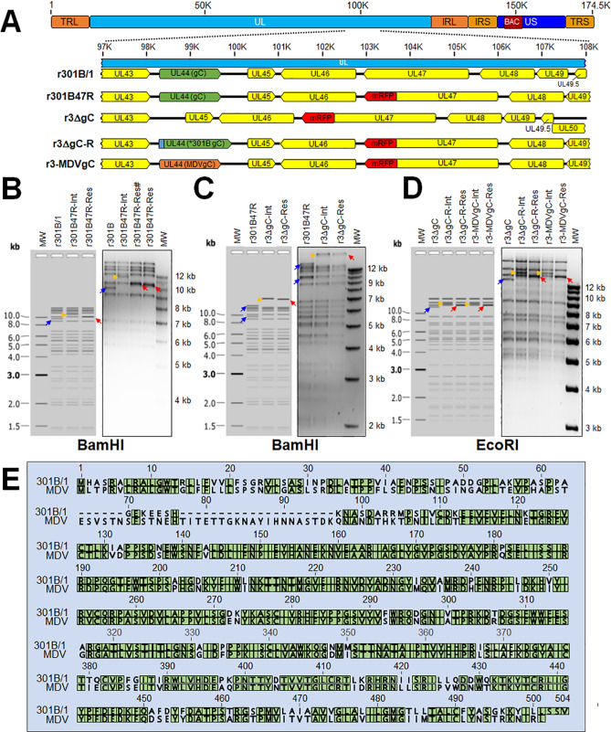
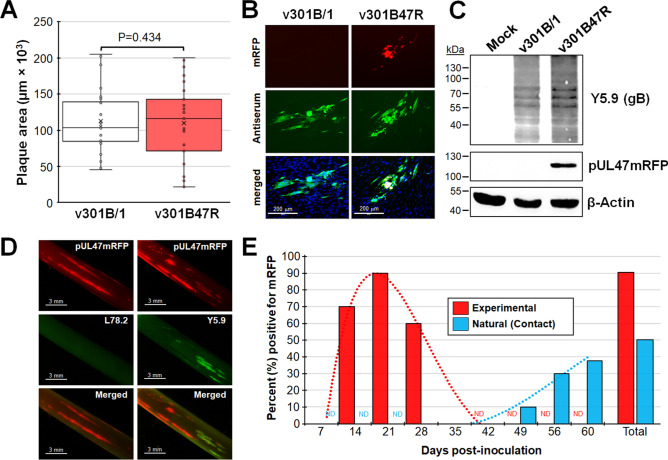
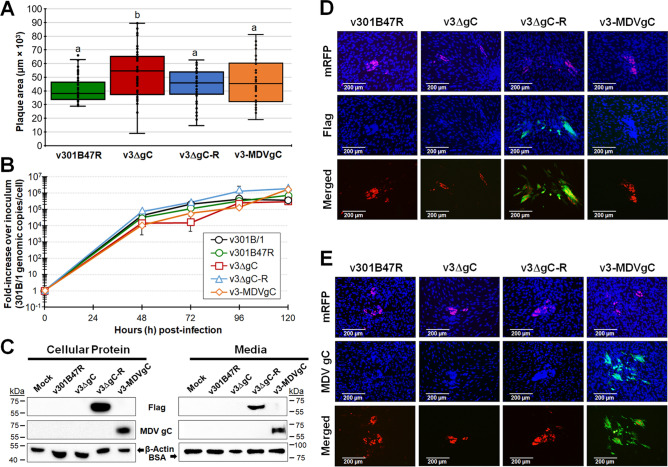
We have formerly shown that glycoprotein C (gC) of Gallid alphaherpesvirus 2, better known as Marek's disease (MD) alphaherpesvirus (MDV), is required for interindividual spread in chickens. Since gC is conserved within the Alphaherpesvirinae subfamily, we hypothesized gC was important for interindividual spread of other alphaherpesviruses. To test this hypothesis, we first generated a fluorescent protein tagged clone of Gallid alphaherpesvirus 3 MD vaccine strain 301B/1 to track virus replication in cell culture and chickens using fluorescent microscopy. Following validation of this system, we removed the open reading frame of 301B/1 gC from the genome and determined whether it was required for interindividual spread using experimental and natural infection studies. Interindividual spread of MD vaccine 301B/1 was abrogated by removal of 301B/1 gC. Rescuent virus in which 301B/1 gC was inserted back into the genome efficiently spread among chickens. To further study the conserved function of gC, we replaced 301B/1 gC with MDV gC and this virus also efficiently spread in chickens. These data suggest the essential function of alphaherpesvirus gC proteins is conserved and can be exploited during the generation of future vaccines against MD that affects the poultry industry worldwide.


Conflict of interest statement

The authors declare no competing interests.

Figures

Figure 1
Figure 1
Generation of r301B/1clones. (A) Schematic representation of the 301B infectious clone genome depicting the locations of the terminal repeat long (TRL) and short (TRS), internal repeat long (IRL) and short (IRS), and unique long (UL) and short (US) regions. The region of the UL spanning UL43 to UL50 is expanded to show the relevant genes within this region and modifications for each r301B/1 clone. (B) Predicted and actual RFLP analysis of r301B/1 clones. BAC DNA obtained for r301B, r301B47R-integrate clone and two resolved clones were digested with BamHI and electrophoresed through a 1.0% agarose gel. Integration of the mRFP + AphAI sequence resulted in an increase in the 9545 bp (blue colour rightwards arrow) fragment to 11,207 bp (yellow colour rightwards arrow). Resolution by removal of the AphAI sequence shifted the 11,207 bp fragment to 10,233 bp (red colour leftwards arrow). One resolved clone (#) was used after this point. (C) Predicted and actual RFLP analysis of r3ΔgC r301B clone. BAC DNA obtained for r301B47R, r3ΔgC-integrate clone and r3ΔgC-resolved clone were digested with BamHI and electrophoresed through a 1.0% agarose gel. Integration of the AphAI sequence into this locus removed a BamHI site combining the 10,223 and 14,854 bp fragments (blue colour rightwards arrow) to 24,671 bp (yellow colour rightwards arrow). Resolution of the AphAI sequence reduced the 23,933 bp fragment by 1028 bp to 23,643 bp (red colour leftwards arrow). (D) Predicted and actual RFLP analysis of r3ΔgC-R and r3-MDVgC clones derived from the r3ΔgC clone. BAC DNA obtained for r3ΔgC, r3ΔgC-R-integrate, r3ΔgC-R-resolved, r3-MDVgC-integrate, and r3-MDVgC-resolved clones were digested with EcoRI and electrophoresed through a 1.0% agarose gel. Integration of 3 × Flag301BgC-AphAI or MDVgC-AphAI sequences into this locus resulted an increase in the 12,879 bp (blue colour rightwards arrow) fragment to 15,420 bp (yellow colour rightwards arrow) or 15,403 bp (yellow colour rightwards arrow), respectively. Removal of the AphAI sequence from r3ΔgC-R-Int reduced the 15,420 bp fragment by 1038 bp to 14,382 bp (red colour leftwards arrow) to generate r3ΔgC-R-Res. Removal of the AphAI sequence from r3-MDVgC-Int reduced the 15,403 bp fragment by 1034 bp to 14,369 bp (red colour leftwards arrow) to generate r3-MDVgC-Res. The molecular weight marker was the 1 kb Plus DNA Ladder from Invitrogen, Inc. (Carlsbad, CA). No extraneous alterations are evident. (E) Alignment of 301B/1 and MDV (RB-1B strain) gC protein using MUSCLE Alignment in Geneious Prime 2021.0.3 (Biomatters, Inc., San Diego, CA). Green highlighted amino acids are conserved between the two proteins.
Figure 2
Figure 2
Replication and fluorescent protein expression in tissue culture cells. (A) Mean plaque areas (n = 25) of viruses reconstituted from r301B/1 and r301B47R were measured and the results are shown as box & whisker plots. There were no significant differences in plaque sizes between the two viruses using Student’s t tests. (B) Representative plaques for v301B/1 and v301B47R are shown. Plaques were stained with polyclonal chicken anti-GaHV-3 antibody and goat anti-chicken-IgY Alexa488 (green) was used as secondary antibody to identify plaques. Fluorescent expression of mRFP (red) was directly visualized and cells were counterstained with Hoechst 33342 to visualize nuclei. (C) Western blotting for pUL47mRFP using anti-mRFP antibody. The anti-GaHV-3 antibody Y5.9 was used to show the relative level of infection in the cultures. Antibody against chicken β-actin is shown as a loading control. (D) Fluorescent protein expression in feather follicles (FFs) infected with v301B47R at 21 dpi. FFs were also stained with anti-HVT L78.2 or -GaHV-3 Y5.9 plus anti-mouse IgG-Alexa488 (green) and images were collected with a fluorescent stereoscope. (E) Percent of chickens positive for pUL47mRFP in experimental and naturally (contact) infected chickens over 60 days. ND not done.
Figure 3
Figure 3
Replication and expression of proteins in cell culture. (A) Mean plaque areas for viruses reconstituted from r301B47R, r3ΔgC, r3ΔgC-R, and r3-MDVgC were measured and shown as box & whisker plots. Significant differences were determined using one-way ANOVA (p < 0.05, n = 200). Mean plaque areas with different letters are significantly different using LSD and Tukey’s post hoc tests (p ≤ 0.05). (B) Multi-step growth kinetics was used to measure virus replication in CEC cultures. The mean fold-change in viral DNA copies over the inoculum is shown for each virus and time point. There were no differences in virus growth (p > 0.05, two-way ANOVA, LSD, n = 3) (C) Western blotting to confirm 301B/1 gC and MDV gC expression. Both total cellular protein and infected cell culture media were used to detect 3 × Flag tagged 301B/1 gC and MDV gC. Anti-Flag M2 was used to detect 301B/1 gC, while anti-MDV gC A6 antibody was used to confirm MDV gC expression. For protein loading control, mouse anti-β-actin was used for total protein, while anti-BSA was used for infected cell media. (D and E) Expression of 301B/1 gC in v3ΔgC-R and MDV gC in v3-MDVgC. Representative plaques for all four viruses were stained with anti-Flag (D) or -MDV gC A6 (E) antibodies with goat anti-mouse-Alexa488 (green) as secondary antibody. Fluorescent expression of mRFP (red) was directly visualized and cells were counterstained with Hoechst 33342 to visualize nuclei. Only anti-Flag (D) or -MDV gC (E) and pUL47mRFP are shown in the merged images.
Figure 4
Figure 4
Replication and interindividual spread of r301B/1 viruses in chickens. Pure Columbian chickens were experimentally infected with v301B47R, v3ΔgC, v3ΔgC-R, or v3-MDVgC as described in the Materials and Methods for 56 days. (A) Replication was monitored in experimentally infected chickens by quantification of 301B/1 genomes in the blood over the first 4 weeks of infection. Shown is the mean 301B/1 genomic copies per 106 blood cells ± standard error of means. No significant differences (p > 0.05, n = 109) were determined between all viruses at the same time point. (B) Quantitative analysis of the percent of birds positive for pUL47mRFP in FFs over the course of the experiment. Using Fisher’s exact test at p < 0.05, there was no significant difference in the total number of chickens positive for experimentally infected chickens with 100% positive by 21 days pi. No naïve contact chickens housed with v3ΔgC were naturally infected, while 88, 75, and 60% of contact chickens were naturally infected with v301B47R, v3-MDVgC, or vΔgC-R, respectively. Using Fisher’s exact test at p < 0.05, there was no significant difference between v3ΔgC-R (p = 0.5105) and v3-MDVgC (p = 1.0000) compared to v301B47R, while v3ΔgC was significantly different (p = 0.0047). (C and D) Feathers were plucked from v301B47R, v3ΔgC, v3ΔgC-R, and v3-MDVgC at 28 dpi, fixed, then stained using anti-Flag M2 (C) or anti- MDV gC (D) antibodies. FFs obtained from v3ΔgC-R-infected birds were positive for 3 × Flag301B gC, while FFs from v3-MDVgC -infected chickens were positive for MDV gC protein. (E) Western blot analysis for 3 × Flag301B and MDV gC in FFEs. Whole-cell protein lysates were collected from FFE cells scraped from infected FFs, electrophoresed through a 10% SDS-PAGE gel, transferred to nitrocellulose membranes, and probed for Flag or MDV gC as described in the Materials and Methods. Anti-β-actin antibody was used as internal cellular control.

References

    1. Lefkowitz EJ, et al. Virus taxonomy: the database of the International Committee on Taxonomy of Viruses (ICTV) Nucl. Acids Res. 2018;46:D708–D717. doi: 10.1093/nar/gkx932. - DOI - PMC - PubMed
    1. Mettenleiter TC. Glycoprotein gIII deletion mutants of pseudorabies virus are impaired in virus entry. Virology. 1989;171:623–625. doi: 10.1016/0042-6822(89)90635-1. - DOI - PubMed
    1. Rue CA, Ryan P. Characterization of pseudorabies virus glycoprotein C attachment to heparan sulfate proteoglycans. J. Gen. Virol. 2002;83:301–309. doi: 10.1099/0022-1317-83-2-301. - DOI - PubMed
    1. Osterrieder N. Construction and characterization of an equine herpesvirus 1 glycoprotein C negative mutant. Virus Res. 1999;59:165–177. doi: 10.1016/s0168-1702(98)00134-8. - DOI - PubMed
    1. Langeland N, et al. Localization on the herpes simplex virus type 1 genome of a region encoding proteins involved in adsorption to the cellular receptor. J. Virol. 1990;64:1271–1277. doi: 10.1128/JVI.64.3.1271-1277.1990. - DOI - PMC - PubMed

Publication types

MeSH terms

Substances

LinkOut - more resources