. 2021 Aug;23(8):1484-1491.
doi: 10.1038/s41436-021-01161-6. Epub 2021 Apr 8.

Clinical spectrum of MTOR-related hypomelanosis of Ito with neurodevelopmental abnormalities

Affiliations

Clinical spectrum of MTOR-related hypomelanosis of Ito with neurodevelopmental abnormalities

Virginie Carmignac et al. Genet Med. 2021 Aug.

Erratum in

  • Correction to: Clinical spectrum of MTOR-related hypomelanosis of Ito with neurodevelopmental abnormalities.
    Carmignac V, Mignot C, Blanchard E, Kuentz P, Aubriot-Lorton MH, Parker VER, Sorlin A, Fraitag S, Courcet JB, Duffourd Y, Rodriguez D, Knox RG, Polubothu S, Boland A, Olaso R, Delepine M, Darmency V, Riachi M, Quelin C, Rollier P, Goujon L, Grotto S, Capri Y, Jacquemont ML, Odent S, Amram D, Chevarin M, Vincent-Delorme C, Catteau B, Guibaud L, Arzimanoglou A, Keddar M, Sarret C, Callier P, Bessis D, Geneviève D, Deleuze JF, Thauvin C, Semple RK, Philippe C, Rivière JB, Kinsler VA, Faivre L, Vabres P. Carmignac V, et al. Genet Med. 2021 Aug;23(8):1585. doi: 10.1038/s41436-021-01217-7. Genet Med. 2021. PMID: 34257424 Free PMC article. No abstract available.

Abstract

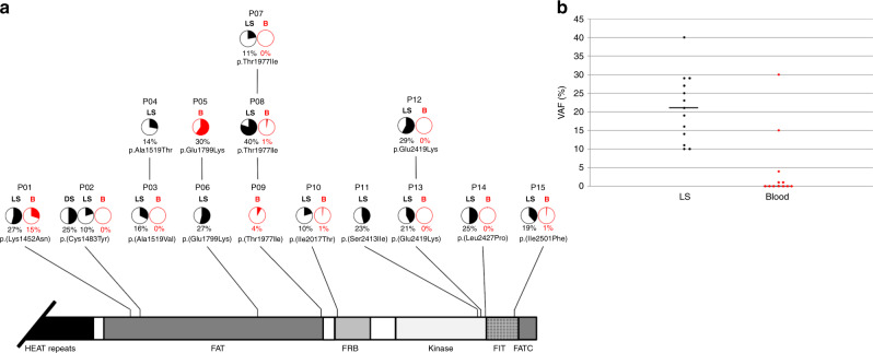
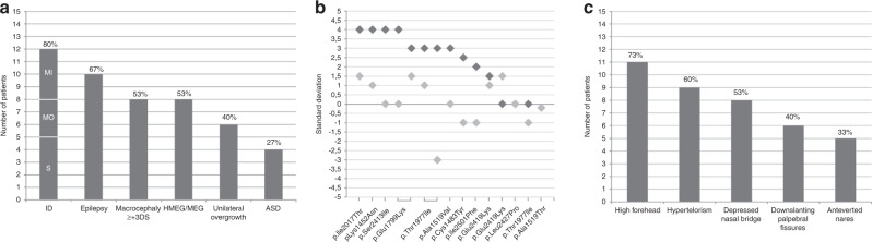
Purpose: Hypomelanosis of Ito (HI) is a skin marker of somatic mosaicism. Mosaic MTOR pathogenic variants have been reported in HI with brain overgrowth. We sought to delineate further the pigmentary skin phenotype and clinical spectrum of neurodevelopmental manifestations of MTOR-related HI.

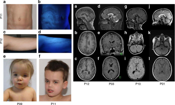
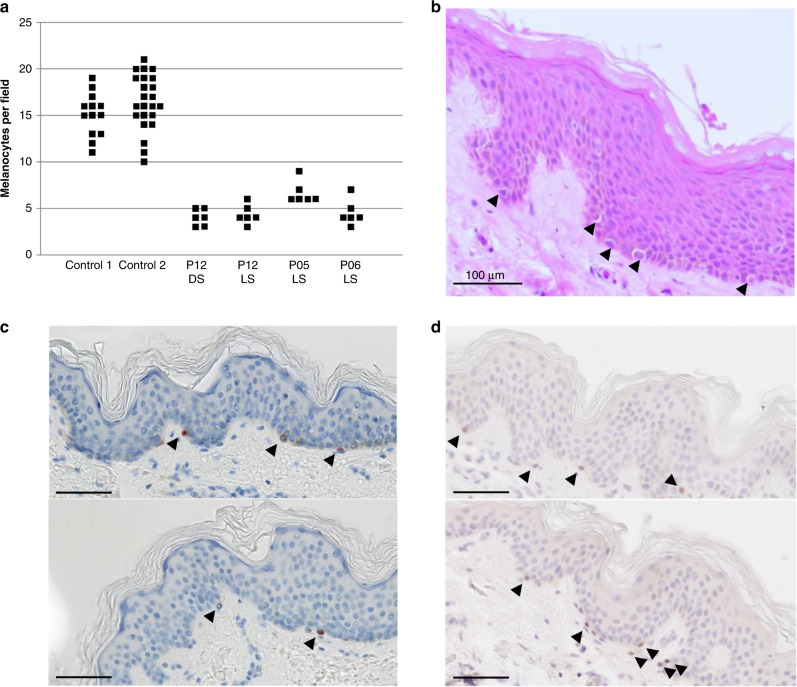
Methods: From two cohorts totaling 71 patients with pigmentary mosaicism, we identified 14 patients with Blaschko-linear and one with flag-like pigmentation abnormalities, psychomotor impairment or seizures, and a postzygotic MTOR variant in skin. Patient records, including brain magnetic resonance image (MRI) were reviewed. Immunostaining (n = 3) for melanocyte markers and ultrastructural studies (n = 2) were performed on skin biopsies.

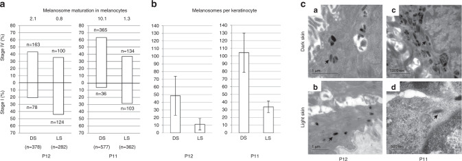
Results: MTOR variants were present in skin, but absent from blood in half of cases. In a patient (p.[Glu2419Lys] variant), phosphorylation of p70S6K was constitutively increased. In hypopigmented skin of two patients, we found a decrease in stage 4 melanosomes in melanocytes and keratinocytes. Most patients (80%) had macrocephaly or (hemi)megalencephaly on MRI.

Conclusion: MTOR-related HI is a recognizable neurocutaneous phenotype of patterned dyspigmentation, epilepsy, intellectual deficiency, and brain overgrowth, and a distinct subtype of hypomelanosis related to somatic mosaicism. Hypopigmentation may be due to a defect in melanogenesis, through mTORC1 activation, similar to hypochromic patches in tuberous sclerosis complex.


Conflict of interest statement

The authors declare no competing interests.

Figures

Fig. 1
Fig. 1. Distribution of postzygotic missense MTOR variations.
(a) Structure of mTOR protein including the Huntingtin, Elongation factor 3, protein phosphatase 2A, and TOR1 (HEAT) repeat; FAT (FRAP, ATM, and TRRAP) domain; FRB (FKBP12-rapamycin binding) domain; kinase (serine-threonine kinase kinase) domain; FATC (FAT, FRAP, ATM and TRRAP carboxy-terminal) domain; and FIT (Found in TOR) domain. Variant allele fractions (VAFs) (%) are shown as pie charts with a maximum allele fraction of 50%. B (red) blood, DS dark skin, LS light skin. Variation details are summarized in Supplementary Table 3. (b) Distribution of VAFs in hypopigmented skin (LS, black dots) and blood (red dots), with median values.
Fig. 2
Fig. 2. Clinical pigmentary skin phenotype and brain imaging.
Clinical pigmentary skin phenotype in two patients (Left). (a, b) P11. (a) Unilateral linear and whorled hypopigmentation in multiple large bands with ragged border and sharp midline limitation on the abdomen. (b) Enhanced contrast on Wood’s lamp illumination. (c, d) P12. (c) Linear hypopigmentation in large bands on left lower limb. (d) Enhanced contrast on Wood’s lamp illumination. (e, f) Facial features of patients P09 and P11. Brain magnetic resonance image (MRI) of subjects P01, P03, P10 and P12 (Right). (ac) P12. Left HMEG with altered ventricle shape (small frontal horn), enlarged left thalamus and caudate nucleus (a), slightly thickened cortical mantle and enlarged white matter with normal signal (b) and enlarged anterior corpus callosum (c). (df) P03. Normal corpus callosum (d), small frontal horns (e, f), and mild overgrowth of the right cerebral hemisphere, with slight enlargement of the right posterior ventricle (c). (gi) P10. Normal corpus callosum on sagittal section (g), overgrowth of the right cerebellar hemisphere (h), right posterior ventricle enlargement, small frontal horn, increased white matter volume and thickened cortical mantle (i). (jl) P01. Thickened corpus callosum (j), with symmetrical enlargement of cerebellar (k) and cerebral hemispheres (l).
Fig. 3
Fig. 3. Microscopy of paraffin embedded skin biopsies.
(a) Numbers of melanocytes per field on whole skin sections from patients P05, P06, and P12 (patients: 6 fields; controls: 13 and 24 fields; hematoxylin eosin [HE] staining). DS dark skin, LS light skin. (b) HE staining from P06 hypomelanotic skin. (c) Subject P12. Melan-A immunohistochemistry on skin biopsy sections from pigmented skin (top) and hypomelanotic skin (bottom). (d) Subject P12. MITF labeling on biopsy sections from pigmented skin (top) and hypomelanotic skin (bottom). Scale bar: 100 µm. Melanocytes are shown by arrowheads.
Fig. 4
Fig. 4. Ultrastructural study.
(a) Melanosome maturation in melanocytes: Melanosome count in melanocytes on skin biopsy sections from pigmented (black) and hypopigmented area (white) in patients P12 (left) and P11 (right). Melanosomes were counted in 15 melanocytes. Upper bars: stage I melanosome. Lower bars: stage IV melanosomes (numbers at top of each bar). Ratio and total number of counted melanosomes are given at top and bottom of each figure. (b) Melanosome quantification in keratinocytes. Mean number of melanosomes per basal layer keratinocyte (n = 50) (c). Transmission electron microscopy (TEM) of melanocytes at the basal epidermal layer in subjects P12 (a, b) and P11 (c, d). Melanosomes (arrows), in dark (ac) and hypopigmented (bd) areas. DS dark skin, LS light skin.
Fig. 5
Fig. 5. Clinical features in hypomelanosis of Ito (HI) individuals with MTOR postzygotic pathogenic variants.
(a) Frequencies of neurological involvement and overgrowth. (b) Standard deviations for occipitofrontal circumference (OFC) (dark gray) and body height (light gray). (c) Frequencies of recurrent facial features. Percentages were calculated with the number of patients with available data as the denominator. ID intellectual disability, MI mild, MO moderate, S severe.

Comment in

Similar articles

Cited by

References

    1. >Ito M. Incontinentia pigmenti achromians. A singular case of nevus depigmentosus systematicus bilateralis. Tohoku J. Exp. Med. 1952;55:57–59.
    1. Schwartz MF, Esterly NB, Fretzin DF, Pergament E, Rozenfeld IH. Hypomelanosis of Ito (incontinentia pigmenti achromians): a neurocutaneous syndrome. J. Pediatr. 1977;90:236–240. doi: 10.1016/S0022-3476(77)80636-7. - DOI - PubMed
    1. Hamada K, et al. Incontinentia pigmenti achromians as part of a neurocutaneous syndrome: a case report. Brain Dev. 1979;1:313–317. doi: 10.1016/S0387-7604(79)80047-9. - DOI - PubMed
    1. Pavone P, Praticò AD, Ruggieri M, Falsaperla R. Hypomelanosis of Ito: a round on the frequency and type of epileptic complications. Neurol. Sci. 2015;36:1173–1180. doi: 10.1007/s10072-014-2049-1. - DOI - PubMed
    1. Glover MT, Brett EM, Atherton DJ. Hypomelanosis of ito: spectrum of the disease. J. Pediatr. 1989;115:75–80. doi: 10.1016/S0022-3476(89)80332-4. - DOI - PubMed

Publication types

Substances

LinkOut - more resources