. 2022 May;29(5):519-532.
doi: 10.1038/s41417-021-00329-1. Epub 2021 Apr 8.

Repurposing cabozantinib with therapeutic potential in KIT-driven t(8;21) acute myeloid leukaemias

Affiliations

Repurposing cabozantinib with therapeutic potential in KIT-driven t(8;21) acute myeloid leukaemias

Kuan-Wei Su et al. Cancer Gene Ther. 2022 May.

Abstract

Cabozantinib is an orally available, multi-target tyrosine kinase inhibitor approved for the treatment of several solid tumours and known to inhibit KIT tyrosine kinase. In acute myeloid leukaemia (AML), aberrant KIT tyrosine kinase often coexists with t(8;21) to drive leukaemogenesis. Here we evaluated the potential therapeutic effect of cabozantinib on a selected AML subtype characterised by t(8;21) coupled with KIT mutation. Cabozantinib exerted substantial cytotoxicity in Kasumi-1 cells with an IC50 of 88.06 ± 4.32 nM, which was well within clinically achievable plasma levels. The suppression of KIT phosphorylation and its downstream signals, including AKT/mTOR, STAT3, and ERK1/2, was elicited by cabozantinib treatment and associated with subsequent alterations of cell cycle- and apoptosis-related molecules. Cabozantinib also disrupted the synthesis of an AML1-ETO fusion protein in a dose- and time-dependent manner. In a mouse xenograft model, cabozantinib suppressed tumourigenesis at 10 mg/kg and significantly prolonged survival of the mice. Further RNA-sequencing analysis revealed that mTOR-mediated signalling pathways were substantially inactivated by cabozantinib treatment, causing the downregulation of ribosome biogenesis and glycolysis, along with myeloid leukocyte activation. We suggest that cabozantinib may be effective in the treatment of AML with t(8;21) and KIT mutation. Relevant clinical trials are warranted.


Conflict of interest statement

The authors declare no competing interests.

Figures
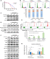
Fig. 1
Fig. 1. Cabozantinib induces G0/G1 cell-cycle arrest and apoptosis in KIT-mutated t(8;21) AML cells.
A Kasumi-1 and SKNO-1 cells were treated with 0.01% DMSO (control) or various concentrations of cabozantinib for 72 h. MTS assay was performed to obtain the IC50 values of cabozantinib for both cell lines. The IC50 values were calculated using the CalcuSyn software. B Flow cytometric analysis after 24-h cabozantinib represents the proportion of cells at different cell-cycle stages. Histograms are one of the representative results. The data represented the averages ± SD of three independent experiments (n = 3). ***p < 0.001, **p < 0.01, *p < 0.05, compared with DMSO control. C Two G1/S transition regulators, p27 and cyclin E, were measured by western blot analysis. β-actin was loaded as a control. Signal intensity was quantified with ImageJ and normalised to β-actin and DMSO controls. D Flow cytometric analysis after 72-h cabozantinib treatment is presented as dot plots and the bar graphs show the proportion of cells at different apoptotic status. Each column represented the averages ± SD of three independent experiments (n = 3). ***p < 0.001, **p < 0.01, *p < 0.05, compared with DMSO control. E Western blot analysis of PARP cleavage, caspase-3 activation and F apoptosis-related proteins, including BAX, BAK, PUMA, Survivin, MCL-1, and BCL-2, in Kasumi-1 cells following 24-h cabozantinib treatment. G BBC3 and survivin relative mRNA expression were analysed by RT-qPCR analysis following 24-h cabozantinib treatment. Each column represented the averages ± SD of three independent experiments (n = 3). ***p < 0.001, **p < 0.01, *p < 0.05, compared with DMSO control.
Fig. 2
Fig. 2. Cabozantinib inhibits the phosphorylation of KIT-mediated signalling molecules and elicits FOXO3a nuclear localisation in Kasumi-1 cells.
Cells were treated with different concentrations of cabozantinib and sorafenib for 4 h. Sorafenib, a known a KIT inhibitor, was used as a positive control. A Phosphorylation of KIT and its downstream STAT3, AKT/mTOR and ERK molecules in Kasumi-1 cells were measured by western blots. Kasumi-1 cells were treated with DMSO or various concentrations of cabozantinib for 4 h. B Western blots of whole-cell lysates and C subcellular fractions of Kasumi-1 cells. Histone H3 (nuclear) and α-tubulin (cytosol) served as loading controls for fractionation. Signal intensity was quantified with ImageJ and normalised to the loading control and control group. D Confocal microscopy images of DMSO-treated or cabozantinib-treated Kasumi-1 cells show signals for DAPI stained nuclei (blue) and FOXO3a (green), and the merged images (overlay). The red arrowheads point to representative cells showing increased nuclear FOXO3a levels. Scale bars, 10 μm.
Fig. 3
Fig. 3. Molecular mechanisms of cabozantinib-induced AML1-ETO downregulation.
A Kasumi-1 cells were treated with DMSO or various concentrations of cabozantinib for 24 h, 48 h, and 72 h before harvesting. Western blot analysis of the time course of changes in protein levels of AML1-ETO. B Western blot analysis of the time course of changes in the protein level of AML1-ETO after DMSO (control), CHX alone or in combination with cabozantinib. Signal intensity was quantified with ImageJ and normalised to β-actin and control (0-h) group. C The protein expression of AML1-ETO was measured in Kasumi-1 cells, which were treated with 500 nM cabozantinib for indicated hours before harvested for western blot analysis. The proteasome inhibitor MG-132 (5 μM) was added 4 h before cell harvest. D Monitoring protein synthesis in Kasumi-1 and OCI-AML3 cells by puromycin labelling. β-actin was loaded as a control. Signal intensity was quantified with ImageJ and normalised to β-actin and DMSO controls. E Co-immunoprecipitation (co-IP) followed puromycin-labelled de novo synthesis protein showed decreased AML1-ETO protein expression level. Mouse IgG was used as a negative control of co-IP.
Fig. 4
Fig. 4. Genome-wide assessment of the anti-leukaemia effects.
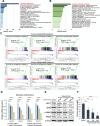
RNA-seq was performed on Kasumi-1 cells that were either untreated or treated with cabozantinib (4 and 24 h at 100 nM). A Gene ontology (GO) analysis using Metascape was performed to analyse genes markedly enriched compared with the DMSO group, after the 4-h cabozantinib treatment and B the 24-h cabozantinib treatment. C Gene set enrichment analysis (GSEA) was also performed to interpret gene expression data (4-h cabozantinib-treated, 24-h cabozantinib-treated). The common downregulated pathways of the two groups were identified by GSEA analysis using the Hallmark collections of the GSEA MSigDB 7.0. In addition, Kasumi-1 cells were treated with DMSO (control) or various concentrations of cabozantinib for 24 h, and harvested for western blot analysis, RT-qPCR, and glucose uptake assay. D RT-qPCR analysis of glycolysis-related genes. Transcript levels were normalised to those of actin, and the relative mRNA expression was calculated using the 2ΔΔCT method. Each column represented the averages ± SD of three independent experiments (n = 3). ***p < 0.001, **p < 0.01, *p < 0.05, compared with DMSO control. E The cell lysates mentioned above were subjected to immunoblotting with c-MYC, LDHA, p-PKM2, PKM2, and β-actin antibodies. β-actin was loaded as control. Signal intensity was quantified with ImageJ and normalised to β-actin and DMSO controls. F Determination of glucose uptake by cabozantinib-treated Kasumi-1 cells. Values represent mean ± SD. ***p < 0.001, **p < 0.01, *p < 0.05, compared with DMSO control.
Fig. 5
Fig. 5. Cabozantinib inhibits AML1-ETO-mediated transcriptome.
A Western blot analysis of the phosphorylation of S6K and 4E-BP1. β-actin was loaded as a control. Signal intensity was quantified with ImageJ and normalised to β-actin and DMSO controls. B GSEA analysis identified genes upregulated following cabozantinib treatment were similar to a pre-defined gene set (DUNNE_TARGETS_OF_AML1_MTG8_FUSION_UP) comprising genes upregulated in Kasumi-1 cells after AML1-ETO knockdown by siRNA. C Relative mRNA expression of AML1-ETO-suppressed genes related to cell differentiation was analysed following 24-h of cabozantinib treatment in Kasumi-1 cells. Actin served as the internal control. Transcript levels were normalised to those of actin and the relative mRNA expression was calculated using the 2−ΔΔCT method. Each column represented the averages SD of three independent experiments (n = 3). ***p < 0.001, **p < 0.01, *p < 0.05, compared with DMSO control.
Fig. 6
Fig. 6. Effects of cabozantinib in a Kasumi-1 xenograft mouse model.
A Cabozantinib causes prominent regression of subcutaneous Kasumi-1 tumours in athymic nude mice. The results were plotted as mean tumour size ± SD of three groups over time. B The tumours were dissected and photographed at 4 h following the last oral administration with cabozantinib. C When the tumour volume reached 1000 mm3 the mice were considered death. Comparisons of survival curves estimated by Kaplan–Meier plots. D No obvious body weight loss was observed during treatments. E Tumours were recovered from Kasumi-1-bearing mice after 4 h of treatment with vehicle or cabozantinib (n = 3/group); then tumour lysates were extracted to analyse phosphorylated KIT, STAT3, AKT/mTOR and ERK molecules by western blot. F H&E staining was performed on Kasumi-1 tumours treated with cabozantinib or vehicle. Scale bars, 50 μm.

Similar articles

Cited by

References

    1. Shipley JL, Butera JN. Acute myelogenous leukemia. Exp Hematol. 2009;37:649–58. doi: 10.1016/j.exphem.2009.04.002. - DOI - PubMed
    1. Short NJ, Rytting ME, Cortes JE. Acute myeloid leukaemia. Lancet. 2018;392:593–606. doi: 10.1016/S0140-6736(18)31041-9. - DOI - PMC - PubMed
    1. Christen F, Hoyer K, Yoshida K, Hou HA, Waldhueter N, Heuser M, et al. Genomic landscape and clonal evolution of acute myeloid leukemia with t(8;21): a international study on 331 patients. Blood. 2019;133:1140–51. doi: 10.1182/blood-2018-05-852822. - DOI - PubMed
    1. Mulloy JC, Cammenga J, MacKenzie KL, Berguido FJ, Moore MA, Nimer SD. The AML1-ETO fusion protein promotes the expansion of human hematopoietic stem cells. Blood. 2002;99:15–23. doi: 10.1182/blood.V99.1.15. - DOI - PubMed
    1. Kelly LM, Gilliland DG. Genetics of myeloid leukemias. Annu Rev Genomics Hum Genet. 2002;3:179–98. doi: 10.1146/annurev.genom.3.032802.115046. - DOI - PubMed

Publication types

MeSH terms