

Zinc Nanoparticles Ameliorate the Reproductive Toxicity Induced by Silver Nanoparticles in Male Rats
- PMID: 33833511
- PMCID: PMC8020588
- DOI: 10.2147/IJN.S307189
Zinc Nanoparticles Ameliorate the Reproductive Toxicity Induced by Silver Nanoparticles in Male Rats
Abstract
Introduction: Silver nanoparticles (Ag-NPs) are among the most commonly used nanoparticles in different fields. Zinc nanoparticles (Zn-NPs) are known for their antioxidant effect. This study was designed to investigate the adverse effects of Ag-NPs (50 nm) on the male reproductive system and also the ameliorative effect of Zn-NPs (100 nm) against these harmful effects.
Methods: Forty adult male rats were used in this study; they were randomly divided into four equal groups: control group, Ag-NPs group, Zn-NPs group, Ag-NPs + Zn-NPs group. Ag-NPs (50 mg/kg) and/or Zn-NPs (30 mg/kg) were administered orally for 90 days.
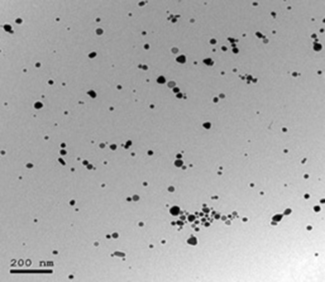
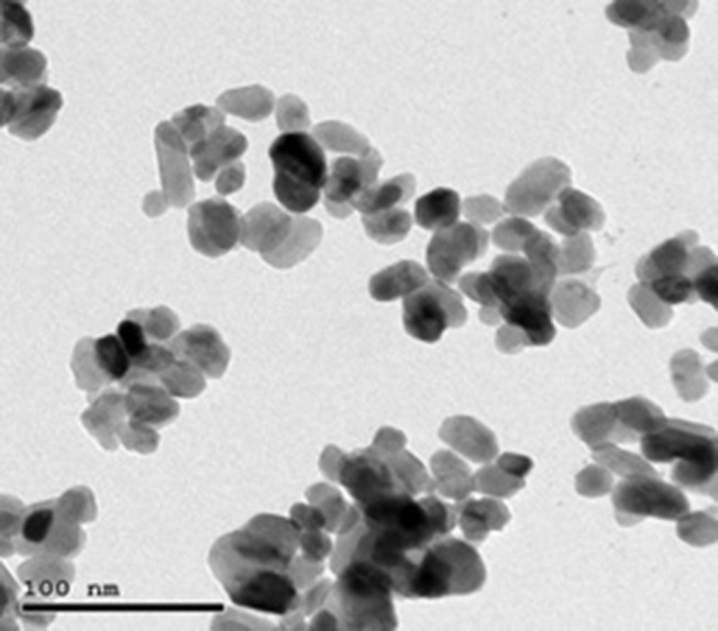
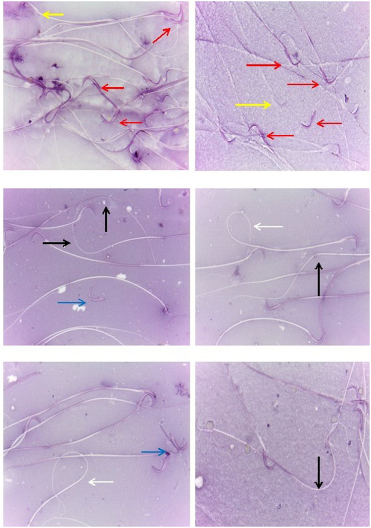
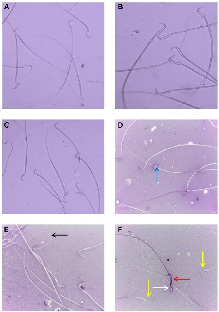
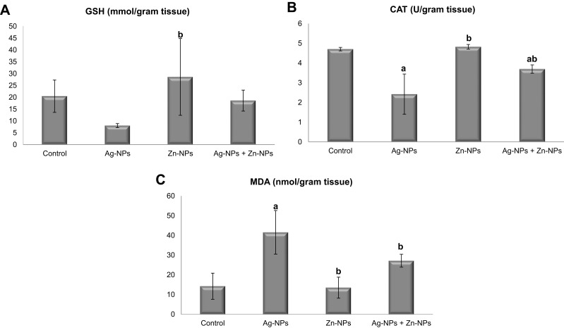
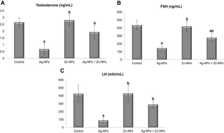
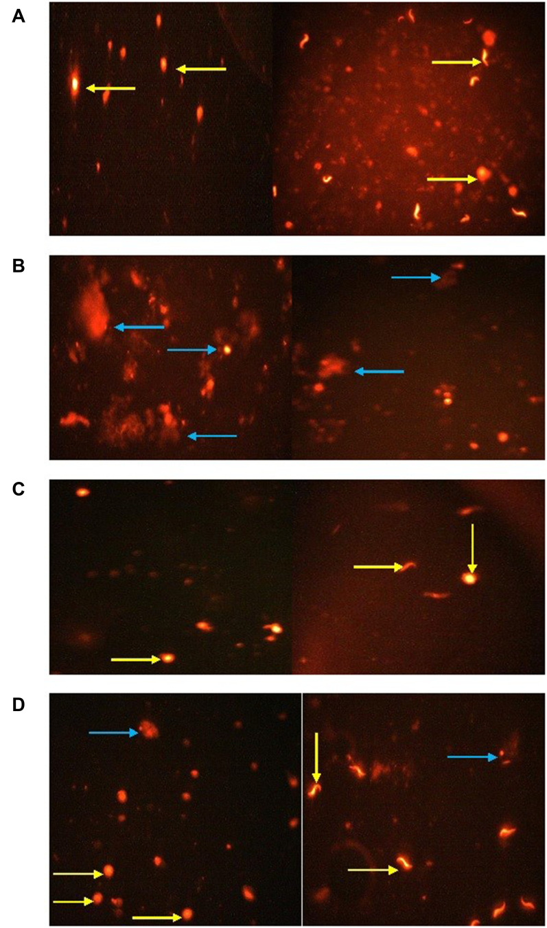
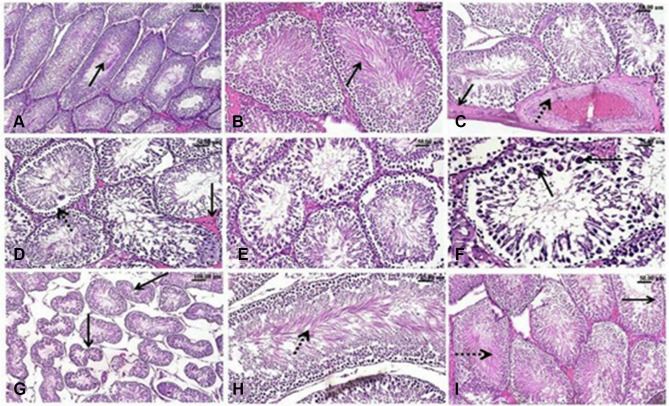
Results: The results revealed that exposure to Ag-NPs adversely affected sperm motility, morphology, viability, and concentration. Ag-NPs also induced oxidative stress and lipid peroxidation in testicular tissue. The exposure to Ag-NPs decreased serum FSH, LH, and testosterone hormones. Additionally, comet assay revealed DNA degeneration in the testicular tissue of rats exposed to Ag-NPs. Histopathological examination showed various histological alterations in the testes of rats intoxicated with Ag-NPs. Furthermore, co-administration of Zn-NPs ameliorated most of the toxic effects of Ag-NPs via their antioxidative capacity.
Keywords: DNA damage; antioxidants; endocrinology; oxidative stress; sperm evaluation; testes.
© 2021 Shehata et al.
Conflict of interest statement
The authors declare no conflicts of interest for this work.
Figures
Similar articles
-
Evaluation of the Ameliorative Effect of Zinc Nanoparticles against Silver Nanoparticle-Induced Toxicity in Liver and Kidney of Rats.Biol Trace Elem Res. 2022 Mar;200(3):1201-1211. doi: 10.1007/s12011-021-02713-2. Epub 2021 Apr 14. Biol Trace Elem Res. 2022. PMID: 33855683
-
Zinc nanoparticles ameliorate oxidative stress and apoptosis induced by silver nanoparticles in the brain of male rats.Neurotoxicology. 2023 Mar;95:193-204. doi: 10.1016/j.neuro.2023.02.005. Epub 2023 Feb 14. Neurotoxicology. 2023. PMID: 36796650
-
Thymol alleviates silica dioxide nanoparticle-induced reproductive performance toxicity via antioxidant and anti-inflammatory mechanisms in male rats.Sci Rep. 2025 Jul 4;15(1):23913. doi: 10.1038/s41598-025-07769-x. Sci Rep. 2025. PMID: 40615456 Free PMC article.
-
Putative adverse outcome pathways for silver nanoparticle toxicity on mammalian male reproductive system: a literature review.Part Fibre Toxicol. 2023 Jan 5;20(1):1. doi: 10.1186/s12989-022-00511-9. Part Fibre Toxicol. 2023. PMID: 36604752 Free PMC article.
-
Fe-NPs and Zn-NPs: Advancing Aquaculture Performance Through Nanotechnology.Biol Trace Elem Res. 2024 Jun;202(6):2828-2842. doi: 10.1007/s12011-023-03850-6. Epub 2023 Sep 18. Biol Trace Elem Res. 2024. PMID: 37723405 Review.
Cited by
-
Modified Spirulina maxima Pectin Nanoparticles Improve the Developmental Competence of In Vitro Matured Porcine Oocytes.Animals (Basel). 2021 Aug 24;11(9):2483. doi: 10.3390/ani11092483. Animals (Basel). 2021. PMID: 34573449 Free PMC article.
-
Nanoparticles-induced potential toxicity on human health: Applications, toxicity mechanisms, and evaluation models.MedComm (2020). 2023 Jul 14;4(4):e327. doi: 10.1002/mco2.327. eCollection 2023 Aug. MedComm (2020). 2023. PMID: 37457660 Free PMC article. Review.
-
Nanotechnology Applications of Flavonoids for Viral Diseases.Pharmaceutics. 2021 Nov 8;13(11):1895. doi: 10.3390/pharmaceutics13111895. Pharmaceutics. 2021. PMID: 34834309 Free PMC article. Review.
-
Do Nanoparticles of Calcium Disodium EDTA Minimize the Toxic Effects of Cadmium in Female Rats?Biol Trace Elem Res. 2024 May;202(5):2228-2240. doi: 10.1007/s12011-023-03842-6. Epub 2023 Sep 18. Biol Trace Elem Res. 2024. PMID: 37721680 Free PMC article.
-
Evaluation of the Ameliorative Effect of Zinc Nanoparticles against Silver Nanoparticle-Induced Toxicity in Liver and Kidney of Rats.Biol Trace Elem Res. 2022 Mar;200(3):1201-1211. doi: 10.1007/s12011-021-02713-2. Epub 2021 Apr 14. Biol Trace Elem Res. 2022. PMID: 33855683
References
-
- Johnston HJ, Hutchison G, Christensen FM, Peters S, Hankin S, Stone V. A review of the in vivo and in vitro toxicity of silver and gold particulates: particle attributes and biological mechanisms responsible for the observed toxicity. Crit Rev Toxicol. 2010;40(4):328–346. - PubMed
-
- Marambio-Jones C, Hoek EMV. A review of the antibacterial effects of silver nanomaterials and potential implications for human health and the environment. J Nanoparticle Res. 2010;12:1531–1551.
-
- Samuel U, Guggenbichler JP. Prevention of catheter-related infections: the potential of a new nano-silver impregnated catheter. Int J Antimicrob Agents. 2004;1:75–78. - PubMed
-
- Kalishwaralal K, Barathmanikanth S, Pandian SR, Deepak V, Gurunathan S. Silver nano- a trove for retinal therapies. J Controlled Release. 2010;145(2):76–90. - PubMed
MeSH terms
Substances
LinkOut - more resources
Full Text Sources
Other Literature Sources
Medical
