. 2021 Feb 1;54(Pt 1):343-355.
doi: 10.1107/S1600576720013412.

ATSAS 3.0: expanded functionality and new tools for small-angle scattering data analysis

Affiliations

ATSAS 3.0: expanded functionality and new tools for small-angle scattering data analysis

Karen Manalastas-Cantos et al. J Appl Crystallogr. .

Abstract

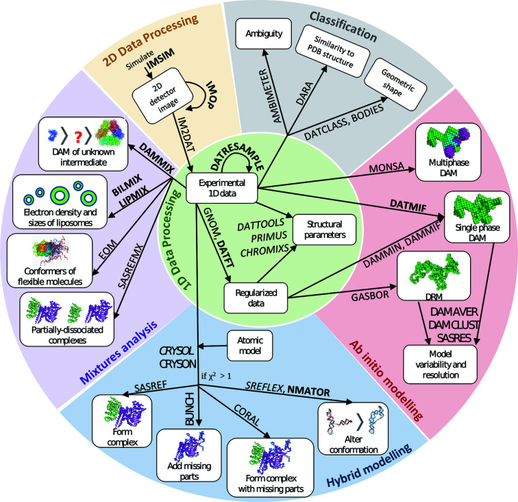
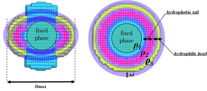
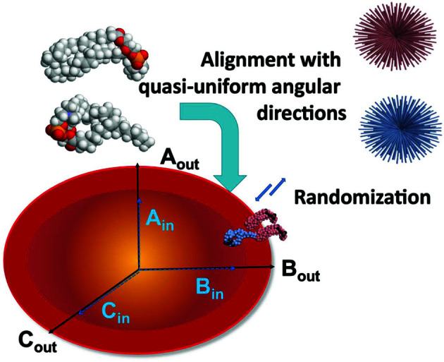
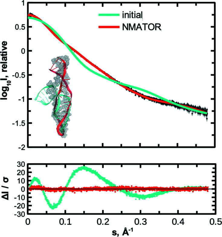
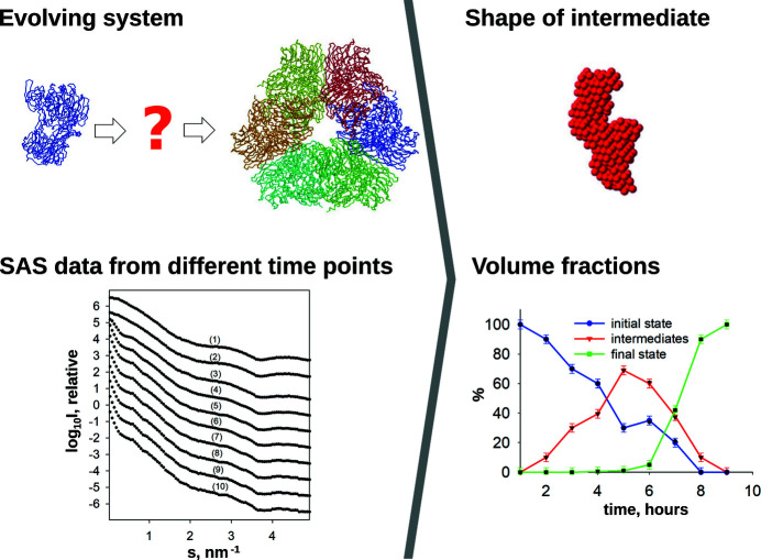
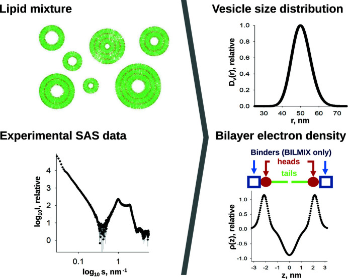
The ATSAS software suite encompasses a number of programs for the processing, visualization, analysis and modelling of small-angle scattering data, with a focus on the data measured from biological macromolecules. Here, new developments in the ATSAS 3.0 package are described. They include IMSIM, for simulating isotropic 2D scattering patterns; IMOP, to perform operations on 2D images and masks; DATRESAMPLE, a method for variance estimation of structural invariants through parametric resampling; DATFT, which computes the pair distance distribution function by a direct Fourier transform of the scattering data; PDDFFIT, to compute the scattering data from a pair distance distribution function, allowing comparison with the experimental data; a new module in DATMW for Bayesian consensus-based concentration-independent molecular weight estimation; DATMIF, an ab initio shape analysis method that optimizes the search model directly against the scattering data; DAMEMB, an application to set up the initial search volume for multiphase modelling of membrane proteins; ELLLIP, to perform quasi-atomistic modelling of liposomes with elliptical shapes; NMATOR, which models conformational changes in nucleic acid structures through normal mode analysis in torsion angle space; DAMMIX, which reconstructs the shape of an unknown intermediate in an evolving system; and LIPMIX and BILMIX, for modelling multilamellar and asymmetric lipid vesicles, respectively. In addition, technical updates were deployed to facilitate maintainability of the package, which include porting the PRIMUS graphical interface to Qt5, updating SASpy - a PyMOL plugin to run a subset of ATSAS tools - to be both Python 2 and 3 compatible, and adding utilities to facilitate mmCIF compatibility in future ATSAS releases. All these features are implemented in ATSAS 3.0, freely available for academic users at https://www.embl-hamburg.de/biosaxs/software.html.

Keywords: ATSAS; biological macromolecules; data analysis; small-angle scattering; structural modelling.


Figures

Figure 1
Figure 1
The number of biological SAS publications per year (1) has steadily increased over the past two decades, accompanied by an increase in the number of biological SAS publications which cite the ATSAS software suite (2). The numbers of unique users per year that downloaded ATSAS (3) and used the web applications in ATSAS online (4) also show a concurrent increase.
Figure 2
Figure 2
General scope of the ATSAS suite, including specific software for different use cases. Names in boldface indicate software newly added to ATSAS 3.0, while names in italics indicate updated programs (DAM: dummy-atom model; DRM: dummy-residue model).
Figure 3
Figure 3
Simulated SAXS data for parvalbumin (PDB ID 1pal), with terbium atoms in two calcium-binding sites of the protein. Regular, wavelength-independent scattering (top panel, black) was computed with CRYSOL in default mode, while anomalous scattering (top panel, red) was evaluated with CRYSOL in anomalous mode, at the L III absorption edge of terbium (7517 eV). Experimental data were simulated with IMSIM at two parvalbumin concentrations, 10 and 50 mg ml−1. DATCMP was used to compare regular and anomalous scattering at the two concentrations, showing greater differences at 50 mg ml−1 (details in Table 1 ▸). Residual plots on the bottom panel more clearly depict the differences between regular and anomalous scattering at 10 (black) and 50 mg ml−1 (red). At both concentrations, there is a reduction in forward scattering at the absorption edge. The difference between regular and anomalous SAXS is partly obscured by noise at 10 mg ml−1 but is more clearly visible at 50 mg ml−1 parvalbumin.
Figure 4
Figure 4
1D scattering data from beta-lactamase (PDB ID 5hw5) simulated by IMSIM and radially averaged with IM2DAT (dark blue), overlaid with the source data calculated by CRYSOL (cyan), and the corresponding fit of the ab initio model from DATMIF (pink). The inset shows the DATMIF bead model superimposed on the source model. The offset residual plots show random distribution of the residuals around zero within the expected bounds (±3). Corresponding goodness-of-fit statistics are reported in Table 1 ▸.
Figure 5
Figure 5
A DAMEMB-generated initial search volume for multiphase modelling of membrane proteins with MONSA. The protein phase, ρ1 (cyan), is defined within a spherical core region, located at the origin of the search volume. The core volume is surrounded by two distinct phases, ρ2 and ρ3, corresponding to the tail (pink) and head group (yellow) regions of a detergent molecule. The thickness of each phase, as well as that of the boundary region Δd, may be specified by the user.
Figure 6
Figure 6
ELLLIP builds a liposome as two nested quasi-ellipsoids corresponding to the inner and outer liposome leaflets. The ellipsoidal shapes can be user specified by defining the lengths of the ellipsoid semi-axes (A out, B out and C out for the outer leaflet, and A in, B in and C in for the inner leaflet). Atomic models of the constituent lipids (grey beads) are placed on angular grids (top right) that define the outer (pink) and inner (blue) leaflets of the liposome. After the grids have been populated with lipids, a randomization step occurs in which the lipid molecules are displaced to account for possible disorder.
Figure 7
Figure 7
NMATOR models conformational changes in RNA structures to fit SAS data, while preserving bond lengths. Both the initial and target models were obtained from the solution NMR ensemble of U65 Box H/ACA snoRNA (35 nt; PDB ID 2pcv; Jin et al., 2007 ▸). The target model is shown as grey spheres in the bottom-left inset, with the initial model superimposed in cyan. SAXS data were simulated from the target model with IMSIM. The conformational differences between the initial and target models are detected as a poor fit between the IMSIM-simulated SAXS data from the target and the scattering data computed by CRYSOL from the initial model (statistics are summarized in Table 1 ▸). The NMATOR model (red) recapitulated the unbending of the short helix, resulting in a better correspondence to the target model and a much better fit to the simulated data. The residuals are shown in the bottom panel.
Figure 8
Figure 8
DAMMIX reconstructs the structure of an unknown intermediate in an evolving system, on the basis of known initial and final states, and experimental SAXS data collected at different time points. The volume fractions of the initial, intermediate and final states at each time point are also derived.
Figure 9
Figure 9
LIPMIX and BILMIX model the size distribution of liposomes [D v(r)] and their electron-density profiles [ρ(z)], based on experimental scattering data. Positioned above the ρ(z) plot is a schematic depicting the location in the lipid bilayer that is being represented.
Figure 10
Figure 10
CHROMIXS updates. (a) Regions in the SEC-SAS data (blue line) which represent the sample (green, on the peak) and buffer (red, on the flat region) can be selected manually or automatically. The R g or MW across the sample region (black correlation, through the sample elution peak) can be calculated. (b) Complementary time-course data (black dots), such as a UV absorbance trace to track protein elution, can be loaded and viewed together with the SEC-SAS data. The third (rightmost) UV absorbance peak corresponds to buffer mismatch, i.e. components in the sample buffer that are not present in the SEC running buffer.

Similar articles

Cited by

References

    1. Acerbo, A. S., Cook, M. J. & Gillilan, R. E. (2015). J. Synchrotron Rad. 22, 180–186. - PMC - PubMed
    1. Adams, P. D., Afonine, P. V., Baskaran, K., Berman, H. M., Berrisford, J., Bricogne, G., Brown, D. G., Burley, S. K., Chen, M., Feng, Z., Flensburg, C., Gutmanas, A., Hoch, J. C., Ikegawa, Y., Kengaku, Y., Krissinel, E., Kurisu, G., Liang, Y., Liebschner, D., Mak, L., Markley, J. L., Moriarty, N. W., Murshudov, G. N., Noble, M., Peisach, E., Persikova, I., Poon, B. K., Sobolev, O. V., Ulrich, E. L., Velankar, S., Vonrhein, C., Westbrook, J., Wojdyr, M., Yokochi, M. & Young, J. Y. (2019). Acta Cryst. D75, 451–454. - PMC - PubMed
    1. Alexandrov, V., Lehnert, U., Echols, N., Milburn, D., Engelman, D. & Gerstein, M. (2005). Protein Sci. 14, 633–643. - PMC - PubMed
    1. Anderson, T. W. & Darling, D. A. (1954). J. Am. Stat. Assoc. 49, 765–769.
    1. Anscombe, F. J. (1948). Biometrika, 35, 246–254.