. 2021 Mar 23:15:633668.
doi: 10.3389/fncel.2021.633668. eCollection 2021.

Hypothesis and Theory: Roles of Arginine Methylation in C9orf72-Mediated ALS and FTD

Affiliations

Hypothesis and Theory: Roles of Arginine Methylation in C9orf72-Mediated ALS and FTD

Anna L Gill et al. Front Cell Neurosci. .

Abstract

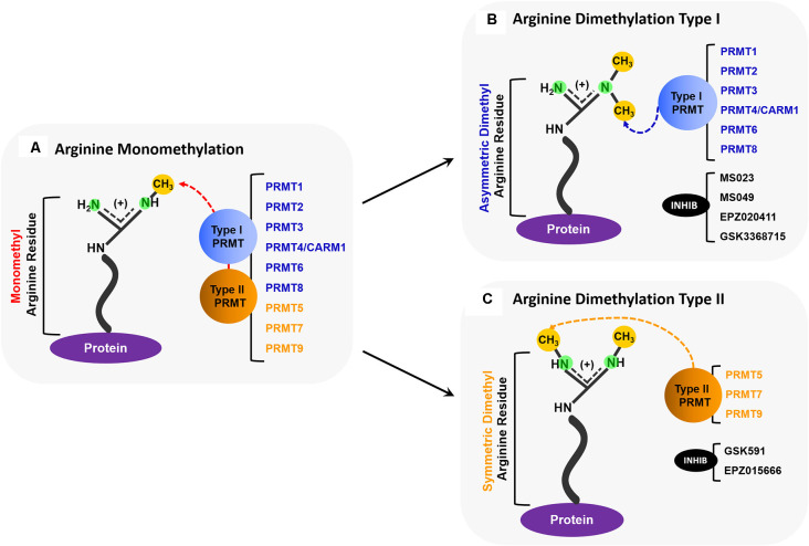
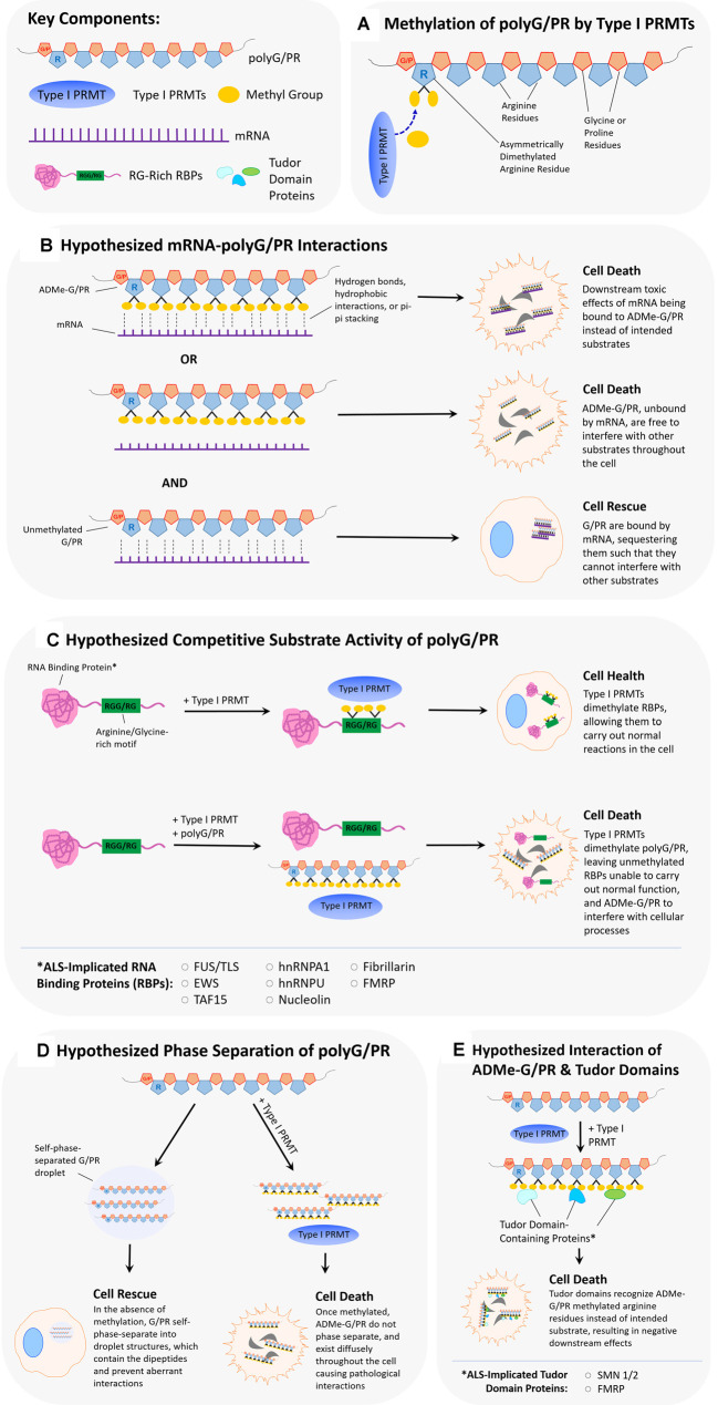
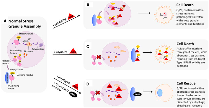
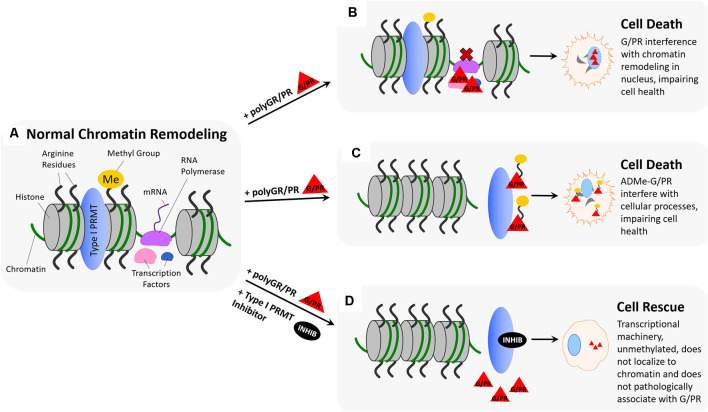
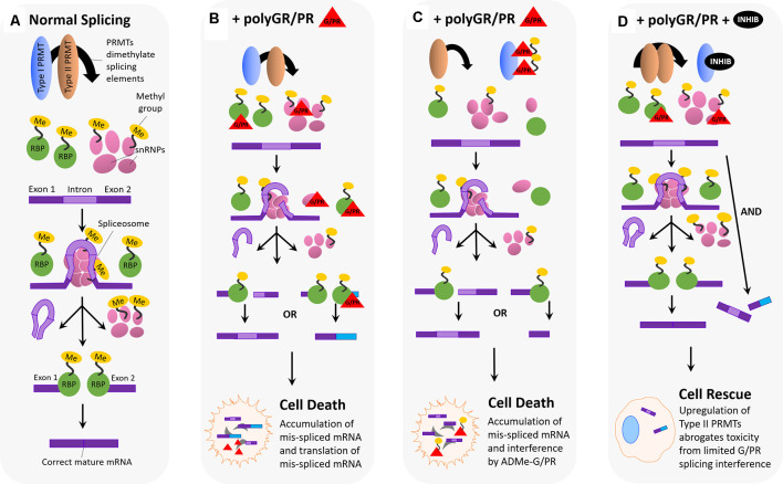
Hexanucleotide repeat expansion (G4C2n) mutations in the gene C9ORF72 account for approximately 30% of familial cases of amyotrophic lateral sclerosis (ALS) and frontotemporal dementia (FTD), as well as approximately 7% of sporadic cases of ALS. G4C2n mutations are known to result in the production of five species of dipeptide repeat proteins (DRPs) through non-canonical translation processes. Arginine-enriched dipeptide repeat proteins, glycine-arginine (polyGR), and proline-arginine (polyPR) have been demonstrated to be cytotoxic and deleterious in multiple experimental systems. Recently, we and others have implicated methylation of polyGR/polyPR arginine residues in disease processes related to G4C2n mutation-mediated neurodegeneration. We previously reported that inhibition of asymmetric dimethylation (ADMe) of arginine residues is protective in cell-based models of polyGR/polyPR cytotoxicity. These results are consistent with the idea that PRMT-mediated arginine methylation in the context of polyGR/polyPR exposure is harmful. However, it remains unclear why. Here we discuss the influence of arginine methylation on diverse cellular processes including liquid-liquid phase separation, chromatin remodeling, transcription, RNA processing, and RNA-binding protein localization, and we consider how methylation of polyGR/polyPR may disrupt processes essential for normal cellular function and survival.

Keywords: C9ORF72 ALS/FTD; PRMT inhibitor; arginine methylation; chromatin remodeling; dipeptide repeat; polyGR; polyPR; splicing.


Conflict of interest statement

The authors declare that the research was conducted in the absence of any commercial or financial relationships that could be construed as a potential conflict of interest.

Figures

Figure 1
Figure 1
Overview of PRMT activity. (A) All PRMTs (PRMTs 1–9) can add one methyl group to a nitrogen atom on a protein arginine residue so that it becomes a monomethyl arginine (MMA). (B) Type I PRMTs (PRMTs 1, 2, 3, 4/CARM1, 6, and 8) can add an additional methyl group to the same nitrogen atom that was monomethylated to form an asymmetrically dimethylated arginine. Some small molecule inhibitors, such as MS023, MS049, EPZ020411, and GSK3368715, inhibit the activity of some Type I PRMTs. (C) Type II PRMTs (PRMTs 5, 7, and 9) can add a methyl group to the second nitrogen to form a symmetrically dimethylated arginine. Some small molecule inhibitors, such as GSK519, and EPZ015666, inhibit the activity of some Type II PRMTs.
Figure 2
Figure 2
Hypothesized scenarios of how arginine dimethylation of polyG/PR affects their interactions with other biomolecules. (A) Asymmetrical dimethylation of the arginine residue of polyG/PR. (B) It is unclear whether methylation of arginine in polyG/PR increases or decreases their propensities to bind to RNAs. In one scenario, arginine dimethylation of polyG/PR may enable binding with RNA through hydrogen bonds, hydrophobic interactions, or pi-pi stacking, leading to downstream cellular toxicity as a result of aberrantly bound RNA. In another scenario, arginine dimethylation of polyG/PR may disrupt binding with RNA, leading to promiscuous interactions with other molecules that may result in cellular toxicity. Toxic effects would possibly be rescued or prevented by the binding and sequestration of polyG/PR by RNA. (C) PolyG/PR may function as competitive substrates of Type I PRMTs. The degree to which G/PR binds PRMTs will depend on their affinity to specific PRMTs and will ultimately influence the methylation-based activity of ALS-associated RNA binding proteins (RBPs) with RG/RGG motifs. (D) If unmethylated GR/PR self-phase separates in cells, they may be prevented from disrupting other cellular processes. Once arginine dimethylated, GR/PR may have more promiscuous cellular interactions resulting in cellular toxicity. (E) Arginine methylation of GR/PR could enable a diverse set of protein:protein interactions. Tudor domain-containing proteins, in particular, can specifically recognize methylated arginine residues, and methylated G/PR may interrupt normal processes that Tudor domain-containing proteins are involved in.
Figure 3
Figure 3
Hypothesized impact of polyGR/PR on stress granules and rescue by PRMT inhibition. (A) Normal stress granule assembly: in response to stress, the cell forms a membrane-less stress granule (SG) that contains important translational machinery crucial to cell survival, such as ribosomal subunits, elongation-initiation factors (eIFs), and RNA binding proteins (RBPs). Methylation of arginine residues on RNA binding proteins results in their recruitment to stress granules, where they can regulate translation to cope with cell stress. (B) Pathological scenario 1: GR and PR localize to stress granules. Methylation of RBPs results in their recruitment to stress granules where harmful interactions with polyGR/PR are facilitated by increased proximity. (C) Pathological scenario 2: GR and PR could act as substrates for type I PRMTs. This could result in competitive inhibition of appropriate RBP methylation, thereby inhibiting protective recruitment to stress granules and impeding stress responses. In a feed-forward scenario, methylated polyG/PR are less predisposed to self-phase separate and may interfere with additional cellular processes. (D) Rescue scenario: PRMT inhibition would reduce recruitment of RBPs to stress granules, thereby reducing the frequency of pathological RBP interactions with polyG/PR. This reduction of RBP stress granule recruitment could also result in the formation of aberrant stress granules containing G/PR, which are recognized by the cell as abnormal and thus degraded. Degradation of stress granules containing G/PR during the period of type I PRMT inhibition could potentially clear enough G/PR from the cell to facilitate cell recovery.
Figure 4
Figure 4
Hypothesized impact of polyGR/PR on chromatin dynamics and rescue by PRMT inhibition. (A) Normal chromatin remodeling: PRMTs methylate arginine residues on histones H3 and H4 resulting in separation of histones and recruitment of RNA polymerase (RNA Pol.), initiating production of immature mRNA from unwound chromatin. (B) Pathological scenario 1: RNA polymerase and transcription factors recruited to chromatin by PRMT methylation of histone arginine residues result in pathological protein:protein interactions between G/PR and transcriptional regulators. As a result, transcription proceeds irregularly or is unable to proceed. (C) Pathological scenario 2: GR/PR act as substrates for Type I PRMTs, resulting in methylation of GR/PR at chromatin instead of histone arginine residue methylation. Non-methylated histones remain tightly coiled, and ADMe-GR/PR are released to interfere with cellular proteins throughout the nucleus and cytoplasm. (D) Rescue scenario: With the introduction of a type I PRMT inhibitors such as MS023 or GSK3368715, Type I PRMT activity is inhibited, preventing histone arginine residue methylation. As a result, histones remain tightly coiled, and no transcriptional regulators are recruited to chromatin, preventing pathological GR/PR interactions with those proteins. Further, no type I PRMTs methylate GR/PR, which in their unmethylated form, have a propensity to form droplets, potentially preventing them from interfering with cellular proteins.
Figure 5
Figure 5
Hypothesized impact of polyGR/PR on splicing dynamics and rescue by PRMT inhibition. (A) Normal splicing activity and spliceosome assembly: for normal splicing activity to occur, type I and type II PRMTs are both involved in recruiting splicing elements to the RNA splice site, and this recruitment occurs via either asymmetric (Type I) or symmetric (Type II) dimethylation of arginine residues on RNA binding proteins (RBPs), small nuclear ribonucleoproteins (snRNPs), splicing factors, and other elements. Five snRNPs self-associate, as the spliceosome, at the site of an intron on premature mRNA, splicing the intron region out and discarding it. Following this step, the spliceosome disassembles, and snRNPs disassociate from the mRNA. RNA binding proteins then associate and join exon pieces to form the correct mature mRNA sequence. (B) Pathological scenario 1: splicing machinery recruited to the splice site by Type I and II PRMTs encounter GR and PR, which associate and interfere with their splicing activity. One possibility is that GR/PR association with snRNPs could result in incorrect spliceosome assembly, and thus incorrect intron excision, leading to mis-spliced mature mRNA containing intron fragments or missing exon fragments. Another possibility is that GR/PR association with RBPs could lead to incorrect exon selection during the splicing process, resulting in a mis-splicing event producing incorrect mature mRNAs. The following accumulation of mis-spliced mRNA, and translation of mis-spliced mRNA, could lead to cell death. (C) Pathological scenario 2: PolyGR/PR act as a sink recruiting Type I PRMTs, reducing asymmetric dimethylation and possibly increasing the proportion of symmetrically dimethylated splicing machinery. Type I PRMTs may instead asymmetrically dimethylate GR/PR to form ADMe-GR/PR, which may interfere with structures and proteins throughout the cell. Limited splicing machinery recruitment may result in impaired splicing with mature mRNA containing intron fragments or missing exon fragments. The accumulation of mis-spliced mRNA, ADMe-GR/PR, and the translation of mis-spliced mRNA could all lead to cell death. (D) Rescue scenario: with the administration of a type I PRMT inhibitor, it is possible that a robust compensatory upregulation of Type II PRMT activity is induced, resulting in more normalized levels of splicing activity such that limited interference by unmethylated GR/PR is not enough to produce detectable cytotoxicity.

Similar articles

Cited by

References

    1. Aalfs J. D., Kingston R. E. (2000). What does “chromatin remodeling” mean? Trends Biochem. Sci. 25, 548–555. 10.1016/s0968-0004(00)01689-3 - DOI - PubMed
    1. Aguzzi A., Altmeyer M. (2016). Phase separation: linking cellular compartmentalization to disease. Trends Cell Biol. 26, 547–558. 10.1016/j.tcb.2016.03.004 - DOI - PubMed
    1. Altmeyer M., Neelsen K. J., Teloni F., Pozdnyakova I., Pellegrino S., Grøfte M., et al. . (2015). Liquid demixing of intrinsically disordered proteins is seeded by poly(ADP-ribose). Nat. Commun. 6:8088. 10.1038/ncomms9088 - DOI - PMC - PubMed
    1. An W., Kim J., Roeder R. G. (2004). Ordered cooperative functions of PRMT1, p300 and CARM1 in transcriptional activation by p53. Cell 117, 735–748. 10.1016/j.cell.2004.05.009 - DOI - PubMed
    1. Andrade N. S., Ramic M., Esanov R., Liu W., Rybin M. J., Gaidosh G., et al. . (2020). Dipeptide repeat proteins inhibit homology-directed DNA double strand break repair in C9ORF72 ALS/FTD. Mol. Neurodegener. 15:13. 10.1186/s13024-020-00365-9 - DOI - PMC - PubMed

LinkOut - more resources