Why Ablation of Sites With Purkinje Activation Is Antiarrhythmic: The Interplay Between Fast Activation and Arrhythmogenesis
- PMID: 33833689
- PMCID: PMC8021688
- DOI: 10.3389/fphys.2021.648396
Why Ablation of Sites With Purkinje Activation Is Antiarrhythmic: The Interplay Between Fast Activation and Arrhythmogenesis
Abstract
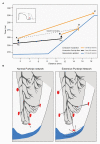
Ablation of sites showing Purkinje activity is antiarrhythmic in some patients with idiopathic ventricular fibrillation (iVF). The mechanism for the therapeutic success of ablation is not fully understood. We propose that deeper penetrance of the Purkinje network allows faster activation of the ventricles and is proarrhythmic in the presence of steep repolarization gradients. Reduction of Purkinje penetrance, or its indirect reducing effect on apparent propagation velocity may be a therapeutic target in patients with iVF.
Keywords: Purkinje; ablation; arrhythmias; early repolarization syndrome; electrophysiology; idiopathic ventricular fibrillation.
Copyright © 2021 Coronel, Potse, Haïssaguerre, Derval, Rivaud, Meijborg, Cluitmans, Hocini and Boukens.
Conflict of interest statement
MC is part-time employed by Philips Research. The remaining authors declare that the research was conducted in the absence of any commercial or financial relationships that could be construed as a potential conflict of interest.
Figures


Similar articles
-
Electrophysiological characterization of BRL-32872 in canine Purkinje fiber and ventricular muscle. Effect on early after-depolarizations and repolarization dispersion.Eur J Pharmacol. 1999 Oct 27;383(2):215-22. doi: 10.1016/s0014-2999(99)00614-7. Eur J Pharmacol. 1999. PMID: 10585537
-
Catheter ablation for patients with ventricular fibrillation.Curr Opin Cardiol. 2009 Jan;24(1):56-60. doi: 10.1097/HCO.0b013e32831c505e. Curr Opin Cardiol. 2009. PMID: 19077817 Review.
-
Mapping and Ablation of Ventricular Fibrillation Associated With Early Repolarization Syndrome.Circulation. 2019 Oct 29;140(18):1477-1490. doi: 10.1161/CIRCULATIONAHA.118.039022. Epub 2019 Sep 23. Circulation. 2019. PMID: 31542949
-
Effects of subendocardial ablation on anodal supernormal excitation and ventricular vulnerability in open-chest dogs.Circulation. 1993 Jan;87(1):216-29. doi: 10.1161/01.cir.87.1.216. Circulation. 1993. PMID: 8419011
-
Proarrhythmic effects of intravenous vasopressors.Ann Pharmacother. 1995 Mar;29(3):269-81. doi: 10.1177/106002809502900309. Ann Pharmacother. 1995. PMID: 7606074 Review.
Cited by
-
Deep neural networks reveal novel sex-specific electrocardiographic features relevant for mortality risk.Eur Heart J Digit Health. 2022 Mar 21;3(2):245-254. doi: 10.1093/ehjdh/ztac010. eCollection 2022 Jun. Eur Heart J Digit Health. 2022. PMID: 36713005 Free PMC article.
-
Purkinje network and myocardial substrate at the onset of human ventricular fibrillation: implications for catheter ablation.Eur Heart J. 2022 Mar 21;43(12):1234-1247. doi: 10.1093/eurheartj/ehab893. Eur Heart J. 2022. PMID: 35134898 Free PMC article.
-
Long-term freedom from ventricular fibrillation despite persistent Purkinje ectopy after catheter ablation.HeartRhythm Case Rep. 2022 Jan 15;8(4):259-263. doi: 10.1016/j.hrcr.2022.01.002. eCollection 2022 Apr. HeartRhythm Case Rep. 2022. PMID: 35497479 Free PMC article. No abstract available.
-
Conduction delays across the specialized conduction system of the heart: Revisiting atrioventricular node (AVN) and Purkinje-ventricular junction (PVJ) delays.Front Cardiovasc Med. 2023 Apr 19;10:1158480. doi: 10.3389/fcvm.2023.1158480. eCollection 2023. Front Cardiovasc Med. 2023. PMID: 37153461 Free PMC article.
-
Functional conduction system mapping in sheep reveals Purkinje spikes in the free wall of the right ventricular outflow tract.Front Physiol. 2025 Jul 1;16:1631426. doi: 10.3389/fphys.2025.1631426. eCollection 2025. Front Physiol. 2025. PMID: 40666117 Free PMC article.
References
-
- Allison J. S., Qin H., Dosdall D. J., Huang J., Newton J. C., Allred J. D., et al. . (2007). The transmural activation sequence in porcine and canine left ventricle is markedly different during long-duration ventricular fibrillation. J. Cardiovasc. Electrophysiol. 18, 1306–1312. 10.1111/j.1540-8167.2007.00963.x, PMID: - DOI - PubMed
LinkOut - more resources
Full Text Sources
Other Literature Sources

