. 2021 Mar 23:8:556776.
doi: 10.3389/fmed.2021.556776. eCollection 2021.

CD107a+ (LAMP-1) Cytotoxic CD8+ T-Cells in Lupus Nephritis Patients

Affiliations

CD107a+ (LAMP-1) Cytotoxic CD8+ T-Cells in Lupus Nephritis Patients

Anika Wiechmann et al. Front Med (Lausanne). .

Abstract

Cytotoxic CD8+ T-cells play a pivotal role in the pathogenesis of systemic lupus erythematosus (SLE). The aim of this study was to investigate the role of CD107a (LAMP-1) on cytotoxic CD8+ T-cells in SLE-patients in particular with lupus nephritis. Peripheral blood of SLE-patients (n = 31) and healthy controls (n = 21) was analyzed for the expression of CD314 and CD107a by flow cytometry. Kidney biopsies of lupus nephritis patients were investigated for the presence of CD8+ and C107a+ cells by immunohistochemistry and immunofluorescence staining. The percentages of CD107a+ on CD8+ T-cells were significantly decreased in SLE-patients as compared to healthy controls (40.2 ± 18.5% vs. 47.9 ± 15.0%, p = 0.02). This was even more significant in SLE-patients with inactive disease. There was a significant correlation between the percentages of CD107a+CD8+ T-cells and SLEDAI. The evaluation of lupus nephritis biopsies showed a significant number of CD107a+CD8+ T-cells mainly located in the peritubular infiltrates. The intrarenal expression of CD107a+ was significantly correlated with proteinuria. These results demonstrate that CD8+ T-cells of patients with systemic lupus erythematosus have an altered expression of CD107a which seems to be associated with disease activity. The proof of intrarenal CD107a+CD8+ suggests a role in the pathogenesis of lupus nephritis.

Keywords: CD107a; LAMP-1; SLE; cytotoxic T-cells; lupus nephritis.


Conflict of interest statement

The authors declare that the research was conducted in the absence of any commercial or financial relationships that could be construed as a potential conflict of interest.

Figures
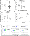
Figure 1
Figure 1
Peripheral circulating CD107a+CD8+ T-cells. (A) The percentages of CD107a+CD8+ T-cells in healthy controls (HC), SLE-patients (SLE), (B) patients with lupus nephritis (with LN) and without lupus nephritis (without LN) are shown. (C) The percentages of CD107a+CD8+ T-cells in healthy controls (HC), active SLE-patients (SLE) and inactive patients are shown. Frequencies of these T-cells are shown. Horizontal lines represent the mean. P-values were calculated using the non-parametric Mann-Whitney U-test. (D) Correlation between percentages of CD107a+CD8+ T-cells and disease activity (n = 30) as assessed by the systemic lupus erythematosus disease activity index (SLEDAI). Spearman analysis was performed to calculate the correlation. A p-value <0.05 was considered significant. (E) A representative dot plot of the flow-cytometry staining is shown for a healthy control (HC), a patient with lupus nephritis (LN) and without LN. The corresponding isotype control is illustrated.
Figure 2
Figure 2
(A) The proportion of CD107a+ CD8+ T-cells we were analyzed according to the immunosuppressive treatment. Patients were subgrouped in (i) no treatment or prednisone alone (ii) prednisone and mycophenolate mofetil, azathioprine or cyclosporine (iii) prednisone and hydroxychloroquine (iv) prednisone and hydroxychloroquine combined with mycophenolate mofetil, azathioprine or cyclosporine. Patients were compared to healthy controls (HC). P-values were calculated using the non-parametric Mann-Whitney U-test. (B) Correlation between percentages of CD107a+CD8+ T-cells for all samples taken (n = 30) daily dose of hydroxychloroquine is shown. Spearman analysis was performed to calculate the correlation. A p-value < 0.05 was considered significant.
Figure 3
Figure 3
CD8+ and CD107a+ T-cell infiltrates in lupus nephritis. This figure shows representative immunohistochemical staining with anti-CD8 of a tonsil which served as positive control (A,B). Immunhistochemical staining of a lupus nephritis renal biopsy shows an overview (C) with one glomerulum and interstitial lymphocytes. Several of these lymphocytes express CD8 as demonstrated in (D). Next, representative immunohistochemical staining with anti-CD107a of a tonsil which served as positive control (E,F). Immunohistochemical staining lupus nephritis renal biopsy shows an overview (G) with one glomerulum and interstitial lymphocytes. Several of these lymphocytes express CD107a as demonstrated in (H).
Figure 4
Figure 4
CD8+ CD107a+ immunofluorescence staining. A staining for CD8 Cy3 (red), CD107a FITC (green) and colocalization of CD8/CD107a/DAPI was performed in a tonsil as positive control (A–C) and a representative renal biopsy of an SLE patient with lupus nephritis (WHO class IV) (D–F). A magnification of a double-positive CD8+CD107a+ kidney infiltrating cell is shown in (G–J). All scales represent 100 μm.
Figure 5
Figure 5
Renal CD8+ and CD107a+ T-cells. Correlation between the CD8+ T-cell count (cells/mm2) in renal biopsies of nine lupus nephritis patients and renal histopathology parameters activity index (AI), chronicity index (CI) and proteinuria (g/d). The same correlation was performed for CD107a+ T-cell count (cells/mm2) in renal biopsies. Spearman analysis was performed to calculate the correlation. A p-value < 0.05 was considered significant.

References

    1. Dolff S, Berden JH, Bijl M. Treatment of lupus nephritis. Expert Rev Clin Immunol. (2010) 6:901–11. 10.1586/eci.10.79 - DOI - PubMed
    1. Dolff S, Witzke O, Wilde B. Th17 cells in renal inflammation and autoimmunity. Autoimmun Rev. (2019) 18:129–36. 10.1016/j.autrev.2018.08.006 - DOI - PubMed
    1. Blanco P, Pitard V, Viallard JF, Taupin JL, Pellegrin JL, Moreau JF. Increase in activated CD8+ T lymphocytes expressing perforin and granzyme B correlates with disease activity in patients with systemic lupus erythematosus. Arthritis Rheum. (2005) 52:201–11. 10.1002/art.20745 - DOI - PubMed
    1. Couzi L, Merville P, Deminiere C, Moreau JF, Combe C, Pellegrin JL, et al. . Predominance of CD8+ T lymphocytes among periglomerular infiltrating cells and link to the prognosis of class III and class IV lupus nephritis. Arthritis Rheum. (2007) 56:2362–70. 10.1002/art.22654 - DOI - PubMed
    1. D'agati VD, Appel GB, Estes D, Knowles DM, II, Pirani CL. Monoclonal antibody identification of infiltrating mononuclear leukocytes in lupus nephritis. Kidney Int. (1986) 30:573–81. 10.1038/ki.1986.223 - DOI - PubMed