Review
. 2021 May 1;320(5):H2080-H2100.
doi: 10.1152/ajpheart.00917.2020. Epub 2021 Apr 9.

Mitochondrial contributions to vascular endothelial dysfunction, arterial stiffness, and cardiovascular diseases

Affiliations
Review

Mitochondrial contributions to vascular endothelial dysfunction, arterial stiffness, and cardiovascular diseases

Danielle L Kirkman et al. Am J Physiol Heart Circ Physiol. .

Abstract

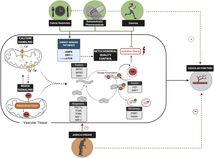
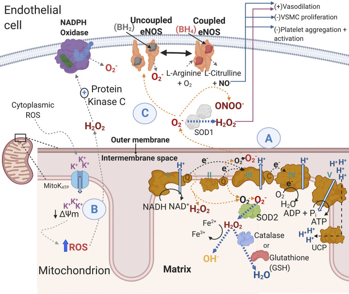
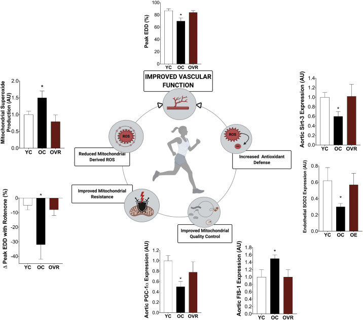
Cardiovascular disease (CVD) affects one in three adults and remains the leading cause of death in America. Advancing age is a major risk factor for CVD. Recent plateaus in CVD-related mortality rates in high-income countries after decades of decline highlight a critical need to identify novel therapeutic targets and strategies to mitigate and manage the risk of CVD development and progression. Vascular dysfunction, characterized by endothelial dysfunction and large elastic artery stiffening, is independently associated with an increased CVD risk and incidence and is therefore an attractive target for CVD prevention and management. Vascular mitochondria have emerged as an important player in maintaining vascular homeostasis. As such, age- and disease-related impairments in mitochondrial function contribute to vascular dysfunction and consequent increases in CVD risk. This review outlines the role of mitochondria in vascular function and discusses the ramifications of mitochondrial dysfunction on vascular health in the setting of age and disease. The adverse vascular consequences of increased mitochondrial-derived reactive oxygen species, impaired mitochondrial quality control, and defective mitochondrial calcium cycling are emphasized, in particular. Current evidence for both lifestyle and pharmaceutical mitochondrial-targeted strategies to improve vascular function is also presented.

Keywords: endothelium; mitochondria; vascular; vascular stiffness.


Conflict of interest statement

No conflicts of interest, financial or otherwise, are declared by the authors.

Figures

Figure 1.
Figure 1.
Optimal mitochondrial redox balance, calcium handling, and quality control are physiologically integrated mitochondrial functions that all play key roles in maintaining vascular function. Age- and disease-related impairments in these aspects of mitochondrial health and functions have adverse implications for vascular function and, consequently, cardiovascular disease risk. Lifestyle and pharmaceutical strategies that reduce mitochondrial-derived oxidative stress and improve mitochondrial quality control via modulation of energy sensing pathways hold promise for improving vascular function. PGC-1α, proliferator-activated receptor-γ coactivator-1α; TFAM, mitochondrial transcription factor A; TFBM, mitochondrial transcription factor B; DRP1, dynamin-related protein 1; FIS1, fission-1; MFN1 and MFN2, transmembrane GTPAses mitofusion-1 and 2; OPA1, optic atrophy protein 1; PINK1, phosphate and tensin homolog-induced kinase protein 1; NRF1- and NRF-2, nuclear respiratory factor 1 and 2; SIRT1, Sirtuin-1; mTOR, mammalian target of rapamycin. Figure was made with biorender.com and published with permission.
Figure 2.
Figure 2.
Efficacy evidence pertaining to improved vascular function mediated by mitochondrial function in aging and disease models and populations. AICAR, 5-aminoimidazole-4-carboxamide ribonucleotide; AMPK, adenosine monophosphate- activated protein kinase; mTOR, mammalian target of rapamycin; NMN, nicotinamide mononucleotide; NR, nicotinamide riboside.
Figure 3.
Figure 3.
Mitochondrial reactive oxygen species ROS generation and consequences in the endothelial cell. A: during normal operation of the electron transport chain electrons are passed from complex I and complex II to ubiquinol, which transfers electrons to complex III. From complex III, electrons are transferred via cytochrome c to complex IV. Hydrogen ions are pumped across the inner mitochondrial membrane creating a proton motive force that complex IV uses to generate ATP. Uncoupling protons facilitate proton leak and dissipation of the proton motive force. However, when electrons are being passed along the electron transport chain, there is electron leak whereby unpaired electrons react with diatomic oxygen to form O2. The primary oxidant produced by complex I in endothelial cells is H2O2 while complex III can generate both H2O2 and O2. The O2 generated from metabolism can be converted to H2O2 in the mitochondria by SOD2, and H2O2 can be further reduced to water by the enzymes’ catalase and the reduced form of glutathione. The H2O2 can also form hydroxyl radicals. Superoxide released into the cytoplasm can be converted into H2O2 by SOD1. B: activation or inhibition of MitoK+ATP influences mitochondrial membrane potential. There are several studies documenting that depolarization and hyperpolarization of the mitochondrial membrane potential are associated with an increase in mitochondrial-derived ROS. Importantly, cytoplasmic ROS may elicit mitochondrial depolarization at least in part through opening MitoK+ATP channels, which results in mitochondrial ROS release by respiratory complexes and the mitochondrial permeability transition pore (not shown here) (24). C: mitochondrial ROS can promote additional ROS generation in the cytoplasm through multiple mechanisms including direct scavenging of NO by superoxide (O2) to form peroxynitrate (ONOO) or by excess mitochondrial ROS “uncoupling” eNOS, which results in eNOS producing O2 in contrast to NO. Additionally, mitochondrial ROS stimulate protein kinase C, which activates the ROS-generating enzyme complex NADPH oxidase to produce O2. The reduction in NO bioavailability has several important actions in maintaining vascular homeostasis (e.g., vasodilation and proliferation). Black arrows indicate “normal” physiology, whereas orange arrows indicate a shift toward mitochondrial ROS production, and blue arrows indicate antioxidant activity. H2O2, hydrogen peroxide; MitoK+ATP, mitochondrial ATP-dependent potassium channels; ONOO, peroxynitrate; NO, nitric oxide; O2, superoxide; ROS, reactive oxygen species; SOD, superoxide dismutase; eNOS, endothelial nitric oxide synthase; VSMC, vascular smooth muscle cells; UCP, uncoupling protein. Figure was made with biorender.com and published with permission.
Figure 4.
Figure 4.
Translational evidence in aging supports a role for exercise training in improving vascular function that is largely mediated by improvements in vascular mitochondrial function. Improvements in mitochondrial quality control, mitochondrial resistance (to mitochondrial stressor, rotenone), and mitochondrial redox balance accompany exercise-related improvements in vascular function. This holds promise for exercise as a mitochondrial-targeted therapeutic strategy to improve vascular function in chronic diseases that have a pathophysiology consistent with accelerated vascular aging. Adapted from our own previously published data (26, 172). EDD, endothelial-dependent dilation; FIS-1, fission- 1; OC, old control; OE, old habitual exercises (human subjects); OVR, old voluntary wheel running (preclinical); PGC-1α, peroxisome proliferator-activated receptor-γ coactivator 1-α; SIRT-3, sirtuin-3; SOD2, superoxide dismutase 2; YC, young control; AU, arbitrary units. *P < 0.05 vs. YC and OVR/OE. Data are means (SE). Figure was made with biorender.com and published with permission.

Similar articles

Cited by

References

    1. Benjamin EJ, Muntner P, Alonso A, Bittencourt MS, Callaway CW, Carson AP, American Heart Association Council on Epidemiology and Prevention Statistics Committee and Stroke Statistics Subcommittee, et al. Heart disease and stroke statistics-2019 update: a report from the American Heart Association. Circulation 139: e56–e528, 2019. [Erratum in Circulation 141: e33, 2020]. - PubMed
    1. Heidenreich PA, Albert NM, Allen LA, Bluemke DA, Butler J, Fonarow GC, Ikonomidis JS, Khavjou O, Konstam MA, Maddox TM, Nichol G, Pham M, Piña IL, Trogdon JG, Stroke Council. Forecasting the impact of heart failure in the United States: a policy statement from the American Heart Association. Circ Heart Fail 6: 606–619, 2013. doi:10.1161/HHF.0b013e318291329a. - DOI - PMC - PubMed
    1. Ward ZJ, Bleich SN, Cradock AL, Barrett JL, Giles CM, Flax C, Long MW, Gortmaker SL. Projected U.S. state-level prevalence of adult obesity and severe obesity. N Engl J Med 381: 2440–2450, 2019. doi:10.1056/NEJMsa1909301. - DOI - PubMed
    1. Boyle JP, Honeycutt AA, Narayan KM, Hoerger TJ, Geiss LS, Chen H, Thompson TJ. Projection of diabetes burden through 2050: impact of changing demography and disease prevalence in the U.S. Diabetes Care 24: 1936–1940, 2001. doi:10.2337/diacare.24.11.1936. - DOI - PubMed
    1. Roth GA, Johnson C, Abajobir A, Abd-Allah F, Abera SF, Abyu G , et al. Global, regional, and national burden of cardiovascular diseases for 10 causes, 1990 to 2015. J Am Coll Cardiol 70: 1–25, 2017. doi:10.1016/j.jacc.2017.04.052. - DOI - PMC - PubMed

Publication types

Substances

LinkOut - more resources