. 2021 May 3;153(5):e202012754.
doi: 10.1085/jgp.202012754.

Structure versus function: Are new conformations of pannexin 1 yet to be resolved?

Affiliations

Structure versus function: Are new conformations of pannexin 1 yet to be resolved?

Carsten Mim et al. J Gen Physiol. .

Abstract

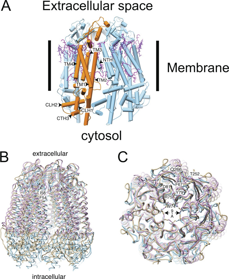
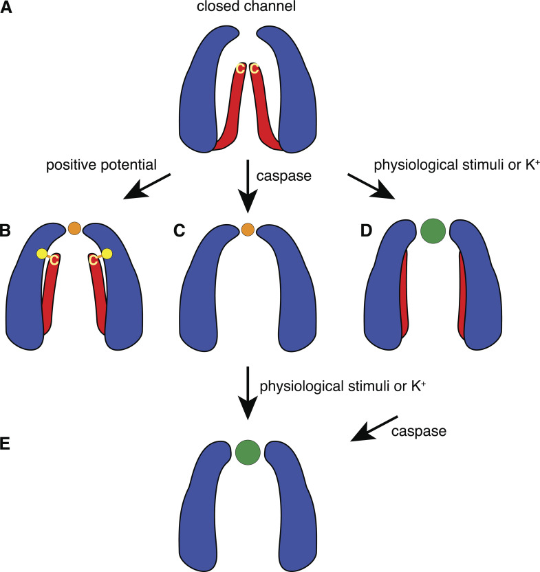
Pannexin 1 (Panx1) plays a decisive role in multiple physiological and pathological settings, including oxygen delivery to tissues, mucociliary clearance in airways, sepsis, neuropathic pain, and epilepsy. It is widely accepted that Panx1 exerts its role in the context of purinergic signaling by providing a transmembrane pathway for ATP. However, under certain conditions, Panx1 can also act as a highly selective membrane channel for chloride ions without ATP permeability. A recent flurry of publications has provided structural information about the Panx1 channel. However, while these structures are consistent with a chloride selective channel, none show a conformation with strong support for the ATP release function of Panx1. In this Viewpoint, we critically assess the existing evidence for the function and structure of the Panx1 channel and conclude that the structure corresponding to the ATP permeation pathway is yet to be determined. We also list a set of additional topics needing attention and propose ways to attain the large-pore, ATP-permeable conformation of the Panx1 channel.


Figures

Figure 1.
Figure 1.
Structural model of Panx1. (A) Cartoon representation of Panx1 WT (PDB accession no. 6wbk) stabilized with detergent. The approximate position of the membrane is indicated by the bars. One protomer of the heptameric assembly is indicated in orange. The lipids resolved in the structure are colored in magenta. It is notable that many of the resolved lipids are found at the interface between subunits. CTH, C-terminal helix; CLH, cytoplasmic linker helix. (B) Overlay of different Panx1 cryo-EM–based structures from different groups (PDB accession no. 6lto = gold, WT hPanx1; PDB accession no. 6wbk = blue, hPanx1 ΔC terminus, ΔN terminus; PDB accession no. 6v6d = lilac, WT hPanx1). The structures diverge very little in the ECD and the membrane domain. However, the variation is larger in the ICD. The approximate positions of F54 and I58 are indicated. (C) Comparison of the extracellular pore. Key residues are annotated using their one-letter abbreviation. All structures are shown as backbone traces except W74, which is part of the constriction ring of the pore. The narrowest constriction is indicated by the arrow.
Figure 2.
Figure 2.
ATP release by oocytes expressing Panx1. (A) Oocytes expressing WT Panx1. All colored bars represent data obtained under voltage-clamp conditions; white bars are data from unclamped cells. ATP in the medium was measured as luciferase luminescence. ATP release from Panx1-expressing oocytes induced by potassium gluconate (KGlu) without and with holding the membrane potential under voltage-clamp conditions at −60, 0, or +40 mV, was determined 20 min after initiating the stimulus. Voltage-clamp conditions are indicated with teal lines below the graph and teal bars in the graph. The presence of 150 mM KGlu (K+ label) is indicated, and the data are displayed as hatched bars. Data are shown as means ± SD; n = 5 for each measurement. Adapted from Wang et al., 2014. (B) Oocytes expressing the truncation mutant Panx1Δ378. Oocytes were not voltage clamped (white bar) or were clamped at −60 or 0 mV (red bars). The cells were exposed to oocyte Ringer solution or to a solution containing 85 mM KGlu (K+ label, hatched bars) as indicated, for 10 min. An aliquot of the supernatant was analyzed for the presence of ATP with the luciferase/luciferin assay. Means ± SE; n as indicated above each bar. Adapted from Wang and Dahl, 2018. Because oocytes expressing Panx1Δ378 have a shortened life span (Jackson et al., 2014), measurements had to be taken in a short time window after injection of mRNA at 80× lower concentration than wtPanx1. Thus, ATP release data cannot be compared between wtPanx1- and Panx1Δ378-expressing cells.
Figure 3.
Figure 3.
Current–voltage relations of mouse WT Panx1 and Panx1Δ378. (A) Replacing extracellular Cl by gluconate in a voltage step protocol applied to HEK cells expressing Panx1 exogenously attenuated the membrane currents and shifted the reversal potential to a positive potential. Adapted with permission from Journal of Cell Science (Romanov et al., 2012). (B) Similarly, in oocytes expressing Panx1 exogenously, replacement of Cl by gluconate (NaGluc) resulted in a shift of the reversal potential to positive values and an attenuation of the currents induced by a voltage ramp from −100 to +100 mV. (C) Voltage ramp–induced currents of wtPanx1-expressing oocytes in KCl solution (black trace) were substantially larger than the currents in uninjected control cells under identical conditions (gray trace). Replacing Cl by gluconate in the bath solution of the same oocyte resulted in attenuation of the currents and a shift of the reversal potential to more negative potential (green trace). (D) Quantitative analysis of reversal potentials after anion replacement shows a shift from positive to negative potentials with K+ as the extracellular cation. Means ± SE; n = 5 (wtPanx1). (E) Voltage ramp–induced membrane currents of WT Panx1 channels (green trace) and of channels formed by the truncation mutant Panx1Δ378, where C-terminal amino acids after aspartate 378 are deleted. Uninjected oocytes served as control (magenta trace). Because Panx1Δ378-expressing cells had a short life span, mRNA was injected 80× diluted as compared with WT, and measurements were performed in a 24-h window. Thus, the current amplitudes of WT Panx1 and Panx1Δ378 cannot be compared. In contrast to WT Panx1 channels, those formed by Panx1Δ378 were active over a wide voltage range, yet currents through both types of channels reversed at the same membrane potential. (F) Similar to WT Panx1, currents through Panx1Δ378 channels reversed at negative membrane potential with extracellular chloride (blue trace) and at positive potential with gluconate solution (red trace). (G) Similar to WT Panx1 channels, replacement of chloride by gluconate ions resulted in the currents reversing at negative potentials when Panx1Δ378-expressing cells were exposed to high extracellular K+. (H) Quantitative analysis of reversal potentials of membrane currents carried by Panx1Δ378 after anion replacement shows a shift from positive to negative potentials with K+ as the extracellular cation. Means ± SE; n = 4. (B–H) Data from Wang and Dahl (2018).
Figure 4.
Figure 4.
Space-filling model of Panx1 without the C terminus. Related to PDB accession no. 6wbg. (A) Top: Top view of the extracellular pore. The space-filling model of ATP (PubChem accession no. 5957, mol wt 507.2) is shown in cyan. Bottom: Side view of the extracellular pore showing the W 74 ring above the ATP. (B) Top: The same model as in A, top view of the extracellular pore. In magenta is the space-filling model of Yo-Pro-1 (PubChem accession no. 6913121, mol wt 375.5). Bottom: Side views of the extracellular pore with a model for Yo-Pro (magenta) placed at the level of the external constriction.
Figure 5.
Figure 5.
Activation mechanisms of Panx1 channels. (A) Based on the presently available structural and functional data, we hypothesize that the closed channel exhibits the external restriction of ∼9 Å and that the C termini reach deep into the pore, occluding it. (B) Depolarization dislocates the C termini, so that the channel pore diameter equals or exceeds that of the external restriction and exposes the terminal cysteine (yellow C) to thiol reagents. The yellow dot indicates the position of Panx1F54 which, when mutated to a cysteine, can be disulfide bonded with the terminal cysteine (Sandilos et al., 2012). In this configuration, the external restriction limits entry by size and selects for chloride (orange dot) over other ions. (C) Cleavage of Panx1 at position 378 removes the “pore gate,” rendering the channel constitutively active. However, as indicated by the published cryo-EM data, caspase cleavage does not affect the external constriction, leaving a chloride-selective channel. (D) Various physiological stimuli initiate a gating mechanism at the external restriction, widening it to accept ATP (green dot) and other molecules in this size range. (E) This mechanism also may move charged amino acids within the external constriction, with the consequence that charge selectivity is attenuated. In the case of K+ stimulation, the terminal cysteine is no further reactive to thiol reagents, while engineered cysteines at the external end of the pore still are. Whether this terminal cysteine concealment also applies to the physiological stimuli remains to be determined. Cleavage of the C terminus by caspase “super stimulates” the channel and irreversibly seals the apoptotic fate of the cell.

Similar articles

Cited by

References

    1. Bao, L., Locovei S., and Dahl G.. 2004. Pannexin membrane channels are mechanosensitive conduits for ATP. FEBS Lett. 572:65–68. 10.1016/j.febslet.2004.07.009 - DOI - PubMed
    1. Billaud, M., Lohman A.W., Straub A.C., Looft-Wilson R., Johnstone S.R., Araj C.A., Best A.K., Chekeni F.B., Ravichandran K.S., Penuela S., et al. . 2011. Pannexin1 regulates α1-adrenergic receptor-mediated vasoconstriction. Circ. Res. 109:80–85. 10.1161/CIRCRESAHA.110.237594 - DOI - PMC - PubMed
    1. Billaud, M., Sandilos J.K., and Isakson B.E.. 2012. Pannexin 1 in the regulation of vascular tone. Trends Cardiovasc. Med. 22:68–72. 10.1016/j.tcm.2012.06.014 - DOI - PMC - PubMed
    1. Boassa, D., Ambrosi C., Qiu F., Dahl G., Gaietta G., and Sosinsky G.. 2007. Pannexin1 channels contain a glycosylation site that targets the hexamer to the plasma membrane. J. Biol. Chem. 282:31733–31743. 10.1074/jbc.M702422200 - DOI - PubMed
    1. Boassa, D., Qiu F., Dahl G., and Sosinsky G.. 2008. Trafficking dynamics of glycosylated pannexin 1 proteins. Cell Commun. Adhes. 15:119–132. 10.1080/15419060802013885 - DOI - PMC - PubMed

Publication types

MeSH terms