. 2021 Nov;140(11):1593-1609.
doi: 10.1007/s00439-021-02279-y. Epub 2021 Apr 9.

In-frame deletion in canine PITRM1 is associated with a severe early-onset epilepsy, mitochondrial dysfunction and neurodegeneration

Affiliations

In-frame deletion in canine PITRM1 is associated with a severe early-onset epilepsy, mitochondrial dysfunction and neurodegeneration

Marjo K Hytönen et al. Hum Genet. 2021 Nov.

Abstract

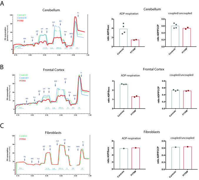
We investigated the clinical, genetic, and pathological characteristics of a previously unknown severe juvenile brain disorder in several litters of Parson Russel Terriers. The disease started with epileptic seizures at 6-12 weeks of age and progressed rapidly to status epilepticus and death or euthanasia. Histopathological changes at autopsy were restricted to the brain. There was severe acute neuronal degeneration and necrosis diffusely affecting the grey matter throughout the brain with extensive intraneuronal mitochondrial crowding and accumulation of amyloid-β (Aβ). Combined homozygosity mapping and genome sequencing revealed an in-frame 6-bp deletion in the nuclear-encoded pitrilysin metallopeptidase 1 (PITRM1) encoding for a mitochondrial protease involved in mitochondrial targeting sequence processing and degradation. The 6-bp deletion results in the loss of two amino acid residues in the N-terminal part of PITRM1, potentially affecting protein folding and function. Assessment of the mitochondrial function in the affected brain tissue showed a significant deficiency in respiratory chain function. The functional consequences of the mutation were modeled in yeast and showed impaired growth in permissive conditions and an impaired respiration capacity. Loss-of-function variants in human PITRM1 result in a childhood-onset progressive amyloidotic neurological syndrome characterized by spinocerebellar ataxia with behavioral, psychiatric and cognitive abnormalities. Homozygous Pitrm1-knockout mice are embryonic lethal, while heterozygotes show a progressive, neurodegenerative phenotype characterized by impairment in motor coordination and Aβ deposits. Our study describes a novel early-onset PITRM1-related neurodegenerative canine brain disorder with mitochondrial dysfunction, Aβ accumulation, and lethal epilepsy. The findings highlight the essential role of PITRM1 in neuronal survival and strengthen the connection between mitochondrial dysfunction and neurodegeneration.


Conflict of interest statement

HL is a paid consultant to Genoscoper Laboratories Ltd providing genetic tests for dogs. JD is an employee of Genoscoper Laboratories Ltd.

Figures

Fig. 1
Fig. 1
Representative forebrain changes in a PITRM1 mutant dog. a Extensive eosinophilic nerve cell necroses are seen within the cerebral cortex (arrowheads) some of which have triggered early microglial neuronophagia (frame). Surrounding non-necrotic changes also show degenerative cytoplasmic and nuclear features such as lobulated giant nuclei (black arrow). b Multiple diencephalic neurons further show central chromatolysis (black arrowhead) and nuclear displacement (white arrowhead). c Immunohistochemistry for Aβ stains some neurons within the necrotic cortical areas (frame). No such staining was identified in age-matched controls (inlet). d Throughout the cortices groups of outer (OPN) and inner pyramidal neurons (IPN) stain immunopositive for PITRM1 antigen similar to control dog sections (upper inlets). This also applies to neurons with degenerate features (arrows). Choroid plexus epithelium (PE), ependymal (Ep) and arterial smooth muscle layers (Art) serve as internal positive control due to constitutive expression of PITRM1 (lower inlet)
Fig. 2
Fig. 2
A pedigree established around the affected dogs. The affected dogs are closely related. Genotype information of the PITRM1 allele is denoted for each genotyped dog. The variant segregates in the pedigree as expected in a recessive disease
Fig. 3
Fig. 3
Homozygosity mapping of two affected and four unaffected dogs. The analysis resulted in a total of 15 case-specific regions of homozygosity (indicated in red)
Fig. 4
Fig. 4
a Multiple alignment of PITRM1 orthologs spanning the predicted deletion site, p.(Leu59_Ser60del) or alternatively p.(Ser60_Leu61del) and surrounding amino acids demonstrates the conservation of the Leu59 as well as alternatively deleted Leu61 across species (Leu highlighted in red and Ser in light red). In human, the corresponding amino acids overlap a junction between a short helix (blue rectangle) and β-strand (green rectangle). b Sequence chromatograms illustrating the PITRM1 deletion allele as homozygous (del/del) in an affected dog, heterozygous (wt/del) in an unaffected obligate carrier dog and homozygous wild-type (wt/wt) allele in an unaffected dog. Deletion site (reference allele CTGTCC) is highlighted in red
Fig. 5
Fig. 5
Comparison of the PITRM1 transcript levels between a case and control dogs. The stability of the PITRM1 transcript was studied by a quantitative PCR in four different tissues (parietal cortex, cerebellum, cardiac muscle and skeletal muscle) in one (~ 2 months old) affected and three unaffected dogs (Control 1 = Wire Fox Terrier; Control 2 = Saluki; Control 3 = Karelian Bear Hound). The results suggest increased expression of the aberrant transcript in the parietal cortex and the skeletal muscle tissues. The error bars refer to variance in experimental tripli- or duplicates and between the three control dogs. The asterisks refer to a P value under 0.05 in a two-sample t test. YWHAZ and GADPH were used for normalization
Fig. 6
Fig. 6
Histopathological findings in a dog affected by PITRM-associated mitochondrial juvenile encephalopathy. a Widespread acute degeneration and necrosis of cerebral grey matter (GM). a HE, WM: white matter. b Shrunken, granular and eosinophilic degenerating neurons (arrows) of the cerebral cortex. HE. c Early inflammatory response to the neuronal necrosis seen as increased cellularity of cortical vessel walls. HE. (d) Vascular wall degeneration with scattered intramural apoptotic cells (arrows). IHC Caspase-3. e Increased intraneuronal amyloid precursor protein (APP) as a sign of degeneration. IHC APP, Hippocampus. Inset: IHC APP control Hippocampus. (F) Accumulation of mitochondria in degeneration neurons. IHC VDAC1/Porin IHC, Hippocampus. Inset: IHC VDAC1/Porin IHC control Hippocampus. g Degenerating neuron with rounded swollen mitochondria. Cerebral cortex, EM. h The mitochondrial cristae are partially lost, consistent with cristolysis (arrows). Cerebral cortex, EM
Fig. 7
Fig. 7
Analysis of mitochondrial function using high-resolution respirometry in the cerebellum (a), frontal cortex (b), and fibroblasts (c). Oxygen consumption traces and quantification in the cerebellum (a) and frontal cortex (b) of the affected dog compared to controls (n = 2). All tissue measurements performed in duplicates. c Oxygen consumption rate traces and quantification in immortalized fibroblasts from an affected dog and age-matched control
Fig. 8
Fig. 8
Respiratory chain complex proteins I–V in unaffected and affected dogs assessed by IHC. Despite mitochondrial dysfunction, expression of respiratory chain complexes I through V in indicator areas as cerebellar cortex is identical between PITRM1 wild-type (a, c, e, g, i) and mutant brains (b, d, f, h, j). ML molecular layer, GCL granular cell layer, PC Purkinje cells, CSG cerebellar synaptic glomeruli
Fig. 9
Fig. 9
In vivo modeling of the mutation in yeast Saccharomyces cerevisiae. The strains harboring the wild-type CYM1HA allele (yeast wt), the cym1HAR41S allele (canine wt), the cym1ΔL40R41 mutant allele (canine mutant) or the empty vector were analyzed. a Spot assay of wt and mutant strains on SC medium supplemented with glucose or ethanol: 1:3 dilutions were performed, starting from 1.5 × 10⁴ cells/spot. b Oxygen consumption rate (OCR) was measured on five independent clones for each strain and normalized for the cells dry weight. Data were then normalized to the OCR of the strain transformed with CYM1HA yeast wt allele and reported as mean ± SD. Statistical analysis was performed by one-way ANOVA followed by a post hoc Bonferroni test. *p < 0.05; **p < 0.01. c Western blot on total protein extracts. Cym1HA signal levels were measured, as reported in “Materials and methods”, and normalized for the Por1 signal and then to the CYM1HA yeast wt strain

Similar articles

Cited by

References

    1. Alikhani N, Guo L, Yan S, Du H, Pinho CM, Chen JX, Glaser E, Yan SS. Decreased proteolytic activity of the mitochondrial amyloid-beta degrading enzyme, PreP peptidasome, in Alzheimer's disease brain mitochondria. J Alzheimers Dis. 2011;27:75–87. doi: 10.3233/JAD-2011-101716. - DOI - PMC - PubMed
    1. Alikhani N, Berglund AK, Engmann T, Spanning E, Vogtle FN, Pavlov P, Meisinger C, Langer T, Glaser E. Targeting capacity and conservation of PreP homologues localization in mitochondria of different species. J Mol Biol. 2011;410:400–410. doi: 10.1016/j.jmb.2011.05.009. - DOI - PubMed
    1. Arumilli M, Layer RM, Hytonen MK, Lohi H. webGQT: A Shiny Server for Genotype Query Tools for Model-Based Variant Filtering. Front Genet. 2020;11:152. doi: 10.3389/fgene.2020.00152. - DOI - PMC - PubMed
    1. Berendt M, Farquhar RG, Mandigers PJ, Pakozdy A, Bhatti SF, De Risio L, Fischer A, Long S, Matiasek K, Munana K, Patterson EE, Penderis J, Platt S, Podell M, Potschka H, Pumarola MB, Rusbridge C, Stein VM, Tipold A, Volk HA. International veterinary epilepsy task force consensus report on epilepsy definition, classification and terminology in companion animals. BMC Vet Res. 2015;11:182–192. doi: 10.1186/s12917-015-0461-2. - DOI - PMC - PubMed
    1. Broeckx BJ, Hitte C, Coopman F, Verhoeven GE, De Keulenaer S, De Meester E, Derrien T, Alfoldi J, Lindblad-Toh K, Bosmans T, Gielen I, Van Bree H, Van Ryssen B, Saunders JH, Van Nieuwerburgh F, Deforce D. Improved canine exome designs, featuring ncRNAs and increased coverage of protein coding genes. Sci Rep. 2015;5:12810. doi: 10.1038/srep12810. - DOI - PMC - PubMed

MeSH terms