. 2021 Apr 9;17(4):e1009501.
doi: 10.1371/journal.ppat.1009501. eCollection 2021 Apr.

Mutations derived from horseshoe bat ACE2 orthologs enhance ACE2-Fc neutralization of SARS-CoV-2

Affiliations

Mutations derived from horseshoe bat ACE2 orthologs enhance ACE2-Fc neutralization of SARS-CoV-2

Huihui Mou et al. PLoS Pathog. .

Abstract

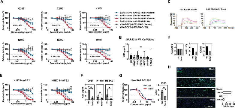
The severe acute respiratory syndrome coronavirus 2 (SARS-CoV-2) spike (S) protein mediates infection of cells expressing angiotensin-converting enzyme 2 (ACE2). ACE2 is also the viral receptor of SARS-CoV (SARS-CoV-1), a related coronavirus that emerged in 2002-2003. Horseshoe bats (genus Rhinolophus) are presumed to be the original reservoir of both viruses, and a SARS-like coronavirus, RaTG13, closely related to SARS-CoV-2, has been identified in one horseshoe-bat species. Here we characterize the ability of the S-protein receptor-binding domains (RBDs) of SARS-CoV-1, SARS-CoV-2, pangolin coronavirus (PgCoV), RaTG13, and LyRa11, a bat virus similar to SARS-CoV-1, to bind a range of ACE2 orthologs. We observed that the PgCoV RBD bound human ACE2 at least as efficiently as the SARS-CoV-2 RBD, and that both RBDs bound pangolin ACE2 efficiently. We also observed a high level of variability in binding to closely related horseshoe-bat ACE2 orthologs consistent with the heterogeneity of their RBD-binding regions. However five consensus horseshoe-bat ACE2 residues enhanced ACE2 binding to the SARS-CoV-2 RBD and neutralization of SARS-CoV-2 pseudoviruses by an enzymatically inactive immunoadhesin form of human ACE2 (hACE2-NN-Fc). Two of these mutations impaired neutralization of SARS-CoV-1 pseudoviruses. An hACE2-NN-Fc variant bearing all five mutations neutralized both SARS-CoV-2 pseudovirus and infectious virus more efficiently than wild-type hACE2-NN-Fc. These data suggest that SARS-CoV-1 and -2 originate from distinct bat species, and identify a more potently neutralizing form of soluble ACE2.


Conflict of interest statement

I have read the journal’s policy and the authors of this manuscript have the following competing interests: M.F., H.M. and B.D.Q had filed a patent for the application of ACE2-Fc variants as a SARS-CoV-2 treatment. M.R.G., C.C.B., M.D.A., and M.F. are all cofounders of, and have an equity interest in Emmune Inc., a biotech company that specializes in the development of antibody-like antiviral therapies.

Figures

Fig 1
Fig 1. The receptor-binding domains (RBD) of SARS-related coronaviruses S proteins bind differentially to ACE2 orthologs.
HEK239T cells transfected to express the ACE2 orthologs of the indicate species, human DPP4 (the receptor of MERS-CoV), or with vector alone (mock) were assayed by flow cytometry for their ability to bind SARS1-RBD-Fc, SARS2-RBD-Fc, RaTG13-RBD-Fc, PgCoV-RBD-Fc, LyRa11-RBD-Fc or MERS-RBD-Fc. Bars display the results of three independent experiments and the mean fluorescence intensity was normalized with relative ACE2 expressing levels shown in S1C Fig that determined by an antibody recognizing an amino-terminal myc-tag. One-way ANOVA was used to test difference across RBDs within each ACE2 ortholog and upon low p value, Dunnett’s multiple comparison test was used to compare each RBD with SARS2-RBD-Fc (*indicates p<0.05).
Fig 2
Fig 2. Surface-plasmon resonance and pseudovirus inhibition studies of SARS1-RBD, SARS2-RBD, PgCoV-RBD, LyRa11-RBD.
(A) Biacore surface plasmon resonsance (SPR) studies were performed with the indicated RBD-Fc (yellow and blue) captured on the sensor chip. The soluble monomeric forms of the indicated ACE2 variants (green, with a four-amino-acid C-terminal tag in red) were injected for analysis as represented in the figure. Biacore X100 sensorgrams representing results from studies in which the indicated RBD-Fc variants were captured by an anti-human Fcγ antibody immobilized on a CM5 chip after instantaneous background depletion. Monomeric forms of tagged soluble human ACE2 (hACE2-CT), human ACE2 lacking its active-site histidines (hACE2-NN-CT), or pangolin ACE2-CT were injected at five concentrations, serially diluted by two-fold from the highest concentration (100 nM for human ACE2 variants). The experiment is representative of two with similar results. (B) The association rate constant (kon), dissociation rate constant (koff), and equilibrium dissociation constant (Kd) were calculated and plotted for comparison from the study shown in (A). (C) 293T-hACE2 cells were pre-incubated with the indicated concentrations of the indicated RBD-Fc variants, and subsequently infected with retroviral pseudoviruses (PV) pseudotyped with the S proteins of SARS-CoV-1 (SARS1-S-PV), SARS-CoV-2 (SAR2-S-PV), or with the G protein of vesicular stomatitis virus (VSV-G-PV), as diagrammed. Nonlinear fit for log(inhibitor) vs. response was calculated for each RBD. In both SARS1-S-PV and SARS2-S-PV panels, SARS-RBD-Fc, SARS2-RBD-Fc, PgCoV-RBD-Fc, LyRa11-RBD-Fc bottom plateaus CI did not overlap with values from VSV-G-PV. Error bars in indicate standard error of the mean (S.E.M) and are representative of results from at least two independent experiments. Asterisks indicate lack of 95% CI overlap of RBD-Fc constructs with respect to VSV-G group.
Fig 3
Fig 3. Sequence comparison of essential ACE2 residues for SARS2-RBD binding in horseshoe bats and pangolin ACE2 orthologs.
(A) Human ACE2 (grey) is shown bound to the SARS-CoV-2 RBD (blue), based on PDB accession number 6M0J (Lan et al., 2020). Yellow indicates the ACE2 surface that is within 5.5 Å of the RBD. Red indicates residues that vary between human and horseshoe bat species. Specific mutations are indicated in the rightmost figure. A glycosylation at asparagine 90, also characterized, is indicated in green. Figure shows successive 90-degree rotations around the vertical and horizontal axes, respectively. The RBD is removed from the rightmost figure for clarity. (B) Sequence of human ACE2 is aligned with the sequence of the pangolin ACE2 as well as selected horseshoe-bat ACE2 orthologs. Residues, directly associating or proximal to, the SARS-CoV-2 RBD are shown. Yellow highlights a residue that directly contacts the RBD. Red indicates human residues that were altered to the indicated pangolin or bat residues shown in gray and characterized in the subsequent figures. Bold indicates a glycosylated asparagine.
Fig 4
Fig 4. Residues from horseshoe-bat ACE2 orthologs improved human ACE2-Fc binding to SARS2-RBD and neutralization of SARS2-S-PV.
(A) SARS1-S-PV (blue), SARS2-S-PV (red), RaTG13-S-PV (yellow) or VSV-G-PV (grey) were pre-incubated with the indicated concentrations of human ACE2-Fc lacking enzymatic activity (hACE2-NN-Fc) or hACE2-NN-Fc variants with the indicated mutations. 293T-hACE2 cells were incubated with these preincubated mixes and infection was analyzed 48 h post infection by measuring luciferase activity. Relative infection is calculated by dividing luciferase signal values to that in the absence of inhibitors. Error bars indicate S.E.M. and are representative of results from at least three independent experiments. Mean half-maximal inhibitory concentration (IC50) to SARS2-S-PV is shown for each variant. (B) SARS2-S-PV IC50 values for three studies shown in (A) are plotted. Error bars indicate the 95% confidence interval for IC50 values observed for each hACE2-NN-Fc variant. n.a. indicates that no reliable IC50 could be determined. (C) The relative percentage of SARS2-S-PV infection in the presence of 16.67 μg/ml indicated hACE2-NN-Fc variant are shown. Error bars indicate 95% confidence intervals and * indicates lack of overlap with Wt value. (D) A diagram is shown illustrating that hACE2-NN-Fc variants were captured and that soluble monomeric SARS2-RBD was injected for the studies showed in (E). (E) SPR analyses showing the kon, koff, and Kd of SARS2-RBD to hACE2-Fc variants. n.d. is short for not detectable or no binding.
Fig 5
Fig 5. A combination of five bat-derived residues improves SARS2-S-PV neutralization.
(A) Experiments similar to those shown Fig 4A except that the indicated ACE2-NN-Fc variant (dark red, blue and grey) is directly compared with wild-type human ACE2-NN-Fc (light red, blue, and grey) for their ability to neutralize SARS1-S-PV (light and dark blue), SARS2-S-PV (light and dark red), or VSV-G-PV (light and dark grey). Mean IC50 values for SARS-CoV-2 of the indicated ACE2-NN-Fc variant over at least two independent studies are provided in each figure. Relative infection is calculated by dividing luciferase signal values to that in the absence of ACE2-Fc variants. Error bars indicate S.E.M. Neutralization data displays at least two independent experiments. (B) SARS2-S-PV IC50 values shown in (A) are plotted. Error bars indicate 95% confidence intervals for IC50 value observed for each hACE2-NN-Fc variant and * indicates lack of overlap with Wt value. (C) Sensorgrams of SPR analyses used to determine binding constants of the SARS2-RBD to wild-type hACE2-Fc (Wt) or hACE2-Fc with the five indicated mutations (5mut). (D) kon, koff, and Kd of SARS2-RBD binding to hACE2-NN-Fc Wt or to hACE2-NN-Fc 5mut. (E) Neutralization potency of hACE2-Fc Wt and hACE2-Fc 5mut were compared with indicated two lung cell lines. Experiments similar to those shown Fig 5A, and color coded according to that figure, with darker colors indicating 5mut and lighter colors indicating ACE2-NN-Fc Wt. Neutralization data represents at least two independent experiments, with error bars indicating standard error of the mean (S.E.M). (F) IC50 values of hACE2-NN-Fc Wt and 5mut obtained from three different cell lines drawn from panels (A) and (E) were plotted, and error bars indicate 95% confidence intervals and * indicates lack of overlap with the Wt value. (G) Neutralization potency of hACE2-NN-Fc Wt and 5mut to SARS-CoV-2 infection were compared on Vero E6-hACE2 cells. Data represents at least two independent experiments, with error bars indicating standard error of the mean (S.E.M). IC50 values were plotted, and error bars indicate 95% confidence intervals and * indicates lack of overlap with the Wt value. (H) Vero E6-hACE2 cells were pre-incubated with PBS, 1 μg/ml hACE2-NN-Fc Wt or the 5mut variant, and subsequently mock-infected or infected with SARS-CoV-2 (MOI = 1). 24 h post infection, cells were fixed and stained with antibody against SARS-CoV-2 Nsp3 (green) and DAPI (blue). The scale bar indicates 100 μm. The infection percentage demonstrated with the bar chart was the percentage of Nsp3-positive cells from a pool of ~12,000 cells in each condition.

Update of

References

    1. Cui J, Li F, Shi ZL. Origin and evolution of pathogenic coronaviruses. Nature reviews Microbiology. 2019;17(3):181–92. 10.1038/s41579-018-0118-9 - DOI - PMC - PubMed
    1. Li W, Moore MJ, Vasilieva N, Sui J, Wong SK, Berne MA, et al.. Angiotensin-converting enzyme 2 is a functional receptor for the SARS coronavirus. Nature. 2003;426(6965):450–4. 10.1038/nature02145 - DOI - PMC - PubMed
    1. Hoffmann M, Kleine-Weber H, Schroeder S, Kruger N, Herrler T, Erichsen S, et al.. SARS-CoV-2 Cell Entry Depends on ACE2 and TMPRSS2 and Is Blocked by a Clinically Proven Protease Inhibitor. Cell. 2020;181(2):271–80 e8. 10.1016/j.cell.2020.02.052 - DOI - PMC - PubMed
    1. Walls AC, Park YJ, Tortorici MA, Wall A, McGuire AT, Veesler D. Structure, Function, and Antigenicity of the SARS-CoV-2 Spike Glycoprotein. Cell. 2020;181(2):281–92 e6. 10.1016/j.cell.2020.02.058 - DOI - PMC - PubMed
    1. Wrapp D, Wang N, Corbett KS, Goldsmith JA, Hsieh CL, Abiona O, et al.. Cryo-EM structure of the 2019-nCoV spike in the prefusion conformation. Science. 2020;367(6483):1260–3. 10.1126/science.abb2507 - DOI - PMC - PubMed

Publication types

MeSH terms

Substances