. 2021 Jun 7;220(6):e202008145.
doi: 10.1083/jcb.202008145.

Spatiotemporal coordination of Greatwall-Endos-PP2A promotes mitotic progression

Affiliations

Spatiotemporal coordination of Greatwall-Endos-PP2A promotes mitotic progression

Myreille Larouche et al. J Cell Biol. .

Abstract

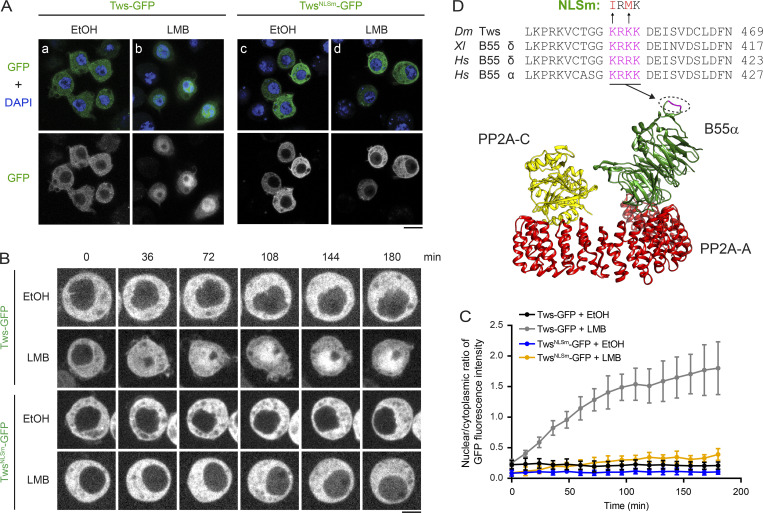
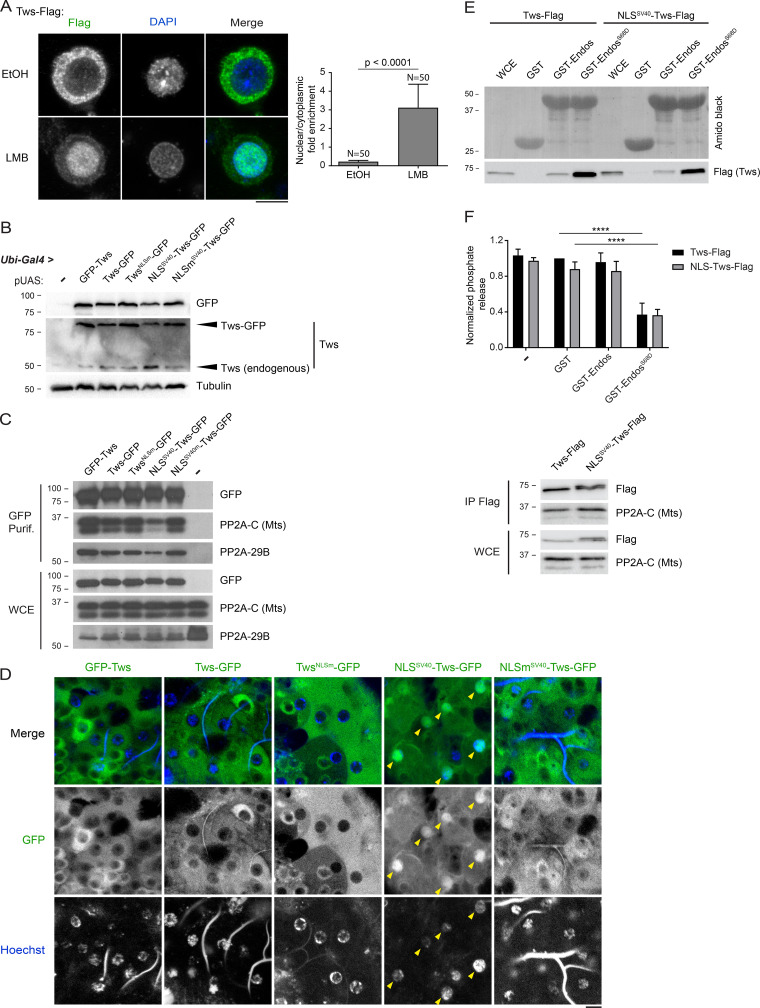
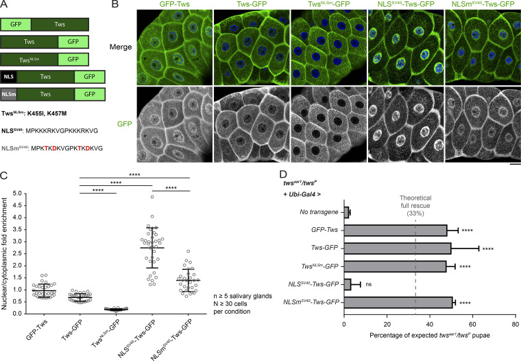
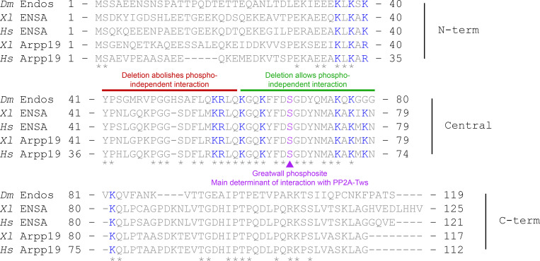
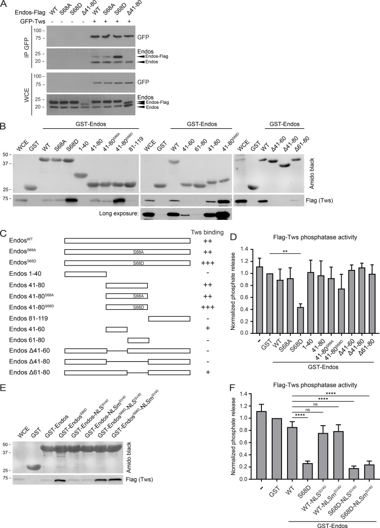
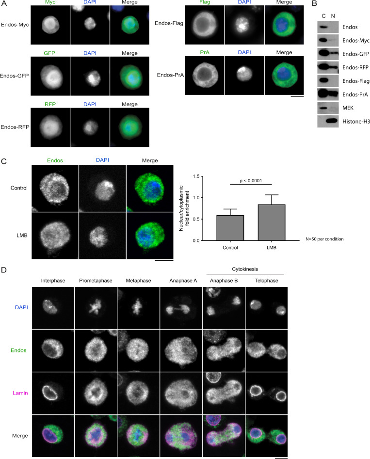
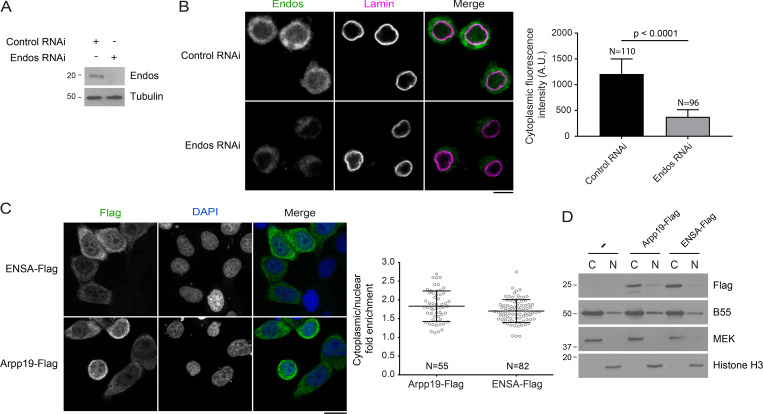
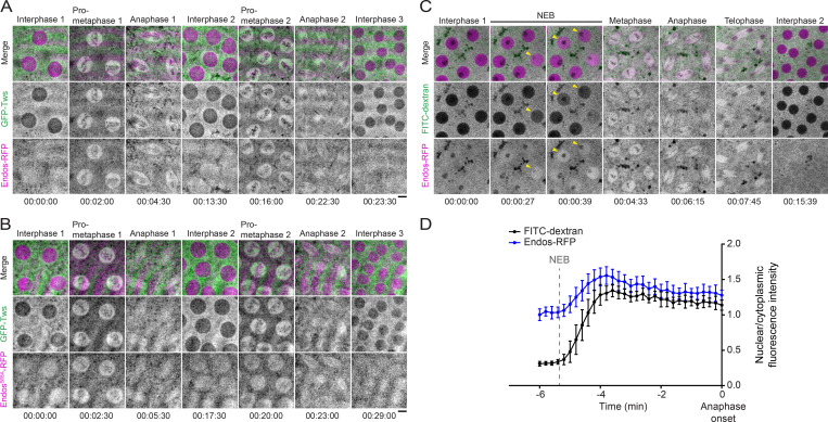
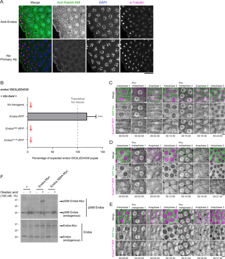
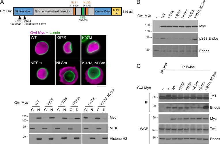
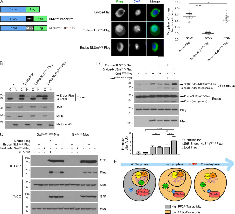
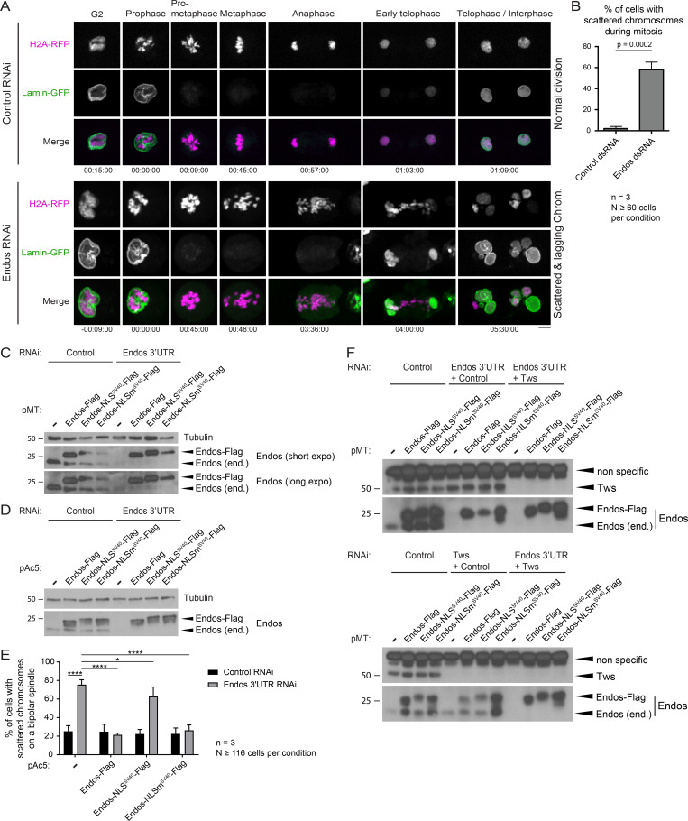
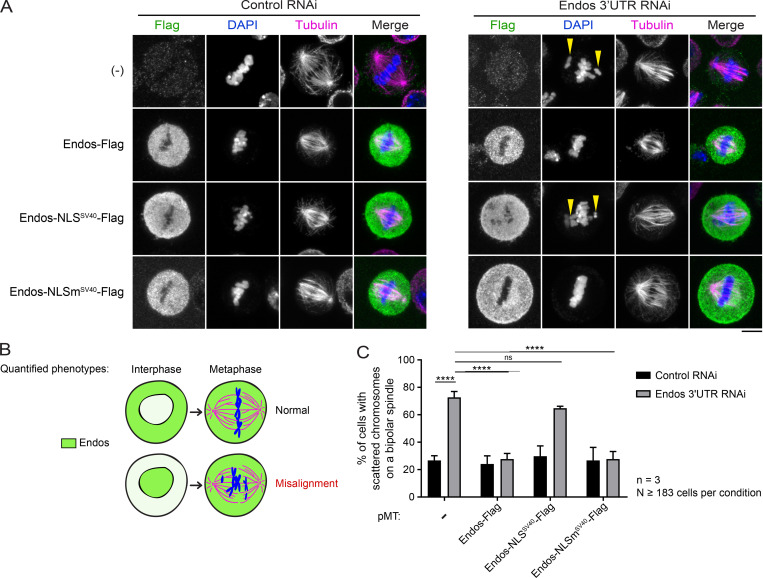
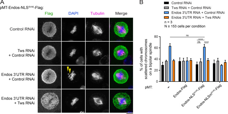
Mitotic entry involves inhibition of protein phosphatase 2A bound to its B55/Tws regulatory subunit (PP2A-B55/Tws), which dephosphorylates substrates of mitotic kinases. This inhibition is induced when Greatwall phosphorylates Endos, turning it into an inhibitor of PP2A-Tws. How this mechanism operates spatiotemporally in the cell is incompletely understood. We previously reported that the nuclear export of Greatwall in prophase promotes mitotic progression. Here, we examine the importance of the localized activities of PP2A-Tws and Endos for mitotic regulation. We find that Tws shuttles through the nucleus via a conserved nuclear localization signal (NLS), but expression of Tws in the cytoplasm and not in the nucleus rescues the development of tws mutants. Moreover, we show that Endos must be in the cytoplasm before nuclear envelope breakdown (NEBD) to be efficiently phosphorylated by Greatwall and to bind and inhibit PP2A-Tws. Disrupting the cytoplasmic function of Endos before NEBD results in subsequent mitotic defects. Evidence suggests that this spatiotemporal regulation is conserved in humans.


Figures

Figure 1.
Figure 1.
Tws is primarily cytoplasmic but shuttles through the nucleus. (A) D-Mel cells expressing Tws-GFP or TwsNLSm-GFP (NLS mutant: K455I, K457M) were treated with 50 nM LMB (b and d) or 0.1% ethanol (EtOH; mock control; a and c) for 2 h before fixation and DNA staining with DAPI. Scale bar: 10 µm. (B) Cells expressing Tws-GFP or TwsNLSm-GFP were treated with LMB or EtOH and imaged. Scale bar: 5 µm. (C) The nuclear/cytoplasmic ratio of GFP fluorescence intensity from video images in B was quantified through time (10 cells for each condition, mean ± SD). (D) Tws contains an NLS that is conserved in vertebrate B55 orthologues. Top: sequence alignment showing the NLS in magenta. Mutations introduced to inactivate the NLS in Tws are shown in red. Bottom: location of the identified NLS in the structure of the human PP2A-B55α complex bound to microcystin-LR (Protein Data Bank accession no. 3DW8; Xu et al., 2008). Dm, Drosophila melanogaster; Hs, Homo sapiens; Xl, Xenopus laevis.
Figure S1.
Figure S1.
Spatiotemporal regulation of Tws localization (complements to Figs. 1 and 2). (A) Left: D-Mel cells expressing Tws-Flag were treated with 50 nM LMB or 0.1% ethanol (EtOH; mock control) for 4 h before fixation, immunostaining, and DNA staining with DAPI. Scale bar: 5 µm. Right: Quantifications of the nuclear/cytoplasmic ratio of signal intensities (mean ± SD). (B) WB from third instar larvae in which expression of the indicated Tws variants was driven by Ubi-Gal4. (C) All GFP-fused Tws variants can associate with PP2A-C (microtubule star [Mts]) and PP2A-29B. 0–2-h-old embryos expressing the indicated proteins were submitted to GFP-affinity purifications. Purification products were analyzed by WB. (D) Images of larval brains (ventral nerve cord) from third instar larvae expressing Tws variants. DNA was stained with Hoechst 33342. Note the higher nuclear/cytoplasmic ratio of NLSmSV40-Tws-GFP (arrows) compared with the other variants. Scale bar: 10 µm. (E) Fusion of NLSSV40 to Tws does not prevent its ability to bind Endos. A GST pulldown was done as in Fig 3 B. (F) Fusion of NLSSV40 to Tws does not prevent its ability to be inhibited by EndosS68D. A phosphatase assay was done as in Fig 3 D. Top: quantification of the phosphatase activity (mean ± SD, n = 3). Bottom: Visualization of the immunoprecipitated (IP) PP2A complexes used in the reactions. Error bar: SD. ****, P ≤ 0.0001. Purif., purification; WCE, whole-cell extract.
Figure 2.
Figure 2.
Tws function is required in the cytoplasm. (A) Tws-GFP variants constructed for transgenic expression. Tws-GFP was fused to two copies of NLSSV40 or to a mutated form (NLSmSV40; substitutions in red). (B) The localization of Tws variants fused to GFP was visualized in third instar larval tissues. Salivary gland cells are shown (see also brain cells in Fig. S1). Transgenes were under the UASp promoter, and expression was driven by Ubi-Gal4. Merge images show GFP (green) and DNA stained with Hoechst 33342 (blue). Scale bar: 20 µm. (C) The nuclear/cytoplasmic ratio of GFP intensity was quantified (mean ± SD). (D) Genetic rescue of twsaar1/twsP mutant flies by the expression of the indicated Tws variants. Values shown correspond to percentages of eclosed twsaar1/twsP pupae (Tb+) relative to the expected number of twsaar1/twsP pupae calculated from the total number of eclosed pupae. See Materials and methods for details. Values are averages of three independent experiments in which between 110 and 1252 eclosed pupae were scored for each cross. Error bars: SD. ****, P ≤ 0.0001.
Figure S2.
Figure S2.
Sequence alignment of Drosophila Endos with vertebrate orthologues. The central region is the most conserved. It contains the Gwl phosphorylation site Ser68, which is the main determinant for Endos interaction with PP2A-Tws. Endos also engages in a phosphorylation-independent interaction with PP2A-Tws that requires a region upstream of the Gwl phosphorylation site. Residues in blue are positively charged and reported to promote ENSA dephosphorylation by PP2A-B55 (Cundell et al., 2016). *, perfectly conserved residues. Based on an alignment obtained using Align from UniProt. Dm, Drosophila melanogaster; Hs, Homo sapiens; term, terminal; Xl, Xenopus laevis.
Figure 3.
Figure 3.
Endos binding and inhibition of PP2A-Tws depends mainly on its Gwl phosphorylation site. (A) D-Mel cells transfected with the indicated proteins were submitted to GFP-Tws immunoprecipitation (IP), and products were analyzed by WB for GFP and Endos. (B) The indicated variants of GST-Endos (or GST alone) were tested for their ability to pull down Tws-Flag from a cell extract (detected with anti-Flag). (C) Summary of Endos variants tested in A and B for their interaction with Tws. (D) The ability of the indicated GST-Endos variants to inhibit PP2A-Tws phosphatase activity toward a phosphopeptide was quantified. (E and F) C-terminal fusion of the SV40 NLS to Endos does not prevent its ability to bind Tws (E, GST pulldown as in B) or to inhibit PP2A-Tws (F, phosphatase assay as in D). Bars: mean ± SD, n = 3. **, P = 0.0022; ****, P ≤ 0.0001. WCE, whole-cell extract.
Figure S3.
Figure S3.
Endos is a cytoplasmic protein, and tags can perturb its nucleocytoplasmic distribution. (A) Fluorescence images of fixed D-Mel cells expressing the indicated tagged forms of Endos. Note that GFP, RFP, and Myc (six copies), which are larger tags, make Endos more nuclear than cytoplasmic. Conversely, Flag (three copies) and the PrA, which are smaller tags, keep Endos in the cytoplasm. (B) Subcellular fractionation and WB show the relative amounts of cytoplasmic and nuclear proteins. MEK and histone H3 are controls as cytoplasmic and nuclear proteins, respectively. (C) Inhibition of Crm1-dependent nuclear export does not cause a strong nuclear accumulation of Endos. Left: D-Mel cells were treated with 50 nM LMB or 0.1% ethanol (control) for 4 h before fixation, immunostaining and DNA staining with DAPI. Scale bar: 5 µm. Right: Quantifications of the nuclear/cytoplasmic ratio of signal intensities (mean ± SD). (D) Endos is mainly cytoplasmic throughout the cell cycle. IF showing the localization of endogenous Endos in the different phases of the mitotic cell cycle. Scale bar: 5 µm. C, cytoplasmic; N, nuclear.
Figure 4.
Figure 4.
Endos and its human orthologues are enriched in the cytoplasm. (A) The specificity of antibodies against Endos is verified by RNAi and WB. (B) IF reveals that Endos is mainly cytoplasmic. Left: Representative images. Scale bar: 5 µm. Right: Quantifications of the cytoplasmic signal. (C) IF reveals that Endos-Flag and Arpp19-Flag are mainly cytoplasmic. Left: Representative images. Scale bar: 20 µm. Right: Quantifications of the cytoplasmic/nuclear ratio of signal intensities. Bars: mean ± SD. N = number of cells quantified.  (D) Subcellular fractionation and WB show the relative amounts of cytoplasmic and nuclear proteins. MEK and histone H3 are controls as cytoplasmic and nuclear proteins, respectively. C, cytoplasmic; N, nuclear.
Figure 5.
Figure 5.
Dynamic localization of Endos-RFP and GFP-Tws in syncytial embryos. (A) Time-lapse imaging reveals that GFP-Tws is enriched in the cytoplasm in interphase and becomes enriched in the nuclear/spindle area simultaneously with Endos-RFP in prometaphase until anaphase. (B) The S68A mutation does not alter the localization dynamics of Endos-RFP. For A and B, images from multiple z-steps were combined in an average intensity projection. (C) Time-lapse imaging of an embryo expressing Endos-RFP and injected with 70 kD FITC-dextran to mark the time of NEBD. Endos-RFP is homogenously dispersed throughout the syncytial embryo in interphase until NEBD, when it becomes enriched in the nuclear/spindle area. Images shown are for a single focal plane. (D) The nuclear/cytoplasmic ratios of FITC-dextran and Endos-RFP fluorescence intensities from Video 2 in C were quantified through time relative to anaphase onset. Values are averages of 14 nuclei. Error bars: SD. Scale bars: 5 µm.
Figure S4.
Figure S4.
Endos-RFP localization to the nuclear/spindle area is independent from interaction with PP2A-Tws. (A) IF against endogenous Endos reveals specific staining in the nuclear area in prometaphase. Scale bar: 20 µm. (B) Endos-RFP but not EndosS68A-RFP or EndosΔ41–80-RFP rescues the development of Endos mutant flies. Transgenes were under the UASp promoter and driven ubiquitously by Ubi-Gal4. Values shown correspond to percentages of eclosed endos1/Df pupae (Tb+) relative to the expected number of endos1/Df pupae calculated from the total number of eclosed pupae. The observed rescue exceeds the expected rescue because of higher stochastic mortality of the other genotypes caused by balancer chromosomes. Values are averages of three independent experiments in which between 193 and 325 eclosed pupae were scored for each cross. Error bars: SD. ****, P < 0.0001. (C–E) Time-lapse imaging of embryos expressing GFP-Tws and variants of Endos-RFP. (C) EndosS68D-RFP (gain of interaction with PP2A-Tws). (D) EndosΔ41–60-RFP (loss of pS68-independent interaction with PP2A-Tws). (E) EndosΔ41–80-RFP (complete loss of interaction with PP2A-Tws). Images from multiple z-steps were combined in an average intensity projection. (F) Validation of the phosphospecific antibody against pS68-Endos. Cells were transfected and treated as indicated and analyzed by WB. Ab, antibody. Scale bars: 5 µm (C–E).
Figure 6.
Figure 6.
Active Gwl in the cytoplasm is necessary and sufficient for the induction of Endos phosphorylation and interaction with PP2A-Tws. (A) Mutations in Gwl used to alter its kinase activity and localization. Top: Location of the mutations in the primary structure. Middle: IF showing the localization of the Gwl-Myc variants. Bottom: Subcellular fractionation and WB showing the relative amounts of cytoplasmic and nuclear proteins. MEK and histone H3 are controls as cytoplasmic and nuclear proteins, respectively. Scale bar: 5 μm. (B) Expression of the constitutively active and cytoplasmic GwlK97M, NLSm-Myc increases levels of pS68-Endos. Cells were transfected with the indicated constructs, treated with 100 nM Okadaic acid for 1 h, and analyzed by WB. (C) Expression of the constitutively active cytoplasmic GwlK97M, NLSm-Myc enhances the interaction between Endos and Tws. Cells were transfected with the indicated constructs and submitted to Tws immunoprecipitation (IP). Products were analyzed by WB. *, IgG. C, cytoplasmic; Dm, Drosophila melanogaster; N, nuclear; NES, nuclear export signal; ter., terminal; WCE, whole-cell extract.
Figure 7.
Figure 7.
Endos must be in the cytoplasm to be phosphorylated by Gwl. (A) Fusion of the SV40 NLS to Endos-Flag targets it to the nucleus. Left: Endos-Flag variants analyzed. Center: Representative IF images. Scale bar: 5 µm. Right: Quantifications of the cytoplasmic/nuclear ratio of signal intensities (bars: mean ± SD). N = number of cells quantified. Error bars: SD. ****, P = 0.0001. (B) Subcellular fractionation and WB show the relative amounts of cytoplasmic and nuclear proteins. MEK and histone H3 are controls as cytoplasmic and nuclear proteins, respectively. (C) Active and cytoplasmic Gwl (K97M, NLSm) enhances Endos interaction with Tws if Endos is in the cytoplasm but not in the nucleus. Cells were transfected with the indicated constructs and submitted to GFP-Tws immunoprecipitation (IP). Products were analyzed by WB. (D) Gwl phosphorylates Endos more efficiently in the cytoplasm than in the nucleus. Cells were transfected with the indicated constructs and analyzed by WB. Quantification of the pS68 Endos-NLS(m)SV40-Flag/total Flag band intensities are shown (mean ± SD, n = 3). *, P < 0.05; **, 0.001 < P < 0.01; ****, P < 0.0001. (E) Model for the spatiotemporal dynamics of the Gwl-Endos-PP2A-Tws module. In prophase, cyclin B-Cdk1 activates Gwl and promotes its export to the cytoplasm where Gwl phosphorylates Endos to induce its binding and inhibition of PP2A-Tws before NEBD. After NEBD, cyclin B-Cdk1 keeps Gwl active, and Gwl keeps Endos phosphorylated and PP2A-Tws inhibited during prometaphase. Spiky shapes: activated proteins. C, cytoplasmic; N, nuclear; WCE, whole-cell extract.
Figure S5.
Figure S5.
RNAi depletion of Endos or its targeting to the nucleus results in Tws-dependent mitotic defects (complements to Figs. 8 and 9). (A) Cells stably expressing H2A-RFP and lamin-GFP were transfected with dsRNA against Endos or the bacterial KAN gene (nontarget control). Cell divisions were then filmed on a spinning-disk microscope. Upon depletion of Endos, mitosis is delayed, and chromosomes become scattered and/or lagging. Lamin-GFP is ultimately recruited aberrantly on dispersed chromatin masses. Scale bar: 5 µm. (B) Quantification of the percentage of mitotic cells displaying scattered chromosomes (mean ± SD). (C) WB validation of protein expression and RNAi depletion for experiment in Fig. 8 A. (D and E) Independent validation with cells constitutively expressing Endos-Flag variants under the pAC5 promoter. (D) WB. (E) Quantification of phenotypes (mean ± SD, n = 3). *, P < 0.05; ****, P < 0.0001. (F) WB validation of protein expression and RNAi depletion for experiments in Fig. 9. chrom., chromosomes; end., endogenous; expo, exposure.
Figure 8.
Figure 8.
Endos must be in the cytoplasm before NEBD to promote correct mitotic entry. (A and B) Expression of the indicated proteins was induced, and cells were transfected with dsRNA targeting endogenous Endos (3′UTR) or nontarget control dsRNA. Scale bar: 5 μm. Representative IF images are shown (A). Arrowheads indicate scattered chromosomes on a bipolar spindle (as illustrated in panel B). (C) Quantification of the percentage of mitotic cells with scattered chromosomes on a bipolar spindle (mean ± SD, n = 3). ****, P ≤ 0.0001.
Figure 9.
Figure 9.
Depletion of Tws rescues mitotic defects resulting from the loss of Endos function in the cytoplasm. (A) Expression of the indicated proteins was induced, and cells were transfected with dsRNA targeting endogenous Endos (3′UTR), Tws, and/or nontarget control dsRNA. Representative IF images are shown. Arrowheads indicate scattered chromosomes on a bipolar spindle. Scale bar: 5 μm. (B) Quantification of the percentage of mitotic cells with scattered chromosomes on a bipolar spindle (mean ± SD, n = 3). ****, P ≤ 0.0001.

References

    1. Álvarez-Fernández, M., and Malumbres M.. 2014. Preparing a cell for nuclear envelope breakdown: spatio-temporal control of phosphorylation during mitotic entry. BioEssays. 36:757–765. 10.1002/bies.201400040 - DOI - PubMed
    1. Álvarez-Fernández, M., Sánchez-Martínez R., Sanz-Castillo B., Gan P.P., Sanz-Flores M., Trakala M., Ruiz-Torres M., Lorca T., Castro A., and Malumbres M.. 2013. Greatwall is essential to prevent mitotic collapse after nuclear envelope breakdown in mammals. Proc. Natl. Acad. Sci. USA. 110:17374–17379. 10.1073/pnas.1310745110 - DOI - PMC - PubMed
    1. Archambault, V., and Glover D.M.. 2009. Polo-like kinases: conservation and divergence in their functions and regulation. Nat. Rev. Mol. Cell Biol. 10:265–275. 10.1038/nrm2653 - DOI - PubMed
    1. Archambault, V., Zhao X., White-Cooper H., Carpenter A.T., and Glover D.M.. 2007. Mutations in Drosophila Greatwall/Scant reveal its roles in mitosis and meiosis and interdependence with Polo kinase. PLoS Genet. 3:e200. 10.1371/journal.pgen.0030200 - DOI - PMC - PubMed
    1. Bergeron-Sandoval, L.P., Safaee N., and Michnick S.W.. 2016. Mechanisms and consequences of macromolecular phase separation. Cell. 165:1067–1079. 10.1016/j.cell.2016.05.026 - DOI - PubMed

Publication types

MeSH terms

Grants and funding

LinkOut - more resources