. 2021 Jun 7;220(6):e202006018.
doi: 10.1083/jcb.202006018.

Borealin directs recruitment of the CPC to oocyte chromosomes and movement to the microtubules

Affiliations

Borealin directs recruitment of the CPC to oocyte chromosomes and movement to the microtubules

Lin-Ing Wang et al. J Cell Biol. .

Abstract

The chromosomes in the oocytes of many animals appear to promote bipolar spindle assembly. In Drosophila oocytes, spindle assembly requires the chromosome passenger complex (CPC), which consists of INCENP, Borealin, Survivin, and Aurora B. To determine what recruits the CPC to the chromosomes and its role in spindle assembly, we developed a strategy to manipulate the function and localization of INCENP, which is critical for recruiting the Aurora B kinase. We found that an interaction between Borealin and the chromatin is crucial for the recruitment of the CPC to the chromosomes and is sufficient to build kinetochores and recruit spindle microtubules. HP1 colocalizes with the CPC on the chromosomes and together they move to the spindle microtubules. We propose that the Borealin interaction with HP1 promotes the movement of the CPC from the chromosomes to the microtubules. In addition, within the central spindle, rather than at the centromeres, the CPC and HP1 are required for homologous chromosome bi-orientation.


Figures

Figure 1.
Figure 1.
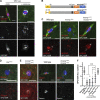
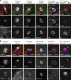
INCENP localization and spindle assembly depends on the N-terminal Borealin/Deterin binding domain. (A) INCENP localization in oocytes after a 60-min colchicine treatment to destabilize the microtubules. The sister centromeres appeared to be separating in colchicine-treated oocytes, the mechanism of which is not known. INCENP is in red, CID/CENP-A is in white, tubulin is in green, and DNA is in blue. (B) A schematic of Drosophila INCENP showing the location of the centromere-targeting BS binding region, SAH, and INbox (IN) domains. The black box (437–441) shows the sequence targeted by the shRNA GL00279 that were mutated to make RNAi-resistant IncenpWT-R. The conserved amino acids 22–30 are deleted in IncenpΔCEN. (C) INCENP (red) localization in Incenp RNAi oocytes and oocytes also expressing the IncenpWT-R RNAi-resistant transgene. Kinetochore protein SPC105R is in white. (D) Spindle and kinetochore assembly defects in Deterin RNAi oocytes. (E) Spindle and kinetochore assembly defects in IncenpΔCEN, Incenp RNAi oocytes, with Deterin or INCENP in red and SPC105R or CID in white. (F) Quantitation of SPC105R localization in RNAi oocytes (n = 7, 14, 32, 12, 22, and 8 oocytes). Error bars indicate 95% confidence intervals; **, P < 0.01 run by Fisher’s exact test. Scale bars, 5 µm (all images).
Figure S1.
Figure S1.
Alignment and domain analysis of Drosophila. The sequence alignment compares Drosophila melanogaster (Dm) INCENP to Drosophila virilis (Dvir) and Xenopus laevis (Xl). The Borealin/Deterin binding domain is from amino acids 1–46 (yellow). CEN and STD deletion mutations are marked in red, two potential HP1 interaction sites are marked in blue, the RNAi mismatch region is marked in black, the SAH domain is marked in orange, and the INbox (IN) is in black.
Figure S2.
Figure S2.
Expression of mis12:INbox in WT oocytes disrupts meiotic progression. (A) MIS12 localization in WT and Incenp RNAi oocytes (arrows). MIS12 is in red, tubulin is in green, DNA is in blue, and CENP-C is in white. Scale bar, 5 µm. (B) Fertilized 0-2-h-old Drosophila embryos were fixed and stained for INCENP (red), tubulin (green), and DNA (blue). Scale bar, 5 µm. (C) Quantitation of SPC105R localization in oocytes with mis12 fusions (n = 6, 9, and 12 oocytes). Error bars indicate 95% confidence intervals; ****, P < 0.0001; **, P < 0.01 run by Fisher’s exact test. (D) Expression of Myc:feo:INbox in WT and mis12:INbox, IncenpRNAi oocytes. Myc:feo:INbox localizes to the central spindle in WT oocytes. (E) Coexpression of FLAG:mis12:INbox and Myc:feo:INbox in Incenp RNAi oocytes. In these images, the Myc tag (FEO:INbox) is red, Aurora B or CID is white, tubulin is green, and DNA is blue. Scale bar, 5 µm.
Figure 2.
Figure 2.
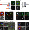
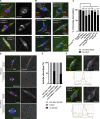
Independent localization of the CPC to the centromere and the central spindle assembles only kinetochore-dependent microtubules. (A) A schematic of the CPC constructs designed to target the CPC to the centromeres. (B) Expression of mis12:Incenp in WT and Incenp RNAi oocytes. (C) Quantitation of the spindle phenotype of MIS12 fusions expressed in Incenp RNAi oocytes (n = 23, 36, and 25 oocytes). (D) Myc:INbox, Flag:mis12:INbox, and Myc:sub:INbox expressed in WT oocytes. (E) Detection of the Aurora B substrate, phosphorylated INCENP (pINCENP). The arrow and inset (3 µm wide, levels increased) show the pINCENP signal at the centromeres when the INbox is targeted by MIS12. INCENP is in red and pINCENP is in white. (F) Flag:mis12:INbox and Myc:sub:INbox expressed in Incenp RNAi oocytes. The arrow points to K-fibers in Flag:mis12:Inbox oocytes. The low-magnification image of Myc:sub:INbox shows microtubule bundles in the cytoplasm instead of around the chromosomes. Transgene proteins are in red, SPC105R in white, DNA is in blue, and tubulin is in green. Scale bars, 5 µm (left four panels) and 10 µm (low-magnification image). (G) Flag:mis12:INbox expressed alone or coexpressed with Myc:sub:INbox in Incenp RNAi oocytes. Merged images showed DNA (blue), tubulin (green), Myc (red), and Aurora B (white). Scale bars, 5 µm.
Figure 3.
Figure 3.
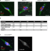
Borealin is sufficient to recruit the CPC for meiotic spindles assembling. (A) A schematic of the Det:Incenp and borr:Incenp fusions compared with WT Incenp. (B) The spindle phenotypes of Det:Incenp (n = 18) and borr:Incenp (n = 28) in Incenp RNAi oocytes. The separate channels show the localization of INCENP and the microtubules. The second borr:Incenp image is an example of a spindle with diffuse INCENP on the spindle. Arrows show K-fibers in Det:Incenp oocytes and central spindle fibers in borr:Incenp oocytes. INCENP is in red, DNA is in blue, tubulin is in green, and CID is in white. (C) Deterin depletion in borr:Incenp oocytes has a spindle phenotype similar to borr:Incenp. (D) Summary of the effect of Deterin depletion in borr:Incenp oocytes (n = 23, 18, 28, and 23). (E) Deterin localization in WT and borr:Incenp, Incenp RNAi oocytes (n = 22). The first borr:Incenp image is an example with a defined central spindle and the second has a diffuse spindle and INCENP. INCENP is in red, Deterin is in white, DNA is in blue, and tubulin is in green. Scale bars, 5 µm (all images).
Figure S3.
Figure S3.
Borealin localization in Det:Incenp, Incenp RNAi oocytes and the localization of the CPC to the chromosomes. (A) Metaphase I oocytes from WT and Det:Incenp, Incenp RNAi females. Borealin is in white, INCENP is in red, tubulin is in green, and DNA is in blue. Scale bar, 5 µm. (B) Expression of Incenp transgenes shown in Fig. 4 C in Incenp RNAi oocytes, including borrΔC:Incenp, IncenpΔHP1, and borrΔC:IncenpΔHP1. The images show SPC105R in white, INCENP in red, DNA in blue, and tubulin in green. Scale bar, 5 µm.
Figure S4.
Figure S4.
Phenotype of haspin, Bub1, and Su(var)205 (HP1) knockdowns. (A) Metaphase I oocytes from haspin HK420 or Bub1 GL00151 single RNAi or haspin, Bub1 double RNAi females. Centromere protein CID is in white, INCENP is in red, tubulin is in green, and DNA is in blue. (B) Fertility and X chromosome NDJ in haspin and Bub1 RNAi and haspin mutant females. (C) Spindle in oocytes depleted of Drosophila HP1 using Su(var)205 GL00531 RNAi oocytes. CID is in white, INCENP is in red, tubulin is in green, and DNA is in blue. Scale bars, 5 µm (all images).
Figure 4.
Figure 4.
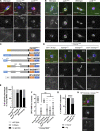
Chromatin–Borealin interaction is critical for CPC-dependent meiotic spindle assembly. (A) Borealin and HP1 localization in WT, Incenp RNAi, and aurB RNAi oocytes. Borealin is in white, HP1 is in red, tubulin is in green, and DNA is in blue. (B) HP1 and H3K9me3 localization in WT and aurB RNAi oocytes. Arrow shows HP1 localization in WT oocytes on the spindle. HP1 or tubulin is in green, INCENP is in red, DNA is in blue, and CID or H3K9me3 are in white. (C) A schematic of Incenp mutant constructs affecting HP1 interactions. Proposed HP1 binding sites are located in the C terminus of Borealin and in INCENP between amino acids 121–232. Full-length Drosophila HP1 was fused to INCENP by substitution for the BS domain. (D) Expression of Incenp transgenes shown in C in Incenp RNAi oocytes, including borrΔC:Incenp, IncenpΔHP1 and borrΔC:IncenpΔHP1. Two different images of IncenpΔHP1 are shown to compare ring-shaped and disorganized (see arrow) localization of the CPC. The images show CID in white, INCENP in red, DNA in blue, and tubulin in green. (E) Quantitation of spindle phenotype in the HP1 interaction–defective mutants shown in D (n = 17, 26, 31, and 49 oocytes, in the order as shown in the graph). (F) SPC105R localization (see Fig. S3) in the HP1 interaction–defective mutant oocytes (n = 8, 32, 9, 15, 14, and 33). Bars indicates 95% confidence intervals; ns, not significant in Fisher’s exact test. (G) Quantitation of INCENP spindle localization in WT (n = 19) and IncenpΔHP1, Incenp RNAi oocytes (n = 17). ****, P < 0.0001. (H) Expressing HP1:Incenp (see C) in Incenp RNAi oocytes. The images show CID in white, INCENP in red, DNA in blue, and tubulin in green.
Figure 5.
Figure 5.
The dissociation of HP1 and the CPC from the chromosomes depends on Aurora B kinase activity and microtubules. (A) WT oocytes were treated with DMSO or BN2 for 1 h to inhibit Aurora B kinase activity. In red are heterochromatic marks HP1 or H3K9me3 and in white are the CPC components INCENP or Deterin. Scale bars, 5 µm. (B) WT oocytes were treated for 60 min with ethanol or colchicine to depolymerize microtubules, and were analyzed for the same markers as in A. In all images, DNA is in blue and tubulin is in green.
Figure 6.
Figure 6.
Analysis of CPC interactions with Subito and microtubules required for central spindle assembly. (A) A schematic showing two INCENP deletions removing regions that promote microtubule interactions. (B) Bipolar spindle assembly when IncenpΔSTD or IncenpΔSAH were expressed in Incenp RNAi oocytes. CID is in white, INCENP is in red, tubulin is in green, and DNA is in blue. (C) Tripolar spindle and mislocalization of the CPC phenotype in subHM26/sub131 oocytes, with tubulin (green), Deterin (white), and HP1 or Subito (red) localization. Scale bars, 5 µm.
Figure 7.
Figure 7.
Disruption of homologue bi-orientation by disruptions of central spindle CPC. (A) Disorganized or mislocalized CPC caused by the Incenp hypomorphic allele Incenp18.197 or the transgenes myc:Incenp and HA:Incenp. The CPC or MYC are in red, CID is in white, tubulin is in green, and DNA is in blue. (B) Incenp mutants examined for homologue bi-orientation using FISH with probes against pericentromeric heterochromatin on the X (359 bp repeat, yellow), second (AACAC, red), and third (dodeca, white) chromosomes. (C) Rates of bi-orientation defects were quantified (n = 57, 37, 50, 30, 69, 60, and 63 in the order of the graph). *, P < 0.05; ****, P < 0.0001 in Fisher’s exact test. (D) WT oocytes and mis12:INbox oocytes treated with colchicine for 30 min. INCENP is in red and CID is in white. (E) Quantitation of spindle assembly after colchicine treatment (n = 5, 10, 7, and 17 in the order of the graph). (F) Localization of HP1 and Deterin in WT and IncenpΔHP1, Incenp RNAi oocytes. HP1 is in green, Deterin is in red, and overlapping region is in yellow.
Figure 8.
Figure 8.
Model for spindle assembly in Drosophila oocytes. After nuclear envelope breakdown, a complex of INCENP, Borealin, and Deterin/Survivin is recruited to the chromosomes. Localization studies suggest that CPC recruitment is enriched in heterochromatic regions containing H3K9me3 and HP1. Aurora B is recruited, which results in kinetochore assembly, limited microtubule (MT) recruitment in the form of K-fibers, and phosphorylation of other targets, including H3S10 and possibly HP1. Aurora B activity also results in Borealin-dependent ejection of HP1 and the CPC from the chromosomes to the microtubules. Once on the microtubules, the kinesin 6 Subito causes enrichment of the CPC and HP1 in the central spindle. HP1 could be involved in a complex pattern of interactions that bring important spindle proteins together (Eissenberg and Elgin, 2014). For example, Aurora B could be brought together with potential phosphorylation substrate Subito, which has a conserved HP1 binding site that is required for its meiotic functions.

Similar articles

Cited by

References

    1. Abad, M.A., Ruppert J.G., Buzuk L., Wear M., Zou J., Webb K.M., Kelly D.A., Voigt P., Rappsilber J., Earnshaw W.C., and Jeyaprakash A.A.. 2019. Borealin-nucleosome interaction secures chromosome association of the chromosomal passenger complex. J. Cell Biol. 218:3912–3925. 10.1083/jcb.201905040 - DOI - PMC - PubMed
    1. Adams, R.R., Maiato H., Earnshaw W.C., and Carmena M.. 2001. Essential roles of Drosophila inner centromere protein (INCENP) and aurora B in histone H3 phosphorylation, metaphase chromosome alignment, kinetochore disjunction, and chromosome segregation. J. Cell Biol. 153:865–880. 10.1083/jcb.153.4.865 - DOI - PMC - PubMed
    1. Adriaans, I.E., Hooikaas P.J., Aher A., Vromans M.J.M., van Es R.M., Grigoriev I., Akhmanova A., and Lens S.M.A.. 2020. MKLP2 Is a Motile Kinesin that Transports the Chromosomal Passenger Complex during Anaphase. Curr. Biol. 30:2628–2637.e9. 10.1016/j.cub.2020.04.081 - DOI - PubMed
    1. Ainsztein, A.M., Kandels-Lewis S.E., Mackay A.M., and Earnshaw W.C.. 1998. INCENP centromere and spindle targeting: identification of essential conserved motifs and involvement of heterochromatin protein HP1. J. Cell Biol. 143:1763–1774. 10.1083/jcb.143.7.1763 - DOI - PMC - PubMed
    1. Alekseyenko, A.A., Gorchakov A.A., Zee B.M., Fuchs S.M., Kharchenko P.V., and Kuroda M.I.. 2014. Heterochromatin-associated interactions of Drosophila HP1a with dADD1, HIPP1, and repetitive RNAs. Genes Dev. 28:1445–1460. 10.1101/gad.241950.114 - DOI - PMC - PubMed

Publication types

MeSH terms

Substances