Review
. 2021 Sep 30;62(6):926-941.
doi: 10.1093/pcp/pcab052.

The Unicellular Red Alga Cyanidioschyzon merolae-The Simplest Model of a Photosynthetic Eukaryote

Affiliations
Review

The Unicellular Red Alga Cyanidioschyzon merolae-The Simplest Model of a Photosynthetic Eukaryote

Shin-Ya Miyagishima et al. Plant Cell Physiol. .

Abstract

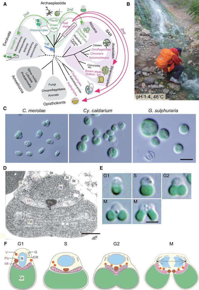
Several species of unicellular eukaryotic algae exhibit relatively simple genomic and cellular architecture. Laboratory cultures of these algae grow faster than plants and often provide homogeneous cellular populations exposed to an almost equal environment. These characteristics are ideal for conducting experiments at the cellular and subcellular levels. Many microalgal lineages have recently become genetically tractable, which have started to evoke new streams of studies. Among such algae, the unicellular red alga Cyanidioschyzon merolae is the simplest organism; it possesses the minimum number of membranous organelles, only 4,775 protein-coding genes in the nucleus, and its cell cycle progression can be highly synchronized with the diel cycle. These properties facilitate diverse omics analyses of cellular proliferation and structural analyses of the intracellular relationship among organelles. C. merolae cells lack a rigid cell wall and are thus relatively easily disrupted, facilitating biochemical analyses. Multiple chromosomal loci can be edited by highly efficient homologous recombination. The procedures for the inducible/repressive expression of a transgene or an endogenous gene in the nucleus and for chloroplast genome modification have also been developed. Here, we summarize the features and experimental techniques of C. merolae and provide examples of studies using this alga. From these studies, it is clear that C. merolae-either alone or in comparative and combinatory studies with other photosynthetic organisms-can provide significant insights into the biology of photosynthetic eukaryotes.

Keywords: Cyanidioschyzon merolae; Cyanidiales; Photosynthetic eukaryotes; Red algae.


Figures

Fig. 1
Fig. 1
Phylogenetic position, habitat and classification of Cyanidiales and the cell cycle-dependent change in the cellular architecture of C. merolae. (A) Position of red algae and Cyanidiales in the eukaryotic tree. The broken lines denote the uncertainty of branch positions in the tree. Red algae and groups possessing chloroplasts (or nonphotosynthetic plastids) of red algal origin are shown in red. Viridiplantae (green algae and land plants) and groups possessing chloroplasts of green algal origin are shown in green. Arrows indicate the primary endosymbiotic event of a cyanobacterium (first) and secondary endosymbiotic events (second). The position and number of red algal secondary endosymbiotic events in the tree remain uncertain at present. This tree is according to Adl et al. (2012). (B) A sulfuric hot spring where cyanidialean red algae dominate (Kusatsu, Japan). (C) Interference-contrast microscopic images of C. merolae 10D, C. caldarium isolated in the hot spring shown in (B), and G. sulphuraria 074 G. Bar = 5 µm. (D) A transmission electron micrograph of C. merolae at the S phase. Bar = 500 nm. (E) Interference-contrast microscopic images of C. merolae showing the cell cycle progression. Bar = 2 µm. The images were reproduced from Miyagishima et al. (2012). (F) Schematic illustrations of C. merolae cells showing organelle inheritance. Cp, chloroplast; G, Golgi apparatus; Mt, mitochondrion; N, nucleus; Po, peroxisome; St, floridean starch granule; V, vacuole.
Fig. 2
Fig. 2
Examples of genetic modification in C. merolae. (A) A schematic diagram of the targeted integration of a transgene and a URA selectable marker into a chromosomal locus and the subsequent removal of the URA marker (Takemura et al. 2018, Takemura et al. 2019). (B) Photos of transformed colonies 2 weeks after the transformation and liquid culture (10 ml) of a clone 2 weeks after the inoculation of a colony. For efficient colony formation, cells were spread on cornstarch beds (white circles) on a gel plate. (C) An inducible/repressive expression system using the NR promoter (Fujiwara et al. 2015). Although the expression of a transgene (GFP) is regulated by the NR promoter, this system is also applicable for an endogenous gene by integrating the NR promoter to the upstream of a gene of interest in the chromosome (Fujiwara et al. 2020). Red is the autofluorescence of the chloroplast, and green is the fluorescence of GFP in the cytosol. Bar = 5 µm.
Fig. 3
Fig. 3
Mechanism of cell cycle progression and its relationship with the chloroplast DNA replication and chloroplast division in C. merolae. (A) A diagram showing the structure of chloroplast division machinery in C. merolae. For reference, the mitochondrial division machinery in C. merolae is also shown. DRP5B, the outer and inner PD rings and two types of FtsZ proteins also participate in chloroplast (plastid) division in plants. DRP3 and MDA1 (called Mdv1p in yeasts) also participate in mitochondrial division in many other eukaryotic lineages. In some lineages of eukaryotes, including C. merolae, the outer and inner MD rings and two types of FtsZ proteins also participate in mitochondrial division. (B) A schematic diagram of the interactive synchronization of the cell cycle progression and the chloroplast division in C. merolae. The host cell restricts the onset of chloroplast division to the S phase by the S phase-specific expression of nucleus-encoded chloroplast division proteins. The formation and constriction of the chloroplast division ring lift the prophase arrest so that the host cell enters into the metaphase only when chloroplast division progresses. (C) A schematic diagram of the G1–S transition, which is regulated by the retrograde signaling from the chloroplast to the host cell cycle and circadian rhythms. The chloroplast DNA replication somehow leads to the accumulation of Mg-Proto in the cell, which binds Fbox3. This binding inhibits the ubiquitination and degradation of G1 cyclin (CYC1) by Fbox3, leading to the accumulation of G1 cyclin-bound CDKA. During the G1 phase, RB-related protein (RBR) inhibits the transcriptional activity of E2F-DP. E2F is phosphorylated in a circadian rhythm-dependent manner, and E2F phosphorylation peaks during the evening. E2F phosphorylation permits CDKA-CYC1 to phosphorylates RBR. RBR phosphorylation inactivates RBR, thereby enabling E2F-DP to transcribe S-phase genes.

Similar articles

Cited by

References

    1. Adl S.M., Simpson A.G., Lane C.E., Lukes J., Bass D., Bowser S.S., et al. (2012) The revised classification of eukaryotes. J. Eukaryot. Microbiol. 59: 429–493. - PMC - PubMed
    1. Albertano P., Ciniglia C., Pinto G., Pollio A. (2000) The taxonomic position of Cyanidium, Cyanidioschyzon and Galdieria: an update. Hydrobiologia 433: 137–143.
    1. Antoshvili M., Caspy I., Hippler M., Nelson N. (2019) Structure and function of photosystem I in Cyanidioschyzon merolae. Photosynth. Res. 139: 499–508. - PubMed
    1. Batista R.A., Kohler C. (2020) Genomic imprinting in plants-revisiting existing models. Genes Dev. 34: 24–36. - PMC - PubMed
    1. Bohuszewicz O., Low H.H. (2018) Structure of a mitochondrial fission dynamin in the closed conformation. Nat. Struct. Mol. Biol. 25: 722–731. - PMC - PubMed