. 2021 Mar 16;118(11):e2023899118.
doi: 10.1073/pnas.2023899118.

Aryl hydrocarbon receptor is essential for the pathogenesis of pulmonary arterial hypertension

Affiliations

Aryl hydrocarbon receptor is essential for the pathogenesis of pulmonary arterial hypertension

Takeshi Masaki et al. Proc Natl Acad Sci U S A. .

Abstract

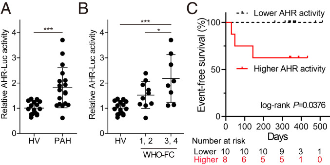
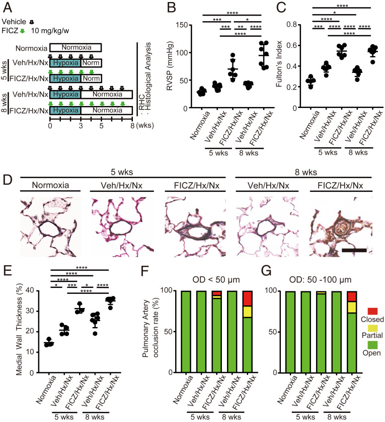
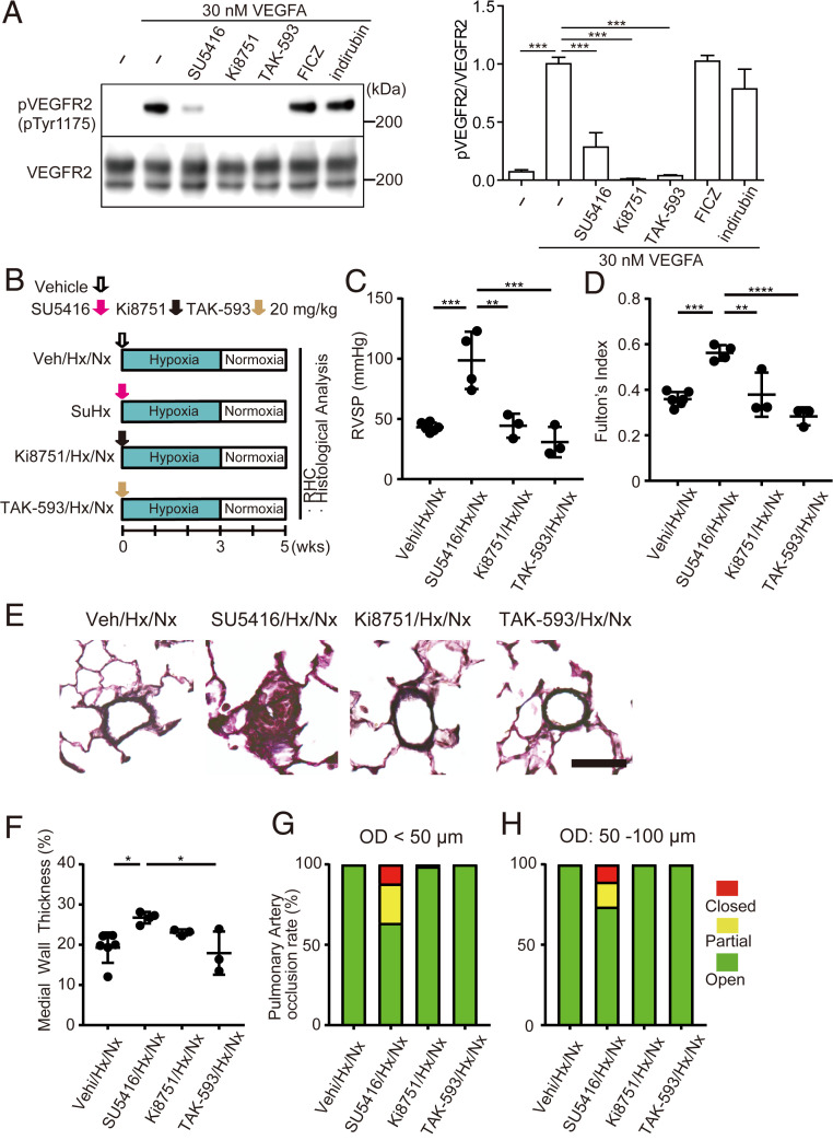
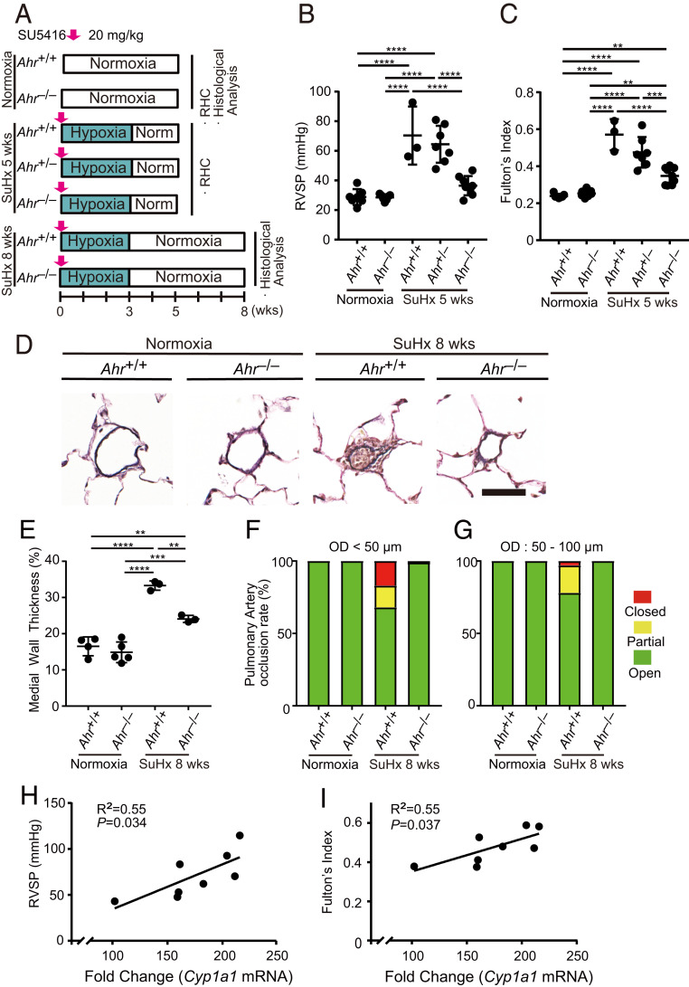
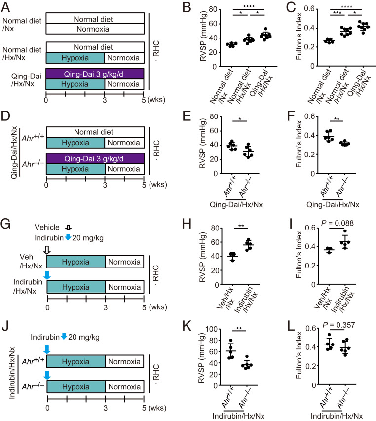
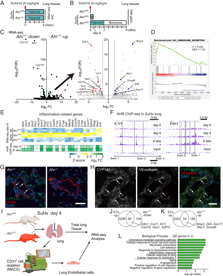
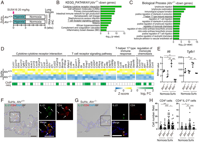
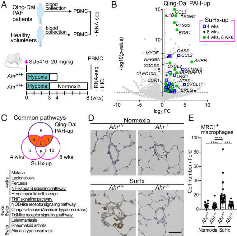
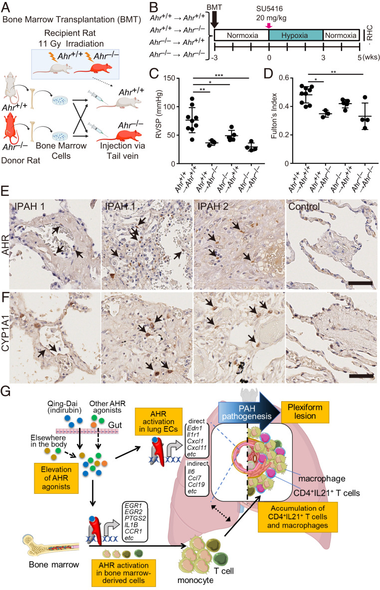
Pulmonary arterial hypertension (PAH) is a devastating disease characterized by arteriopathy in the small to medium-sized distal pulmonary arteries, often accompanied by infiltration of inflammatory cells. Aryl hydrocarbon receptor (AHR), a nuclear receptor/transcription factor, detoxifies xenobiotics and regulates the differentiation and function of various immune cells. However, the role of AHR in the pathogenesis of PAH is largely unknown. Here, we explore the role of AHR in the pathogenesis of PAH. AHR agonistic activity in serum was significantly higher in PAH patients than in healthy volunteers and was associated with poor prognosis of PAH. Sprague-Dawley rats treated with the potent endogenous AHR agonist, 6-formylindolo[3,2-b]carbazole, in combination with hypoxia develop severe pulmonary hypertension (PH) with plexiform-like lesions, whereas Sprague-Dawley rats treated with the potent vascular endothelial growth factor receptor 2 inhibitors did not. Ahr-knockout (Ahr-/- ) rats generated using the CRISPR/Cas9 system did not develop PH in the SU5416/hypoxia model. A diet containing Qing-Dai, a Chinese herbal drug, in combination with hypoxia led to development of PH in Ahr+/+ rats, but not in Ahr-/- rats. RNA-seq analysis, chromatin immunoprecipitation (ChIP)-seq analysis, immunohistochemical analysis, and bone marrow transplantation experiments show that activation of several inflammatory signaling pathways was up-regulated in endothelial cells and peripheral blood mononuclear cells, which led to infiltration of CD4+ IL-21+ T cells and MRC1+ macrophages into vascular lesions in an AHR-dependent manner. Taken together, AHR plays crucial roles in the development and progression of PAH, and the AHR-signaling pathway represents a promising therapeutic target for PAH.

Keywords: Qing-Dai; aryl hydrocarbon receptor (AHR); inflammation; pulmonary arterial hypertension; transcription factor.


Conflict of interest statement

Competing interest statement: R.A. and T.O. belong to a department endowed by funding from Nippon Shinyaku Co., Ltd.

Figures

Fig. 1.
Fig. 1.
AHR agonistic activity in sera is up-regulated in the patients with PAH and reflects PAH severity. (A) AHR-Luc activity in sera from HV and PAH patients, determined by AHR luciferase reporter assay (HV: n = 16, PAH: n = 18). (B) Distribution of AHR-Luc activity classified by WHO functional class (WHO-FC) (HV: n = 16; WHO-FC 1, 2: n = 10; WHO-FC 3, 4: n = 8). (C) Kaplan–Meier analysis of event-free survival of patients with lower AHR-Luc activity (n = 10) or higher AHR-Luc activity (n = 8) (P = 0.0376, two-sided log-rank test). The major clinical events were defined as death, lung transplantation, and hospitalization for right heart failure. Values are means ± SD; ***P < 0.001, *P < 0.05.
Fig. 2.
Fig. 2.
The endogenous AHR ligand FICZ induces severe PH in rats in combination with hypoxia. (A) Experimental protocol for determining whether FICZ treatment can induce PH in rats in combination with hypoxia (FICZ/Hx/Nx model). FICZ or vehicle was subcutaneously administered to rats every week. The rats were housed in a state of hypoxia (10% oxygen) for the first 3 wk, followed by normoxia for 2 or 5 wk. RHC: right heart catheterization. (B and C) Assessment of FICZ/Hx/Nx rats (Veh: vehicle, Hx: hypoxia, Nx: normoxia; normoxia: n = 5, Veh/Hx/Nx 5 wk: n = 7, FICZ/Hx/Nx 5 wk: n = 6, Veh/Hx/Nx 8 wk: n = 6, FICZ/Hx/Nx 8 wk: n = 7). RVSP (B), Fulton’s index (C). (D) Representative images of the vascular remodeling of distal acinar arterioles in lung sections subjected to Elastica van Gieson (EVG) staining in normoxia, Veh/Hx/Nx 5-wk, FICZ/Hx/Nx 5-wk, Veh/Hx/Nx 8-wk, and FICZ/Hx/Nx 8-wk rats. (Scale bar, 30 μm.) (E) Medial wall thickness index of FICZ/Hx/Nx rats (normoxia: n = 3, Veh/Hx/Nx 5 wk: n = 4, FICZ/Hx/Nx 5 wk: n = 3, Veh/Hx/Nx 8 wk: n = 6, FICZ/Hx/Nx 8 wk: n = 6). (F and G) Pulmonary arterial occlusions were graded as open (no luminal occlusion; green), partial (<50% occlusion; yellow), or closed (≥50% occlusion; red). Percentages of open, partial, and closed pulmonary arteries of outer diameter (OD) < 50 μm (F) and OD: 50–100 μm (G) in FICZ/Hx/Nx rats (normoxia: n = 3, Veh/Hx/Nx 5 wk: n = 4, FICZ/Hx/Nx 5 wk: n = 3, Veh/Hx/Nx 8 wk: n = 6, FICZ/Hx/Nx 8 wk: n = 6). Values are means ± SD; ****P < 0.0001, ***P < 0.001, **P < 0.01, *P < 0.05.
Fig. 3.
Fig. 3.
Potent VEGFR2 inhibitors do not induce PH in rats even in combination with hypoxia. (A) Western blotting of pVEGFR2 and total VEGFR2 (Left) and quantification of pVEGFR2 (Right). SU5416, Ki8751, and TAK-593, but not FICZ and indirubin, inhibited phosphorylation of VEGFR2 by VEGFA. (B) Experimental protocol for comparing the effect of Ki8751 and TAK-593 treatment with that of SU5416, in combination with hypoxia in rats. SU5416, Ki8751, TAK-593, or vehicle was subcutaneously administered once on day 0. (C and D) Assessment of SuHx, Ki8751/Hx/Nx, and TAK-593/Hx/Nx rats (Veh/Hx/Nx: n = 6, SuHx: n = 4, Ki8751/Hx/Nx: n = 3, TAK-593/Hx/Nx: n = 3). RVSP (C), Fulton’s index (D). (E) Representative images of distal acinar arterioles in lung sections of SuHx, Ki8751/Hx/Nx, or TAK-593/Hx/Nx rats subjected to EVG staining. (Scale bar, 30 μm.) (F) Medial wall thickness index of rats in E (Veh/Hx/Nx: n = 6, SuHx: n = 4, Ki8751/Hx/Nx: n = 3, TAK-593/Hx/Nx: n = 3). (G and H) Percentages of open, partial, and closed pulmonary arteries with OD < 50 μm (G) and OD: 50–100 μm (H) of rats in E (Veh/Hx/Nx: n = 6, SuHx: n = 4, Ki8751/Hx/Nx: n = 3, TAK-593/Hx/Nx: n = 3). Values are means ± SD; ****P < 0.0001, ***P < 0.001, **P < 0.01, *P < 0.05.
Fig. 4.
Fig. 4.
Ahr −/− rats are resistant to PH in the SuHx rat model. (A) Experimental protocol for examining the role of the AHR signal pathway in the SuHx rat model. SU5416 or vehicle was subcutaneously administered to Ahr+/+, Ahr+/−, and Ahr−/− rats once on day 0. (B and C) Assessment of the SuHx model at 5 wk (normoxia Ahr+/+: n = 7, normoxia Ahr−/−: n = 7, SuHx 5-wk Ahr+/+: n = 3, SuHx 5-wk Ahr+/−: n = 7, SuHx 5-wk Ahr−/−: n = 8). RVSP (B), Fulton’s index (C). (D) Representative images of vascular remodeling of distal acinar arterioles in lung sections subjected to EVG staining in normoxia and SuHx 8-wk Ahr+/+ or Ahr−/− rats. (Scale bar, 30 μm.) (E) Medial wall thickness index of SuHx 8-wk rats (normoxia Ahr+/+: n = 4, normoxia Ahr−/−: n = 5, SuHx 8-wk Ahr+/+: n = 3, SuHx 8-wk Ahr−/−: n = 3). (F and G) Percentages of open, partial, and closed pulmonary arteries of OD < 50 μm (F) and of OD: 50–100 μm (G) in SuHx 8-wk rats (normoxia Ahr+/+: n = 4, normoxia Ahr−/−: n = 5, SuHx 8-wk Ahr+/+: n = 3, SuHx 8-wk Ahr−/−: n = 3). (H and I) Relationships between RVSP and Cyp1a1 mRNA levels (H) and between Fulton’s index and Cyp1a1 mRNA levels (I) in the lungs in SuHx 5-wk Ahr+/+ rats. Cyp1a1 mRNA levels are expressed as fold change relative to those of Ahr+/+ rats’ lung in normoxia. Values are means ± SD; ****P < 0.0001, ***P < 0.001, **P < 0.01.
Fig. 5.
Fig. 5.
Oral administration of Qing-Dai induces PH in rats in combination with hypoxia. (A) Experimental protocol for examining the effect of Qing-Dai–containing diet in combination with hypoxia on PH phenotype in rats (Qing-Dai/Hx/Nx rats). Qing-Dai–containing diet (3 g/kg/d) or control diet was fed to rats every day. (B and C) Assessment of Qing-Dai/Hx/Nx rats (normal diet/Nx: n = 5, normal diet/Hx/Nx: n = 8, Qing-Dai/Hx/Nx: n = 8). RVSP (B), Fulton’s index (C). (D) Experimental protocol for examining the effect of Ahr deletion on Qing-Dai/Hx/Nx rats. Qing-Dai–containing diet (3 g/kg/d) was fed to Ahr+/+ or Ahr−/− rats every day. (E and F) Assessment of the effect of Ahr deletion on the PH phenotype of Qing-Dai/Hx/Nx rats (Qing-Dai/Hx/Nx Ahr+/+: n = 6, Qing-Dai/Hx/Nx Ahr−/−: n = 6). RVSP (E), Fulton’s index (F). (G) Experimental protocol for examining the effect of indirubin in combination with hypoxia (Indirubin/Hx/Nx rats). Indirubin or vehicle was subcutaneously administered once on day 0. (H and I) Assessment of Indirubin/Hx/Nx rats (Veh/Hx/Nx: n = 3, Indirubin/Hx/Nx: n = 5). RVSP (H), Fulton’s index (I). (J) Experimental protocol for examining the effect of Ahr deletion on Indirubin/Hx/Nx rats. (K and L) Assessment of Indirubin/Hx/Nx rats (Indirubin/Hx/Nx Ahr+/+: n = 5, Indirubin/Hx/Nx Ahr−/−: n = 6). RVSP (K), Fulton’s index (L). Values are means ± SD; ****P < 0.0001, ***P < 0.001, **P < 0.01, *P < 0.05.
Fig. 6.
Fig. 6.
Activation of AHR induces inflammation-related genes in endothelial cells of SuHx rats. (A) Experimental protocol for immunohistofluorescent analysis and RNA-seq analysis in the early stage (day 4) of SuHx rats. SU5416 was subcutaneously administered to Ahr+/+ and Ahr−/− rats once on day 0 (n = 3 for each group). (B) Experimental protocol for ChIP-seq analysis of SuHx rats. SU5416 was subcutaneously administered to Ahr+/+ rats once on day 0 (n = 3 pooled samples for each group). (C, Left) Volcano plot of AHR-dependent gene expression in SuHx rat lung on day 4. Typical AHR target genes are depicted in the plot. (C, Right) Enlarged diagram of the dashed box in the Left. PAH-related genes are depicted in the plot. (D) GSEA analysis of genes differentially expressed between Ahr+/+ and Ahr−/− rats in the SuHx model with normalized P value and enrichment score (NES) for chemokine secretion. (E) Z-score of representative inflammation-related genes up-regulated in SuHx and AHR binding on these genes expressed as log2 fold change. (F) Representative ChIP-seq data of AHR in SuHx rat lung (days 0, 4, and 8 wk). (G) Representative immunofluorescence images of pulmonary arteries stained for CYP1A1 (green) and αSMA (red) in lung tissues of Ahr+/+ (Left) and Ahr−/− (Right) on day 4 after SU5416 administration. Arrows indicate ECs. (Scale bar, 30 μm.) (H) Representative images of pulmonary arteries stained for CYP1A1 (Left) and VE-cadherin (Middle) in lung tissues of Ahr+/+ SuHx rat on day 4; merged image is shown at Right. Arrows indicate representative cells double-stained for CYP1A1 and VE-cadherin. (Scale bar, 30 μm.) (I) Illustrative scheme of preparation of rat total lung tissue and CD31+ ECs for RNA-seq analysis. (J) Venn diagram of genes up-regulated in ECs (ECs-up) and genes down-regulated in Ahr−/− (Ahr−/−-down). (K) Venn diagram of ECs-up and genes up-regulated in Ahr−/− (Ahr−/−-up). (L) GO enrichment analysis of the 60 genes indicated in J.
Fig. 7.
Fig. 7.
AHR induces up-regulation of inflammatory signals and accumulation of CD4+IL-21+ T cells in vascular lesions in the advanced stage of SuHx rats. (A) Experimental protocol for RNA-seq (n = 3 for each group) and ChIP-seq (n = 3 pooled samples for each group) in the advanced stage of SuHx rats. SU5416 was subcutaneously administered to Ahr+/+ and Ahr−/− rats once on day 0. (B) KEGG pathway enrichment analysis of 702 Ahr−/−-down genes identified by RNA-seq analysis in SuHx rat lung at 8 wk. (C) GO enrichment analysis of the 702 genes. (D) Z-score of RNA-seq data and log2 fold change of enriched genes of ChIP-seq data about cytokine–cytokine receptor interaction, T cell receptor signaling pathway, T helper 17 type immune response, and regulation of monocyte chemotaxis in B and C. (E) RPKM values calculated in RNA-seq of representative genes about T helper 17 type immune response, Il6 and Tgfb1. (F) Representative immunohistofluorescence images of pulmonary arterioles of SuHx 8-wk Ahr+/+ rats stained for IL-21 and CD4. Arrows indicate cells double-positive for IL-21 and CD4 (CD4+IL-21+). Arrowheads indicate CD4+ cells with attenuated expression of IL-21. (Scale bar, 30 μm.) (G) Representative immunohistofluorescence images of pulmonary arteries of SuHx 8-wk Ahr−/− rats stained with IL-21 and CD4. (H) Number of CD4+ cells and CD4+IL-21+ cells of SuHx 8-wk Ahr+/+ and Ahr−/− rats in 724 mm × 541 mm fields captured around arteries (number of tested rats: normoxia Ahr+/+: n = 3, normoxia Ahr−/−: n = 3, SuHx 8-wk Ahr+/+: n = 5, SuHx 8-wk Ahr−/−: n = 4). Values are means ± SD; ****P < 0.0001, **P < 0.01, *P < 0.05.
Fig. 8.
Fig. 8.
Activation of AHR induces inflammation-related genes in PBMCs of SuHx rats and Qing-Dai–induced PAH. (A) Experimental protocol for RNA-seq of PBMCs in Qing-Dai–induced PAH patients (n = 2) and PBMCs in SuHx rat lungs (n = 3 for each group). (B) Volcano plot of gene expression of PBMCs in Qing-Dai–induced PAH patients vs. HV. Genes up-regulated in PBMCs of both patients and SuHx rats at 4 and 8 wk, identified by RNA-seq experiments, are indicated by blue open squares (4 wk), black circles (8 wk), and green circles (4 and 8 wk), respectively. (C) Pathway analysis of genes up-regulated by Qing-Dai and SuHx. Orange indicates the 12 common pathways, which are shown in the lower panel. (D) Representative immunohistochemical images of pulmonary arterioles stained for MRC1 in Ahr+/+ and Ahr−/− rats. Arrows indicate MRC1+ macrophages. (Scale bar, 50 μm.) (E) Number of MRC1+ macrophages in 724 mm × 541 mm fields captured around arteries (number of tested rats: normoxia Ahr+/+: n = 3, normoxia Ahr−/−: n = 3, SuHx 8-wk Ahr+/+: n = 4, SuHx 8-wk Ahr−/−: n = 3). Values are means ± SD; ****P < 0.0001, ***P < 0.001.
Fig. 9.
Fig. 9.
The AHR signaling pathway both in ECs and in bone marrow-derived immune cells contributes to the pathogenesis of PAH. (A) Illustrative scheme of BMT experiment using Ahr+/+ and Ahr−/− rats. Ahr+/+ or Ahr−/− BM was transferred to Ahr+/+ or Ahr−/− rats, respectively, after lethal irradiation (11 Gy). (B) Experimental protocol for RHC experiment for SuHx rats after BMT. Three weeks after BMT, SU5416 was subcutaneously administered to the rats. (C and D) Assessment of the SuHx rat model of BMT rats (Ahr+/+ BM-transplanted Ahr+/+: n = 9; Ahr+/+ BM-transplanted Ahr−/−: n = 3; Ahr−/− BM-transplanted Ahr+/+: n = 5; Ahr−/− BM-transplanted Ahr−/−; n = 4). RVSP (C), Fulton’s index (D). (E) Representative images immunostained for AHR. Arrows indicate nuclear localization of AHR in ECs (IPAH 1, Left) and in infiltrating cells in a plexiform lesion (IPAH 1, right, and IPAH 2). No nuclear localization was seen around arteries in the control (Control). (F) Representative images immunostained for CYP1A1. Arrows indicate positive signals in some ECs (IPAH 1, Left) and in infiltrating cells in plexiform lesions (IPAH 1, right, and IPAH 2). No positive staining was seen around arteries in the control (Control). (Scale bar, 50 μm.) (G) Schematic illustration of development of PAH via AHR activation. This illustration was created using Servier Medical Art (https://smart.servier.com) and BioRender (https://biorender.com). Values are means ± SD; ***P < 0.001, **P < 0.01, *P < 0.05.

References

    1. Humbert M., et al. ., Cellular and molecular pathobiology of pulmonary arterial hypertension. J. Am. Coll. Cardiol. 43(12, suppl. S)13S–24S (2004). - PubMed
    1. Rabinovitch M., Molecular pathogenesis of pulmonary arterial hypertension. J. Clin. Invest. 122, 4306–4313 (2012). - PMC - PubMed
    1. Humbert M., et al. ., Pathology and pathobiology of pulmonary hypertension: State of the art and research perspectives. Eur. Respir. J. 53, 1801887 (2019). - PMC - PubMed
    1. Schermuly R. T., Ghofrani H. A., Wilkins M. R., Grimminger F., Mechanisms of disease: Pulmonary arterial hypertension. Nat. Rev. Cardiol. 8, 443–455 (2011). - PMC - PubMed
    1. Wu D., et al. ., Activation of aryl hydrocarbon receptor induces vascular inflammation and promotes atherosclerosis in apolipoprotein E-/- mice. Arterioscler. Thromb. Vasc. Biol. 31, 1260–1267 (2011). - PMC - PubMed

Publication types

MeSH terms