. 2021 Apr 9;13(1):76.
doi: 10.1186/s13195-021-00809-4.

A comparative study of the effects of Aducanumab and scanning ultrasound on amyloid plaques and behavior in the APP23 mouse model of Alzheimer disease

Affiliations

A comparative study of the effects of Aducanumab and scanning ultrasound on amyloid plaques and behavior in the APP23 mouse model of Alzheimer disease

Gerhard Leinenga et al. Alzheimers Res Ther. .

Abstract

Background: Aducanumab is an anti-amyloid-β (Aβ) antibody that achieved reduced amyloid pathology in Alzheimer's disease (AD) trials; however, it is controversial whether it also improved cognition, which has been suggested would require a sufficiently high cumulative dose of the antibody in the brain. Therapeutic ultrasound, in contrast, has only begun to be investigated in human AD clinical trials. We have previously shown that scanning ultrasound in combination with intravenously injected microbubbles (SUS), which temporarily and safely opens the blood-brain barrier (BBB), removes amyloid and restores cognition in APP23 mice. However, there has been no direct testing of how the effects of SUS compare to immunotherapy or whether a combination therapy is more effective.

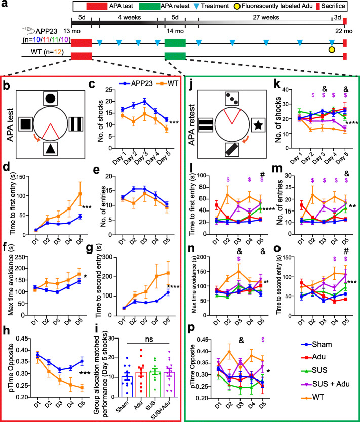
Methods: In a study comprising four treatment arms, we tested the efficacy of an Aducanumab analog, Adu, both in comparison to SUS, and as a combination therapy, in APP23 mice (aged 13-22 months), using sham as a control. The active place avoidance (APA) test was used to test spatial memory, and histology and ELISA were used to measure amyloid. Brain antibody levels were also determined.

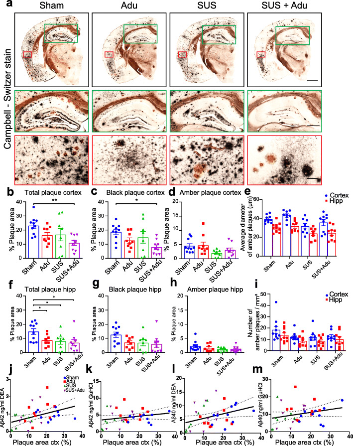
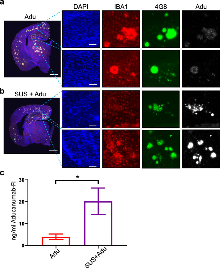
Results: We found that both Adu and SUS reduced the total plaque area in the hippocampus with no additive effect observed with the combination treatment (SUS + Adu). Whereas in the cortex where there was a trend towards reducing the total plaque area from either Adu or SUS, only the combination treatment yielded a statistically significant decrease in total plaque area compared to sham. Only the SUS and SUS + Adu groups included animals that had their plaque load reduced to below 1% from above 10%. There was a robust improvement in spatial memory for the SUS + Adu group only, and in this group the level of Adu, when measured 3 days post-treatment, was 5-fold higher compared to those mice that received Adu on its own. Together, these findings suggest that SUS should be considered as a treatment option for AD. Alternatively, a combination trial using Aducanumab together with ultrasound to increase brain levels of the antibody may be warranted.

Keywords: Alzheimer’s disease; Amyloid-β; Blood–brain barrier; Dementia; Focused ultrasound; Immunotherapy.


Conflict of interest statement

No competing interests exist.

Figures

Fig. 1
Fig. 1
Study overview and results of APA test and retest. Overview of the study with timeline (a). In the active place avoidance test (APA) mice must use spatial cues to avoid a shock zone (indicated as a red triangle) (b). APP23 mice had impaired performance in the APA test in terms of number of shocks received (c), and time to first entry to the shock zone (d) as determined by a two-way ANOVA. Although the APP23 mice did not show significant impairment in the measure number of entries (e) or maximum time avoidance (of the shock zone) (f), they were impaired on the measures time to second entry (g) and proportion of time spent in the opposite quadrant to the shock zone (h). The mice were then assigned to treatment groups based on matching performance on day 5 of the APA test (i). The APA retest was performed after four once-per-week treatments with changes to room cues, shock zone location, and the direction of rotation (j). In the post-treatment APA retest an effect of SUS + Adu treatment on number of shocks compared to sham-treated mice was revealed (k), whereas treatment with SUS improved time to first entry (l). SUS + Adu improved the performance of the mice on the measures number of entries (m), and maximum time avoidance (n). Time to second entry was improved by SUS (o), while SUS + Adu improved the proportion of time spent in the opposite quadrant to the shock zone (p). Data are represented as mean ± SEM. Statistical differences: *p < 0.05, **p < 0.01, ***p < 0.001, ****p < 0.0001, $ = simple effect comparing wild-type vs sham p < 0.05, # = simple effect comparing SUS vs sham p < 0.05, & = simple effect comparing SUS + Adu vs sham p < 0.05. Sham N = 10, Adu N = 11, SUS N = 11, SUS + Adu N = 10, WT N = 12. Data were analyzed with a two-way ANOVA and follow-up Holm-Sidak tests for simple effects
Fig. 2
Fig. 2
Treatment strategies reduce plaques in APP23 mice. a Representative Campbell-Switzer silver staining for amyloid plaques in the four treatment groups. Plaques stained black are more diffuse, whereas amber plaques are compact and discrete. The black box shows the entire hemisphere (scale bars 1 mm). Insert outlined in green shows higher magnification view of dorsal hippocampus (scale bars 500 μm), and the red inset shows higher magnification image of cortex overlying the hippocampus (scale bars, 100 μm). b There was a significant reduction of plaque burden in the cortex of SUS + Adu-treated mice, driven largely by reduction in black plaques (c) as area, number and size of amber plaque was less affected by treatment (d, e, i). Plaque load in the hippocampus was reduced by Adu, SUS and SUS + Adu (f), with hippocampal black plaques (g) and amber plaques (h) analyzed separately. A significant correlation was found between amyloid plaque burden measured by histology and Aβ levels in cortical lysate measured by ELISA (j–m). Data are represented as mean ± SEM. Statistical differences: *p < 0.05, **p < 0.01, ***p < 0.001. Data were analyzed with a one-way ANOVA and follow-up Holm-Sidak tests. Sham N = 10, Adu N = 9, SUS N = 8, SUS + Adu N = 9
Fig. 3
Fig. 3
Aducanumab analog does not affect levels of cerebral amyloid angiopathy (CAA) or microhemorrhages in APP23 mice. a Representative Campbell-Switzer silver staining shows CAA in the cortex identified by a rod-like appearance, as well as meningeal CAA identified as open circles on top of the cortex. b Adu, whether administered with or without SUS, had no effect on the number of CAA-affected vessels, average size, or percent area occupied by CAA. c Adu, whether administered by itself or with SUS, had no effect on the number of microhemorrhages detected by Prussian blue staining in 22-month old APP23 mice. Data are represented as mean ± SEM. Statistical differences: *p < 0.05. Data were analyzed with a one-way ANOVA and t test. Scale bar 200 μm
Fig. 4
Fig. 4
Scanning ultrasound (SUS) increases the levels of the Aducanumab analog in the brain. a Fluorescently labeled Adu is detectable in the whole brain (scale bars 1 mm) and when visualized at higher magnification in the cortex and hippocampus (scale bars 100 μm). In APP23 mice treated with Adu alone, the fluorescent Adu is bound to plaques, which were immunolabeled with 4G8 antibody. b The levels of Adu were higher when Adu was delivered together with SUS in SUS + Adu-treated mice. The amyloid plaques in SUS + Adu-treated mice were decorated all over with Adu, whereas in mice treated with Adu alone the Adu is mainly confined to the outsides of the plaques. Microglia as identified by IBA1 immunostaining were located near plaques which have Adu bound to them. c The levels of fluorescent antibody in the cortical brain lysate was greatly increased in the SUS + Adu group compared to the Adu group. Data are represented as mean ± SEM. Statistical differences: *p < 0.05. Data were analyzed with t test. Adu N = 9, SUS + Adu N = 9

References

    1. Long JM, Holtzman DM. Alzheimer disease: an update on pathobiology and treatment strategies. Cell. 2019;179(2):312–339. doi: 10.1016/j.cell.2019.09.001. - DOI - PMC - PubMed
    1. Polanco JC, Li C, Bodea LG, Martinez-Marmol R, Meunier FA, Götz J. Amyloid-beta and tau complexity - towards improved biomarkers and targeted therapies. Nat Rev Neurol. 2018;14(1):22–39. doi: 10.1038/nrneurol.2017.162. - DOI - PubMed
    1. Cummings J, Lee G, Ritter A, Sabbagh M, Zhong K. Alzheimer’s disease drug development pipeline: 2019. Alzheimers Dement (N Y) 2019;5(1):272–293. doi: 10.1016/j.trci.2019.05.008. - DOI - PMC - PubMed
    1. Arndt JW, Qian F, Smith BA, Quan C, Kilambi KP, Bush MW, Walz T, Pepinsky RB, Bussière T, Hamann S, Cameron TO, Weinreb PH. Structural and kinetic basis for the selectivity of aducanumab for aggregated forms of amyloid-beta. Sci Rep. 2018;8(1):6412. doi: 10.1038/s41598-018-24501-0. - DOI - PMC - PubMed
    1. Sevigny J, Chiao P, Bussière T, Weinreb PH, Williams L, Maier M, Dunstan R, Salloway S, Chen T, Ling Y, O’Gorman J, Qian F, Arastu M, Li M, Chollate S, Brennan MS, Quintero-Monzon O, Scannevin RH, Arnold HM, Engber T, Rhodes K, Ferrero J, Hang Y, Mikulskis A, Grimm J, Hock C, Nitsch RM, Sandrock A. The antibody aducanumab reduces Abeta plaques in Alzheimer’s disease. Nature. 2016;537(7618):50–56. doi: 10.1038/nature19323. - DOI - PubMed

Publication types