. 2021 Apr 9;23(1):109.
doi: 10.1186/s13075-021-02483-1.

Synovial fluid neutrophils in oligoarticular juvenile idiopathic arthritis have an altered phenotype and impaired effector functions

Affiliations

Synovial fluid neutrophils in oligoarticular juvenile idiopathic arthritis have an altered phenotype and impaired effector functions

Sabine Arve-Butler et al. Arthritis Res Ther. .

Abstract

Background: Neutrophils are the most prevalent immune cells in the synovial fluid in inflamed joints of children with oligoarticular juvenile idiopathic arthritis (JIA). Despite this, little is known about neutrophil function at the site of inflammation in JIA and how local neutrophils contribute to disease pathogenesis. This study aimed to characterize the phenotype and function of synovial fluid neutrophils in oligoarticular JIA.

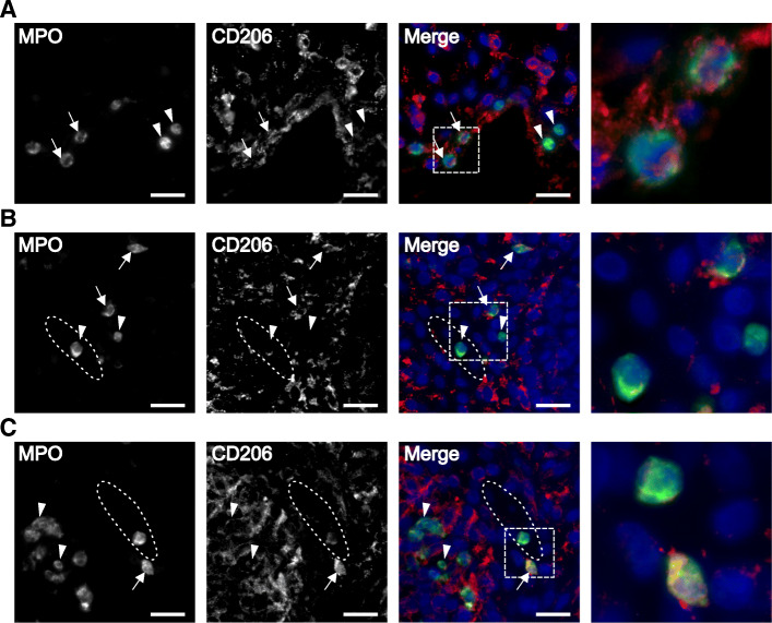
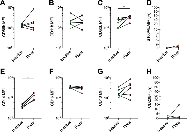
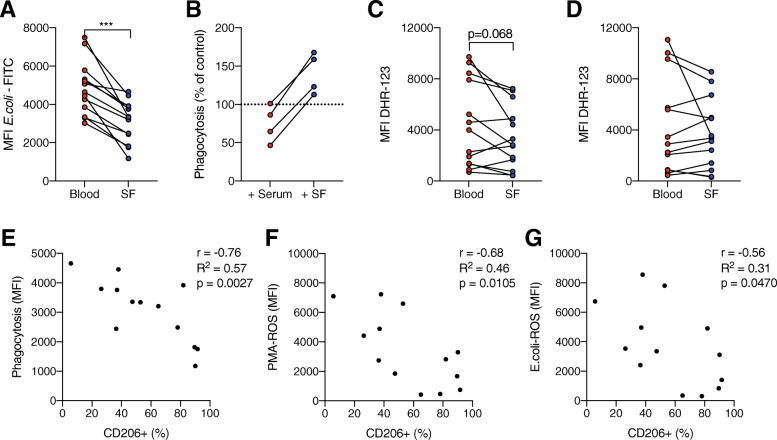
Methods: Neutrophils obtained from paired blood and synovial fluid from patients with active oligoarticular JIA were investigated phenotypically (n = 17) and functionally (phagocytosis and oxidative burst, n = 13) by flow cytometry. In a subset of patients (n = 6), blood samples were also obtained during inactive disease at a follow-up visit. The presence of CD206-expressing neutrophils was investigated in synovial biopsies from four patients by immunofluorescence.

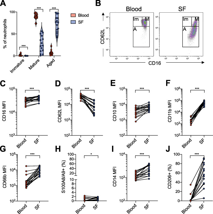
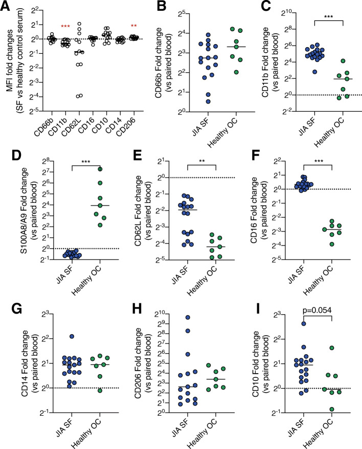
Results: Neutrophils in synovial fluid had an activated phenotype, characterized by increased CD66b and CD11b levels, and most neutrophils had a CD16hi CD62Llowaged phenotype. A large proportion of the synovial fluid neutrophils expressed CD206, a mannose receptor not commonly expressed by neutrophils but by monocytes, macrophages, and dendritic cells. CD206-expressing neutrophils were also found in synovial tissue biopsies. The synovial fluid neutrophil phenotype was not dependent on transmigration alone. Functionally, synovial fluid neutrophils had reduced phagocytic capacity and a trend towards impaired oxidative burst compared to blood neutrophils. In addition, the effector functions of the synovial fluid neutrophils correlated negatively with the proportion of CD206+ neutrophils.

Conclusions: Neutrophils in the inflamed joint in oligoarticular JIA were altered, both regarding phenotype and function. Neutrophils in the synovial fluid were activated, had an aged phenotype, had gained monocyte-like features, and had impaired phagocytic capacity. The impairment in phagocytosis and oxidative burst was associated with the phenotype shift. We speculate that these neutrophil alterations might play a role in the sustained joint inflammation seen in JIA.

Keywords: Juvenile idiopathic arthritis; Neutrophil; Oxidative burst; Phagocytosis; Phenotype; Reactive oxygen species; Synovial fluid.


Conflict of interest statement

The authors declare that they have no competing interests.

Figures

Fig. 1
Fig. 1
Synovial fluid neutrophils have an aged and activated phenotype and express monocyte-related markers. Neutrophils from paired samples of blood and synovial fluid from patients with oligoarticular JIA were analyzed by flow cytometry. a Neutrophil phenotypical maturities in blood and synovial fluid based on CD62L and CD16 expression. b Representative plot of neutrophil maturity phenotype gating. Mature neutrophils (M) are CD16hi CD62Lhi, immature neutrophils (Im) are CD16mid CD62Lhi, and aged A neutrophils are CD62Llow. cg, i Median fluorescence intensity (MFI) of neutrophil expression of c CD16, d CD62L, e CD10, f CD11b, g CD66b, and i CD14. h, j Proportion of neutrophils, presented as % of total neutrophils, with cell surface expression of h S100A8/A9 and j CD206. *p < 0.05, ***p < 0.001, Wilcoxon signed-rank test
Fig. 2
Fig. 2
CD206-expressing neutrophils are found in synovial tissue. Synovial tissue biopsies from three patients, stained for MPO (green), CD206 (red), and DAPI (blue). Representative images of a patient 4, b patient 8, and c patient 18. Neutrophils expressing CD206, indicated with arrows, and neutrophils without CD206 expression, indicated with arrow heads, were found in all three biopsies. The synovial vessels are indicated with dotted ellipses. Neutrophils were found both scattered in the synovial tissue and inside the synovial blood vessels. The fourth image in each panel represents a magnification of the area indicated in the merged image. Scale bar is 20 μm. All images are taken at × 40 magnification
Fig. 3
Fig. 3
Neutrophil phenotypes in JIA blood are similar during flare and inactive disease. Neutrophil surface marker levels in paired blood samples taken during arthritis flare or inactive disease without arthritis. ac, eg Median fluorescence intensity (MFI) of neutrophil expression of a CD66b, b CD11b, c CD62L, e CD14, f CD16, and g CD10. d, h Proportion of neutrophils, presented as % of total neutrophils, with cell surface expression of d S100A8/A9 and h CD206. *p < 0.05, Wilcoxon signed-rank test
Fig. 4
Fig. 4
The neutrophil phenotype shift is not explained by exposure to synovial fluid or transmigration. a Healthy control blood neutrophils were challenged with 20% JIA synovial fluid (n = 12) or 20% serum from the healthy blood donor for 1 h followed by the analysis of surface marker expression. Control serum-stimulated neutrophils are represented by the dotted line. Data are presented as fold change of median fluorescence intensity (MFI) in synovial fluid stimulated compared to control neutrophils. bi Neutrophils in JIA synovial fluid (SF) were compared to neutrophils from the oral cavity (OC) of healthy controls, to investigate the alterations in surface markers between the blood and tissue in paired samples of blood and SF or blood and OC. Blood values are represented by the dotted line. Data are presented as fold change of MFI values of b CD66b, c CD11b, d S100A8/A9, e CD62L, f CD16, g CD14, h CD206, and i CD10 in tissue neutrophils compared to paired blood neutrophils. Line at the median. **p < 0.01, ***p < 0.001, Mann-Whitney U test
Fig. 5
Fig. 5
Neutrophil effector functions are impaired in JIA blood and synovial fluid. a Phagocytosis of fluorescently labeled opsonized E. coli in neutrophils from paired samples of JIA blood and synovial fluid (SF). b Phagocytosis of fluorescently labeled opsonized E. coli in healthy control neutrophils in a mixture of 10% whole blood and 90% serum or cell-free synovial fluid from four JIA patients. The results are presented as phagocytosis, measured as MFI, in serum/SF-treated samples compared to non-treated whole blood. c, d Neutrophil ROS production, quantified using DHR-123 fluorescence, after stimulation with c PMA or d opsonized E. coli. eg Pearson r correlation analysis of the proportion of CD206+ neutrophils in the synovial fluid with e phagocytosis of opsonized E. coli, f ROS production by PMA-stimulation, and g ROS production by opsonized E. coli stimulation. ***p < 0.001, Wilcoxon signed-rank test. MFI, median fluorescence intensity

References

    1. Glerup M, Rypdal V, Arnstad ED, Ekelund M, Peltoniemi S, Aalto K, Rygg M, Toftedal P, Nielsen S, Fasth A, Berntson L, Nordal E, Herlin T, the Nordic Study Group of Pediatric Rheumatology Long-term outcomes in juvenile idiopathic arthritis: eighteen years of follow-up in the Population-Based Nordic Juvenile Idiopathic Arthritis Cohort. Arthritis Care Res (Hoboken) 2020;72(4):507–516. doi: 10.1002/acr.23853. - DOI - PubMed
    1. Matsumoto T, Matsui T, Hirano F, Tohma S, Mori M. Disease activity, treatment and long-term prognosis of adult juvenile idiopathic arthritis patients compared with rheumatoid arthritis patients. Mod Rheumatol. 2020;30(1):78–84. doi: 10.1080/14397595.2018.1554228. - DOI - PubMed
    1. Prakken B, Albani S, Martini A. Juvenile idiopathic arthritis. Lancet. 2011;377(9783):2138–2149. doi: 10.1016/S0140-6736(11)60244-4. - DOI - PubMed
    1. Berthold E, Månsson B, Kahn R. Outcome in juvenile idiopathic arthritis: a population-based study from Sweden. Arthritis Res Ther. 2019;21(1):218. doi: 10.1186/s13075-019-1994-8. - DOI - PMC - PubMed
    1. Macaubas C, Nguyen K, Milojevic D, Park JL, Mellins ED. Oligoarticular and polyarticular JIA: epidemiology and pathogenesis. Nat Rev Rheumatol. 2009;5(11):616–626. doi: 10.1038/nrrheum.2009.209. - DOI - PMC - PubMed

Publication types