

Detection of SARS-CoV-2 genomic and subgenomic RNA in retina and optic nerve of patients with COVID-19
- PMID: 33836988
- PMCID: PMC8042582
- DOI: 10.1136/bjophthalmol-2020-318618
Detection of SARS-CoV-2 genomic and subgenomic RNA in retina and optic nerve of patients with COVID-19
Erratum in
-
Correction: Detection of SARS-CoV-2 genomic and subgenomic RNA in retina and optic nerve of patients with COVID-19.Br J Ophthalmol. 2023 May;107(5):e2. doi: 10.1136/bjophthalmol-2020-318618.corr1. Br J Ophthalmol. 2023. PMID: 37080602 Free PMC article. No abstract available.
Abstract
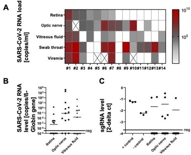
Purpose: Presence of SARS-CoV-2 RNA in human retinal biopsies (RBs) was previously reported by us. In this consecutive study, we analysed RB and optic nerve biopsies (ONBs) in deceased patients with confirmed COVID-19 assessing viral RNA load, possible virus replication and infectivity.
Patients and methods: In this case series, 14 eyes of 14 deceased patients with COVID-19 were enucleated during autopsy. RB and ONB were subjected to molecular detection of viral RNA, virus cultivation and immunohistochemistry. SARS-CoV-2 RNA loads were compared with RNA loads in the respective throat swabs, vitreous humour and blood samples.
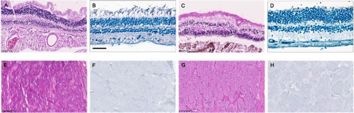
Results: SARS-CoV-2 RNA was detected in 7/14 RBs and in 10/13 ONBs. While virus isolation failed and immunohistochemistry of SARS-CoV-2 spike protein was negative, subgenomic RNA (sgRNA) was detectable (40% RB; 60% ONB).
Conclusion: SARS-CoV-2 RNA is detectable in RB and ONB of patients with COVID-19. Presence of sgRNA could point to a SARS-CoV-2 infection of neuronal tissue, but as virus isolation failed and immunohistochemistry of SARS-CoV-2 spike protein was negative, an active infection seems unlikely.
Keywords: COVID-19; microbiology; optic nerve; retina.
© Author(s) (or their employer(s)) 2022. Re-use permitted under CC BY-NC. No commercial re-use. See rights and permissions. Published by BMJ.
Conflict of interest statement
Competing interests: MS reported receiving grants from Novartis, IDxDR and Boehringer Ingelheim; and personal fees from Bayer, Oxurion, Roche, Allergan, Alcon, Neurogene and GSK outside the submitted work.
Figures
References
Publication types
MeSH terms
Substances
LinkOut - more resources
Full Text Sources
Other Literature Sources
Medical
Miscellaneous