. 2021 Apr 9;12(4):384.
doi: 10.1038/s41419-021-03600-3.

WBP2 negatively regulates the Hippo pathway by competitively binding to WWC3 with LATS1 to promote non-small cell lung cancer progression

Affiliations

WBP2 negatively regulates the Hippo pathway by competitively binding to WWC3 with LATS1 to promote non-small cell lung cancer progression

Qiang Han et al. Cell Death Dis. .

Abstract

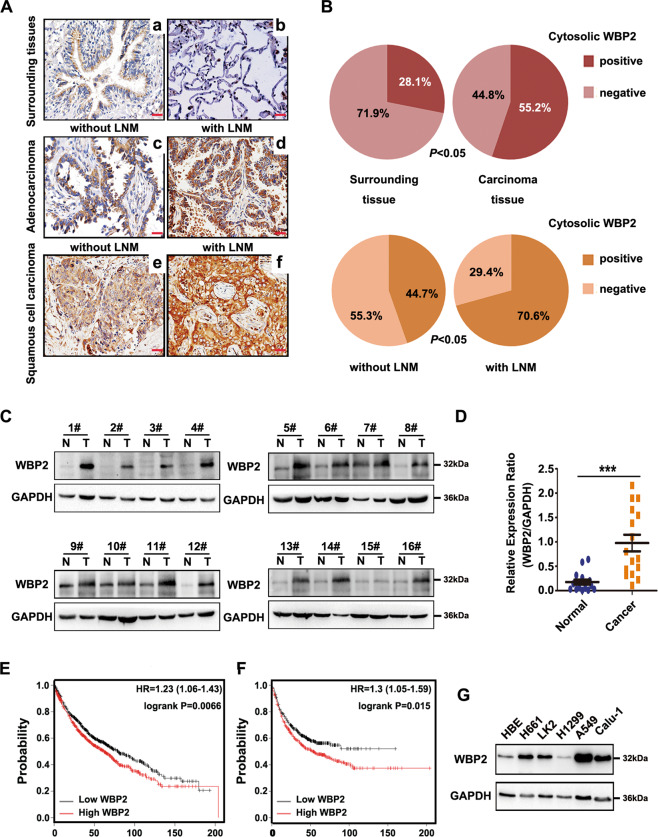
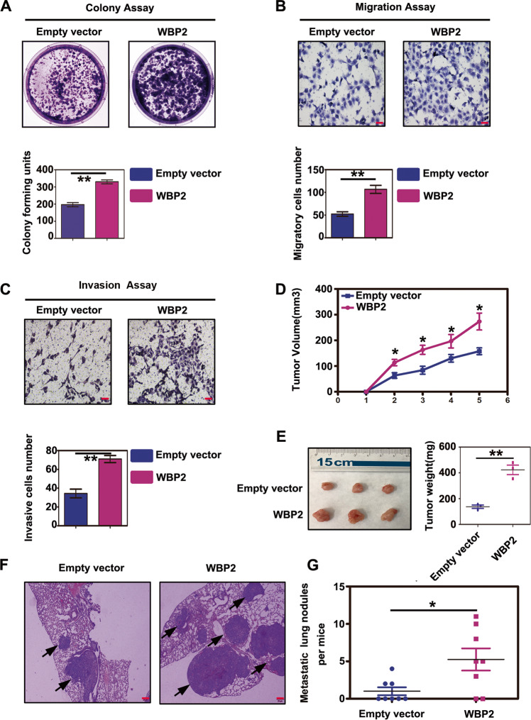
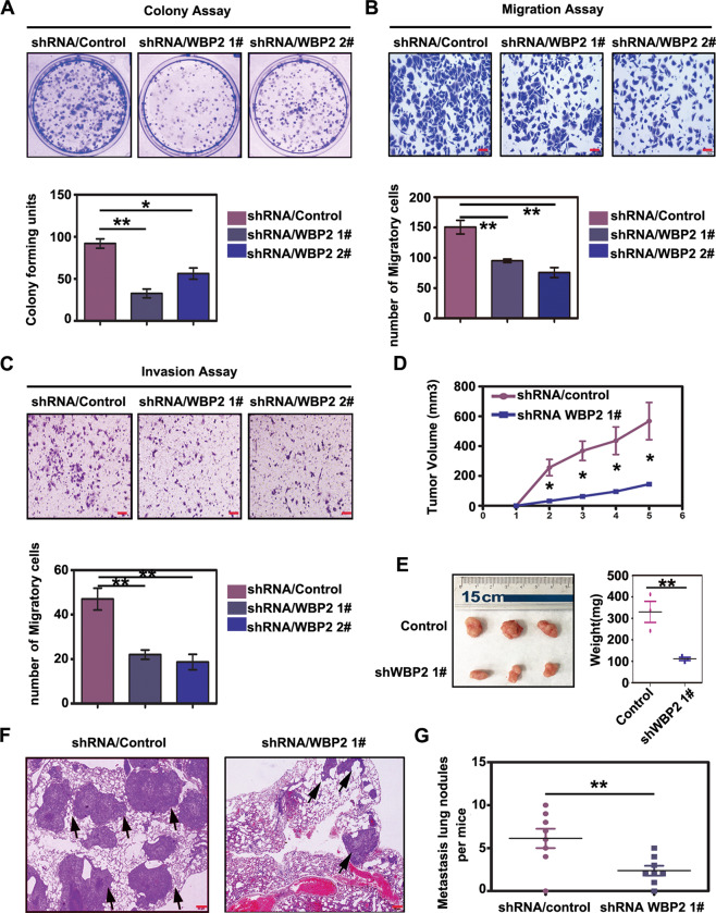
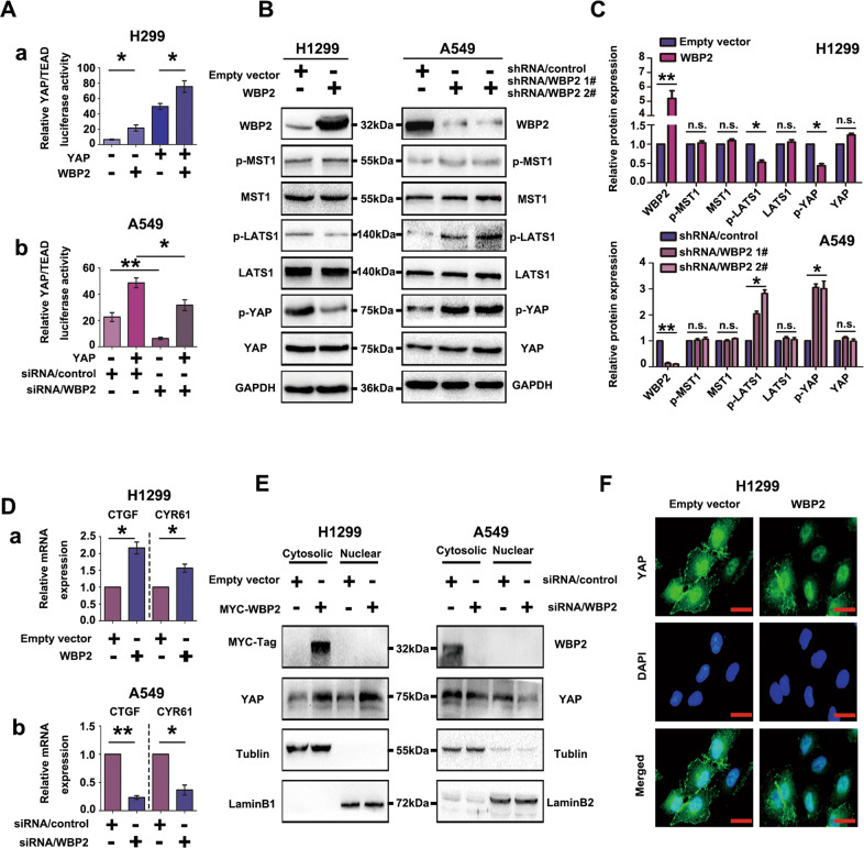
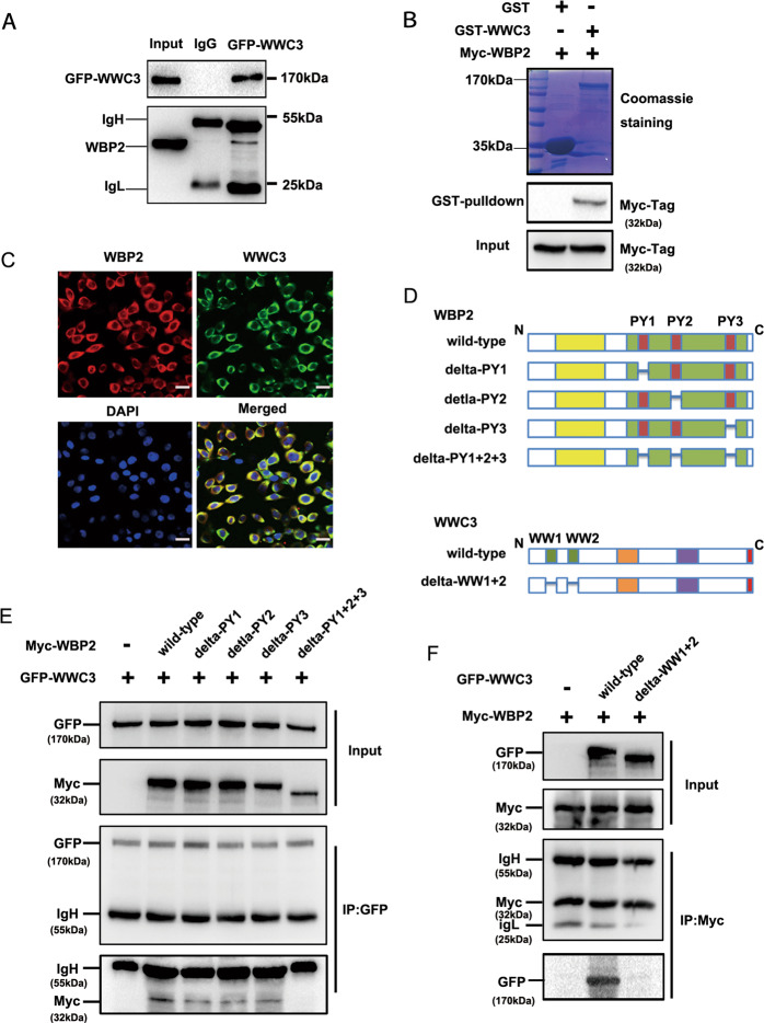
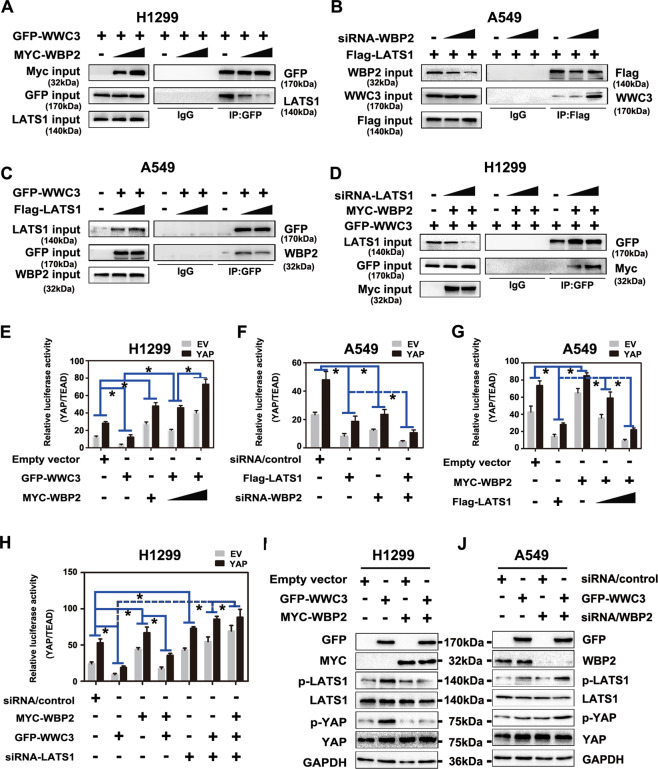
WW domain binding protein-2 (WBP2) can function as a Yes-associated protein/transcriptional co-activator with PDZ-binding motif (YAP/TAZ) co-activator and has a crucial role in promoting breast cancer progression. However, the expression and potential molecular mechanisms of WBP2 in the context of lung cancer are not fully understood. We determined that WBP2 was highly expressed in lung cancer specimens and cell lines and that this expression was closely related to the advanced pTNM stage, lymph node metastasis, and poor prognosis of patients. In addition, gain- and loss-of-function experiments revealed that WBP2 could significantly promote the proliferation and invasion of lung cancer cells both in vivo and in vitro. To elucidate the underlying molecular mechanism, we determined that wild-type WBP2 could competitively bind to the WW domain of WWC3 (WW and C2 domain-containing-3) with LATS1 (Large tumor suppressor-1) through its PPxY motifs, thus inhibiting the formation of the WWC3-LATS1 complex, reducing the phosphorylation level of LATS1, suppressing the activity of the Hippo pathway, and ultimately promoting YAP nuclear translocation. Therefore, from the aspect of upstream molecules of Hippo signaling, WBP2 promotes the malignant phenotype of lung cancer cells in a unique manner that is not directly dependent upon YAP, thus providing a corresponding experimental basis for the development of targeted therapeutic drugs for lung cancer.


Conflict of interest statement

The authors declare no competing interests.

Figures

Fig. 1
Fig. 1. High expression levels of WBP2 in non-small cell lung cancer correlate with poor prognosis.
A, B The expression of WBP2 is low or negative in normal bronchial epithelium (Aa) and alveolar epithelium (Ab), positive (+−++) in adenocarcinoma (Ac) and squamous cell carcinoma (Ae) without lymph node metastasis (LNM), and strongly positive (++−+++) in adenocarcinoma (Ad) and squamous cell carcinoma (Af) with LNM. Magnification: ×400, scale bar: 50 μm. LNM: lymph node metastasis. C, D Western blot analysis revealed that the expression level of WBP2 in lung cancer tissues is significantly higher than that in adjacent normal lung tissues. GAPDH was used as the loading control. E, F Kaplan–Meier plotter network database analysis indicates that the overall survival (OS; E) and progression-free survival (PFS; F) of patients with lung cancer presenting high WBP2 expression are significantly lower than those of patients with low WBP2 expression. G Western blot analysis of WBP2 expression in the human immortalized bronchial epithelial cell line HBE and in a panel of non-small cell lung cancer cell lines (n = 5). GAPDH was used as the loading control. P < 0.05 indicates statistical significance, and *** represents P < 0.001.
Fig. 2
Fig. 2. Overexpression of WBP2 promotes proliferation, invasion, and metastasis of lung cancer cells.
A–C, In vivo experiments: the plasmid WBP2 was transfected into the H1299 cell line, and stable expression monoclonal cells were screened using G418 (800 μg/mL). Colony-forming assays and transwell assays revealed that overexpression of WBP2 protein significantly enhances the proliferation (A control vs WBP2: 196 ± 12 vs. 330 ± 12, P < 0.01), migration (B control vs. WBP2: 52 ± 5 vs. 106 ± 8, P < 0.01), and invasiveness (C control vs. WBP2: 34 ± 4 vs. 71 ± 3, P < 0.01) of lung cancer cells. DG Subcutaneous tumor transplantation and lung metastasis experiments via tail vein injections in nude mice demonstrate that the volumes and weights of subcutaneous transplanted tumors in nude mice are both significantly increased following WBP2 overexpression (D, E Volume:control vs WBP2: 157.7 ± 13.0 vs 273.3 ± 32.8 [mm3], P < 0.05; weight:control vs WBP2: 137.3 ± 13.1 vs 423.3 ± 36.5 [mg], P < 0.01). The number of lung cancer metastasis foci is also markedly increased (F, G control vs WBP2: 1.0 ± 0.5 vs 5.2 ± 1.5, P < 0.05). P < 0.05 indicates statistical significance, *P < 0.05, **P < 0.01.
Fig. 3
Fig. 3. Knockdown of WBP2 weakens the proliferation, invasion, and metastatic abilities of lung cancer cells.
Lentivirus-coated shRNA-WBP2 was added to the A549 cell line, and stable cells with WBP2 knockdown were screened for use of puromycin (5 μg/mL). The colony-forming and Transwell assay results reveal that the reduction in WBP2 protein significantly weakens proliferation (A control vs shWBP2-1 vs shWBP2-2: 92 ± 5 vs 32 ± 5 vs 56 ± 6, P < 0.05), migration (B control vs shWBP2-1 vs shWBP2-2: 150 ± 11 vs 75 ± 8 vs 95 ± 2, P < 0.01), and invasiveness (C control vs shWBP2-1 vs shWBP2-2: 47 ± 5 vs 22 ± 2 vs 18 ± 3, P < 0.01) of lung cancer cells. In vivo: subcutaneous tumor transplantation and lung metastasis experiments via tail vein injection in nude mice reveal that volumes and weights of subcutaneously transplanted tumors in nude mice are significantly decreased (D, E control vs shWBP2-1: volume: 568.0 ± 125.3 vs 145.0 ± 9.3 [mm3], P < 0.05; weight: control vs shWBP2-1: 330.3 ± 49.2 vs 111.3 ± 7.5 [mg], P < 0.05), and the number of lung cancer metastasis foci is also significantly reduced (F, G control vs shWBP2-1: 6.1 ± 1.1 vs 2.4 ± 0.6, P < 0.01). P < 0.05 indicates statistical significance, *P < 0.05, **P < 0.01.
Fig. 4
Fig. 4. WBP2 is a negative regulator of the Hippo pathway in lung cancer cells.
A (a, b) WBP2 upregulates the transcriptional activity of the TEAD promoter. The WBP2 plasmid and siRNA-WBP2 were transfected into H1299 and A549 cell lines, respectively. After 48 h, the cells were collected and lysed. Dual-luciferase reporter gene detection shows that the ectopic expression of WBP2 can significantly upregulate the transcriptional activity of TEAD (Aa), whereas it decreases after WBP2 knockdown (Ab). Transfection with YAP was used as the stimulus. TK was used as the internal reference. B, C WBP2 downregulates LATS1 phosphorylation and YAP phosphorylation through an MST-independent pathway. After overexpression and knockdown of WBP2 in H1299 and A549 cells, respectively, the phosphorylation and total amount of key proteins in the Hippo pathway were analyzed by western blotting. GAPDH was used as the loading control. Differences were analyzed using the Image J software. *P < 0.05, **P < 0.01. D (a, b), RT-qPCR analysis reveals that the mRNA levels of CTGF and CYR61 are upregulated after WBP2 overexpression in H1299 (Da), and the mRNA levels of these two target genes are downregulated (Db) after WBP2 silencing in A549 cells. P < 0.05 indicates statistical significance, *P < 0.05, **P < 0.01. E Nuclear and cytosolic fractionation assays indicate that WBP2 overexpression promotes YAP nuclear translocation in H1299 cells, whereas WBP2 silencing in A549 exerts the opposite effect. Beta-tubulin and LaminB1 were used as the cytosolic and nuclear loading controls, respectively. F Immunofluorescence assay results reveal that the level of YAP nuclear translocation increases in response to WBP2 overexpression in H1299 cells. Magnification: ×400, scale bar: 50 μm.
Fig. 5
Fig. 5. WBP2 interacts with the WW domain of WWC3 via PPxY motifs.
A GFP-WWC3 was transfected into H1299 cells, and after 48 h, the cells were collected and lysed. A GFP monoclonal antibody was used for pulldown. The presence of WBP2 in the precipitate was detected by western blot analysis using a WBP2 antibody. B After incubation with purified GST or GST coupled-WWC3 protein for 6 h at 4° C, the binding status of two proteins was examined using Coomassie brilliant blue staining and western blot analysis. GST, glutathione-S-transferase. C Immunofluorescence assay results indicate that WBP2 and WWC3 colocalize within the cytoplasm of A549 cells. Magnification: ×400, scale bar: 50 μm. D Schematic diagram of WBP2 and WWC3 splicing mutants. E GFP-WWC3 and Myc-WBP2 wild-type or a series of mutants were co-transfected into H1299 cells. After 48 h, the cells were collected and lysed. GFP antibody was used for precipitation, and the presence of WBP2 was detected by immunoblotting using a Myc antibody. F Similarly, Myc-WBP2 and GFP-WWC3 wild-type or GFP-WWC3-△WW mutants were co-transfected into H1299 cells. GFP antibody was used for precipitation, and the presence of WBP2 was detected by immunoblotting using a Myc antibody.
Fig. 6
Fig. 6. WBP2 and LATS1 competitively bind to WWC3 to inhibit Hippo pathway activity.
A WBP2 overexpression attenuates the binding between WWC3 and LATS1. In the H1299 cell line, the WBP2 plasmid (1 μg and 2.5 μg) was transfected in a dose gradient manner. After 48 h, the cells were collected and lysed. A GFP monoclonal antibody was used for immunoprecipitation. The combination change in WWC3 and LATS1 protein in the sediment was detected by western blotting. B The binding of WWC3 to LATS1 increases after WBP2 knockdown. siRNA-WBP2 (5 pmol and 10 pmol) was transfected into A549 cells with high WBP2 expression. After cell collection, binding was detected by anti-FLAG immunoprecipitation followed by anti-WWC3 immunoblotting. C, D Following transfection of LATS1 into A549 cells with low LATS1 expression, the binding of WBP2 to WWC3 gradually decreases with an increase in LATS1 expression (C). In contrast, the binding of WBP2 to WWC3 gradually increases with the downregulation of LATS1 after siRNA-LATS1 was transfected into H1299 cells with high LATS1 expression (D). E, F Forty-eight hours post-transfection with WBP2 plasmid or siRNA-WBP2 into H1299 cells and A549 cells, the cells were collected and lysed. The results of the dual-luciferase reporter assay reveal that WBP2 overexpression reverses the decrease in TEAD transcriptional activity induced by WWC3 (E). Conversely, WBP2 knockdown further enhances the decrease in TEAD transcriptional activity induced by WWC3 (F). G, H Similarly, dual-luciferase reporter gene detection assays reveal that LATS1 overexpression reverses the WBP2-induced increase in TEAD transcriptional activity (G). Moreover, silencing of LATS1 further enhances the WBP2-induced increase in TEAD transcriptional activity (H). I, J Western blot analysis indicates that overexpression of WBP2 reverses the increase in LATS1 and YAP phosphorylation levels induced by WWC3 (I). Conversely, knockdown of WBP2 further increases the phosphorylation levels of LATS1 and YAP (J). P < 0.05 indicates statistical significance, *P < 0.05.

Similar articles

Cited by

References

    1. Meng Z, Moroishi T, Guan KL. Mechanisms of Hippo pathway regulation. Genes Dev. 2016;30:1–17. doi: 10.1101/gad.274027.115. - DOI - PMC - PubMed
    1. Misra JR, Irvine KD. The Hippo signaling network and its biological functions. Annu. Rev. Genet. 2018;52:65–87. doi: 10.1146/annurev-genet-120417-031621. - DOI - PMC - PubMed
    1. Harvey KF, Zhang X, Thomas DM. The Hippo pathway and human cancer. Nat. Rev. Cancer. 2013;13:246–257. doi: 10.1038/nrc3458. - DOI - PubMed
    1. Ma S, Meng Z, Chen R, Guan KL. The hippo pathway: biology and pathophysiology. Annu Rev. Biochem. 2019;88:577–604. doi: 10.1146/annurev-biochem-013118-111829. - DOI - PubMed
    1. Yu FX, Zhao B, Guan KL. Hippo pathway in organ size control, tissue homeostasis, and cancer. Cell. 2015;163:811–828. doi: 10.1016/j.cell.2015.10.044. - DOI - PMC - PubMed

Publication types

LinkOut - more resources