Review
. 2021 Jul;22(7):483-504.
doi: 10.1038/s41580-021-00356-8. Epub 2021 Apr 9.

Intracellular mRNA transport and localized translation

Affiliations
Review

Intracellular mRNA transport and localized translation

Sulagna Das et al. Nat Rev Mol Cell Biol. 2021 Jul.

Erratum in

Abstract

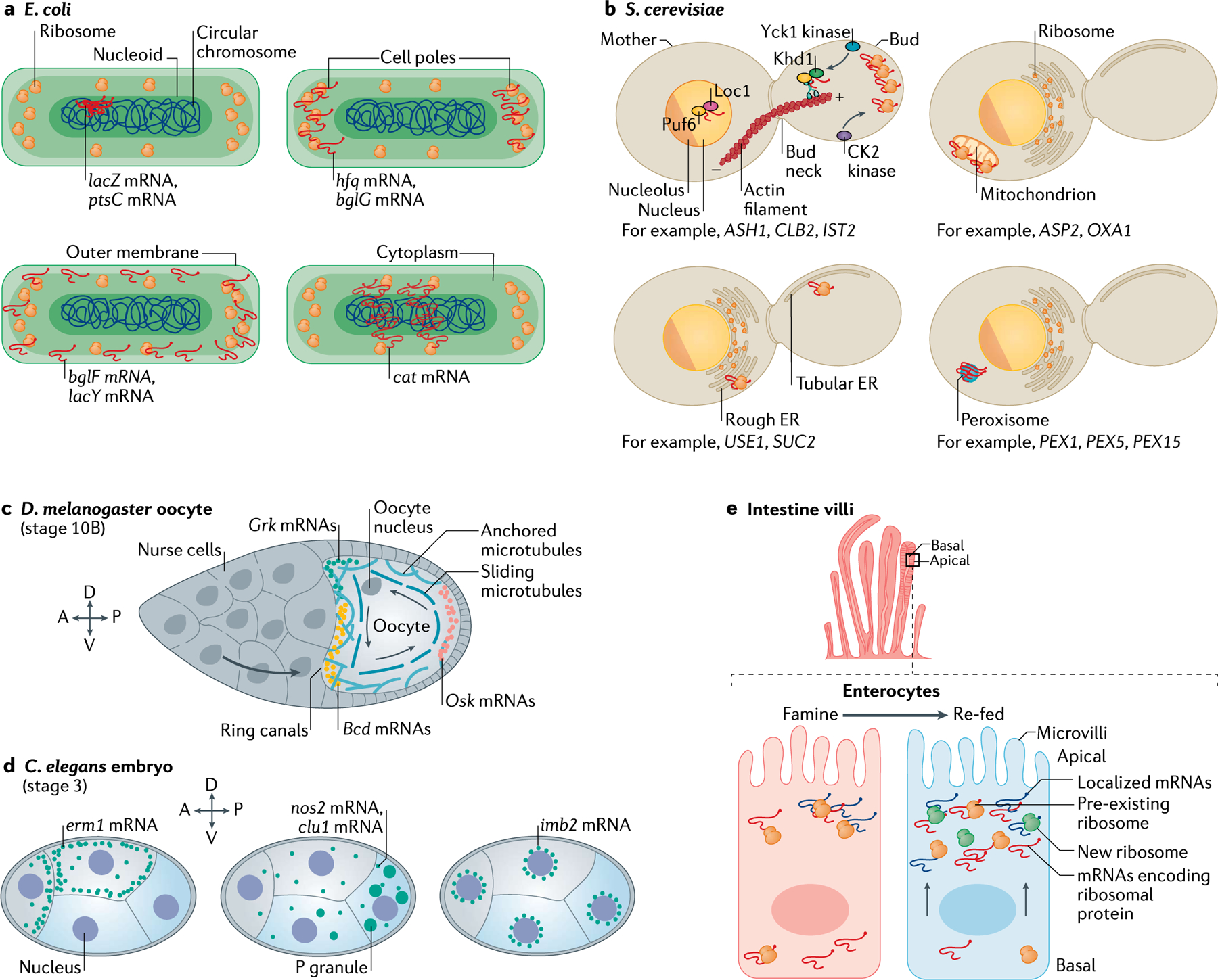
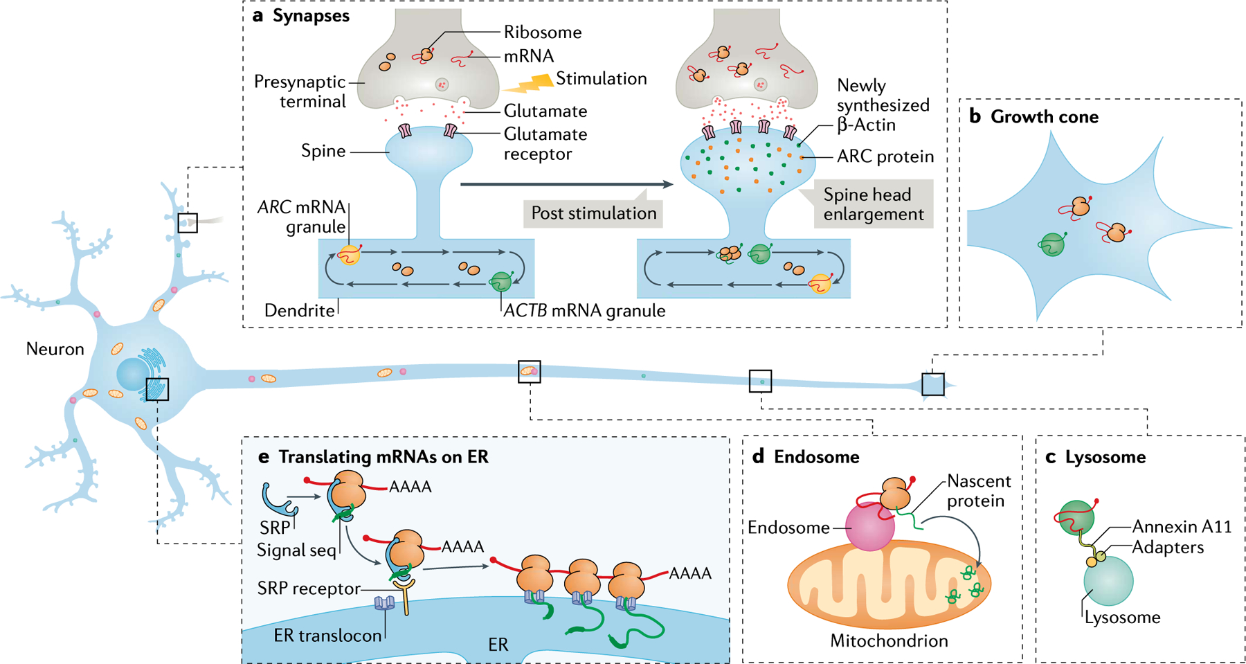
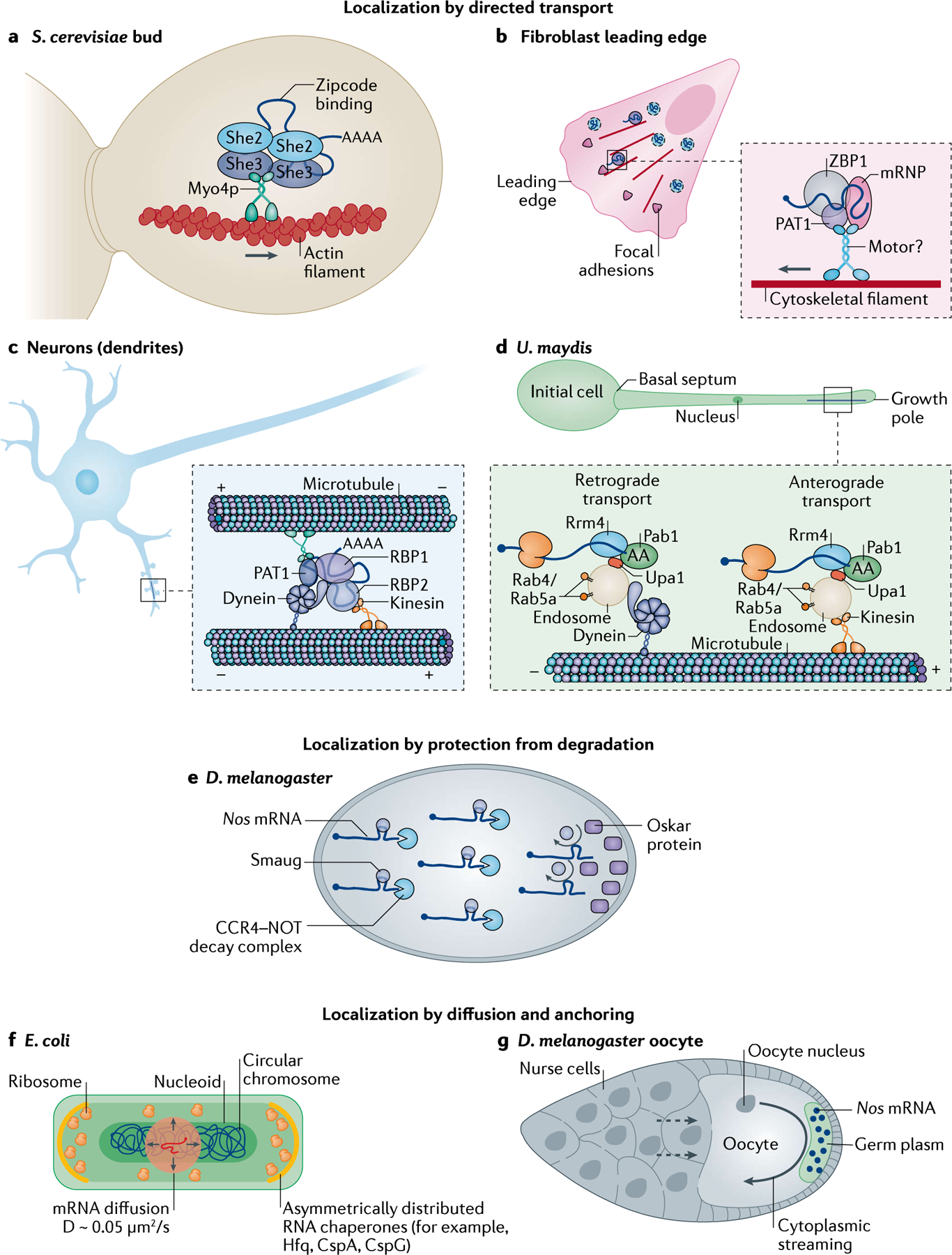
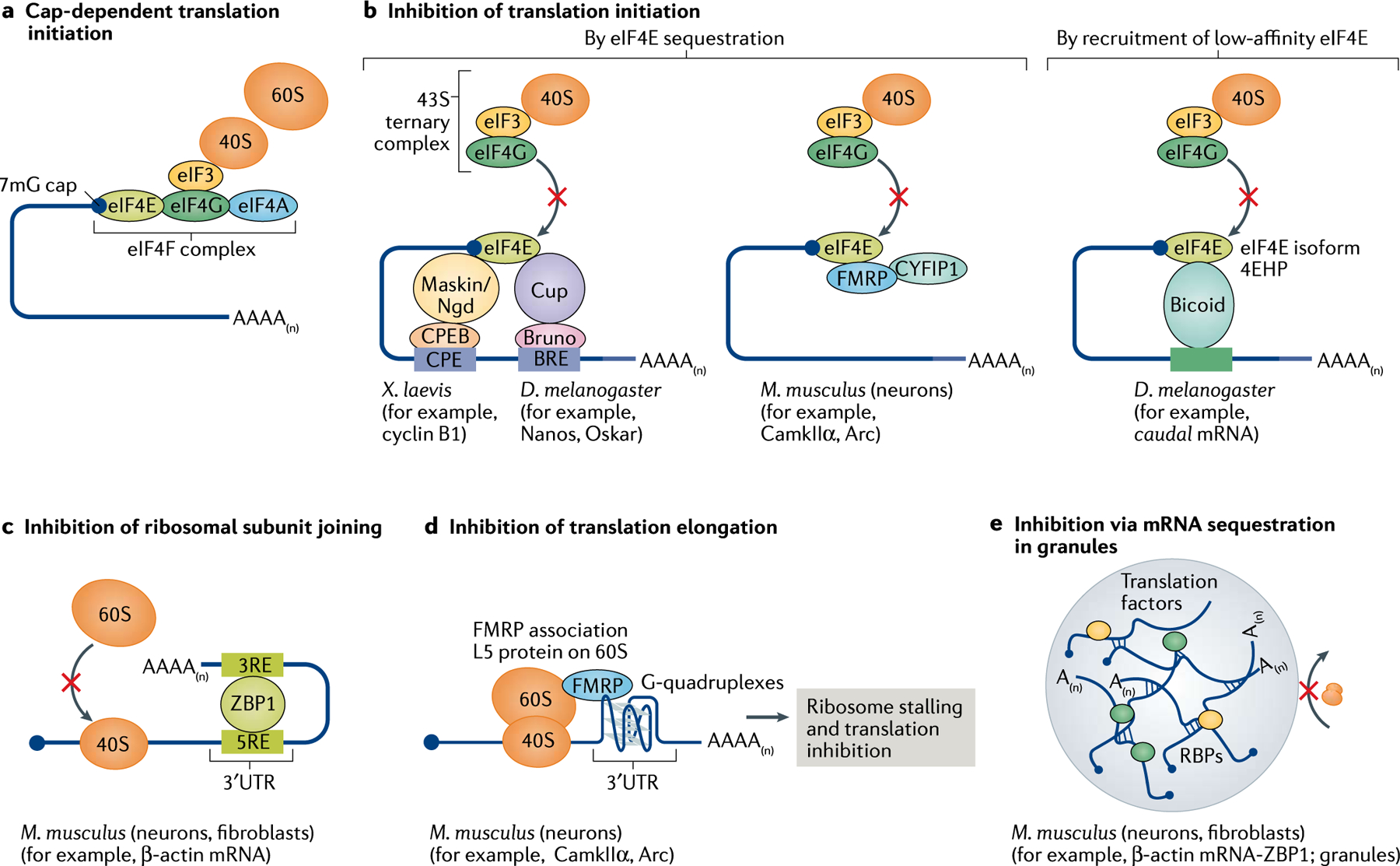
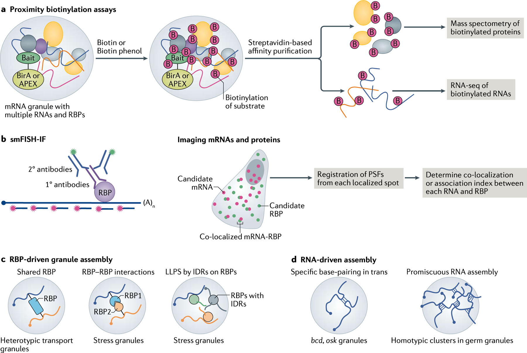
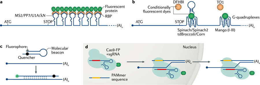
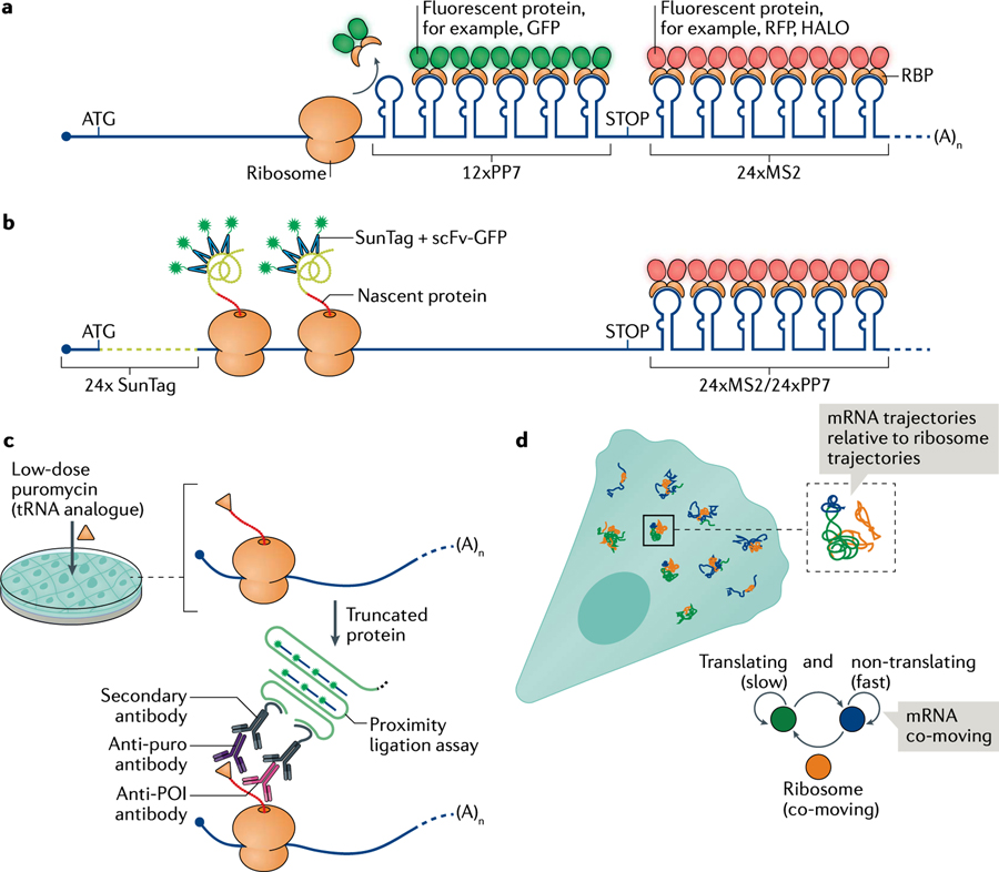
Fine-tuning cellular physiology in response to intracellular and environmental cues requires precise temporal and spatial control of gene expression. High-resolution imaging technologies to detect mRNAs and their translation state have revealed that all living organisms localize mRNAs in subcellular compartments and create translation hotspots, enabling cells to tune gene expression locally. Therefore, mRNA localization is a conserved and integral part of gene expression regulation from prokaryotic to eukaryotic cells. In this Review, we discuss the mechanisms of mRNA transport and local mRNA translation across the kingdoms of life and at organellar, subcellular and multicellular resolution. We also discuss the properties of messenger ribonucleoprotein and higher order RNA granules and how they may influence mRNA transport and local protein synthesis. Finally, we summarize the technological developments that allow us to study mRNA localization and local translation through the simultaneous detection of mRNAs and proteins in single cells, mRNA and nascent protein single-molecule imaging, and bulk RNA and protein detection methods.


Conflict of interest statement

Competing interests

The authors declare no competing interests.

Figures

Fig. 1 |
Fig. 1 |. mRNA localization and local translation in single-cell and multi-cellular organisms.
a | In prokaryotes, such as Escherichia coli, the cell is divided into specific sub-compartments, namely the nucleoid (where the circular DNA molecule resides), the cell poles (where ribosomes accumulate) and the outer membrane (where both ribosomes and transporters reside), to which several mRNAs have been shown to localize. ptsC and lacZ mRNAs localize to the nucleoid, hfq and bglG mRNAs localize at the cell poles, bglF and lacY mRNAs localize to the outer membrane, and cat mRNA has a characteristic helical distribution in the cytoplasm. b | In unicellular eukaryotes like Saccharomyces cerevisiae, mRNA is asymmetrically distributed in multiple subcellular compartments. In the growing bud, mRNAs such as ASH1, CLB2 and IST2 are actively transported on actin filaments by the She2–She3–Myo4 complex. Sequences in the 3′ untranslated region (3′UTR) of ASTP2 and OXA1 mRNAs localize these mRNAs to the outer mitochondrial membrane. USE1 and SUC2 mRNAs are localized to the endoplasmic reticulum (ER). PEX1, PEX5 and PEX15 mRNAs are found in peroxisomes. c | During mid-oogenesis in Drosophila melanogaster, the microtubule cytoskeleton of the oocyte is reorganized by cytoplasmic streaming (sliding microtubules) to localize the mRNAs that determine body plan. While Bcd and Grk mRNAs are positioned on the anterior side, Osk mRNAs primarily occupy the posterior side; all three mRNAs are locally translated at their respective positions. d | In Caenorhabditis elegans, maternally inherited transcripts display distinct localization patterns. Transcripts in anterior-biased cells (grey) tend to localize to the cell periphery, where the encoded protein localizes (for example, erm1). mRNAs enriched in posterior cells (blue), such as nos2 and clu1, form clustered granules that overlap with P granules. The imb2 mRNA localizes at the perinuclear region. e | In the intestine, enterocytes lining the villi are polarized cells with distinct apical and basal sides. Components of the translation machinery change their apical–basal distribution in response to nutrient availability. As mRNAs encoding ribosomal proteins move from the basal to the apical side via microtubules, the translation of mRNAs localized at the apical side is boosted to favour nutrient absorption. A, anterior; D, dorsal; P, posterior; V, ventral.
Fig. 2 |
Fig. 2 |. Subcellular mRNA localization and local translation in neurons.
In neurons, mRNA localization and translation occur in processes (dendrites and axons). a | Neuronal transport granules, such as those containing ACTB and ARC mRNA, are trafficked along microtubules in dendrites like a conveyor belt patrolling multiple spines. The activation of specific synapses by stimulating the presynaptic terminal or by direct stimulation of postsynaptic spines using glutamate uncaging increases the binding of glutamate to the glutamate receptors. Synaptic stimulation leads to the capture of the moving mRNAs to the base of the stimulated spine, resulting in the localization and translation of mRNAs (for example ACTB mRNA). The newly synthesized proteins (green and orange dots) participate in enlarging the spine head and strengthening the synapse. Many dendritic mRNAs are localized following activity but it is unknown whether they all move and localize with similar kinetics. mRNA localization and local translation is also observed in the presynaptic compartment in response to stimulation. b | mRNAs such as ACTB mRNA are trafficked along axons to localize and translate in growth cones; this localization has critical roles in development and synaptogenesis. c | Long-distance mRNA transport in axons may also occur via the hitchhiking of mRNAs on lysosomes. The tethering of mRNAs to the lysosomal membrane occurs via proteins such as Annexin A11. d | Endosomes are often closely associated with the mitochondria and may behave as translation platforms for axonal mRNAs such as those encoding Lamin-B2 and VDAC2. The newly synthesized proteins are imported into and contribute to the function of mitochondria. e | mRNAs encoding secretory and membrane proteins are proposed to localize and translate using ribosomes on the endoplasmic reticulum (ER). Translation begins in the cytosol and the ER signal sequence on the nascent peptide gets bound by the signal recognition particle (SRP), which in turn binds to the SRP receptor on the ER membrane. Translation, often engaging polysomes, is resumed on the ER membrane and the nascent protein remains within the ER lumen, where it undergoes further processing.
Fig. 3 |
Fig. 3 |. Modes of mRNA transport and localization in cells and organisms.
a | Several mRNAs are localized to the bud of Saccharomyces cerevisiae. She2 dimerizes and binds these mRNAs via their zipcodes, before binding She3, which bridges the interaction of the complex with the type V myosin motor Myo4. The ribonucleoparticles are actively transported along actin filaments. b | In mammalian fibroblasts, mRNAs such as those encoding β-actin are localized to the leading edge by RNA binding proteins (RBPs) such as ZBP1, which binds to the zipcode on the 3′ untranslated region (3′UTR) of the mRNAs to form messenger ribonucleoproteins (mRNPs) that associate with unidentified motors. PAT1 acts as a direct adapter between ZBP1 and the motor. This represents a small percentage of mRNA movement as the majority of mRNAs undergo corralled cytoplasmic diffusion (indicated by the dashed boundaries). c | Localization to distal spines is achieved by packaging mRNAs involved in synaptic remodelling into transport granules composed of RBPs, the minus-end-directed motor dynein and the plus-end-directed motor kinesin. Due to the mixed polarity of microtubules in dendrites and the presence of both motors, these granules move bi-directionally (that is, in anterograde and retrograde motion). The net movement is proposed to occur by a ‘tug-of-war’ between the motors determined by their stoichiometry. d | In Ustilago maydis, cells switch from yeast to filamentous growth to promote plant invasion. To sustain asymmetric growth, protein, ribosomes and mRNA are transported to the growth pole. mRNAs are bound to endosomes via the endosome membrane-binding protein Upa1, which mediates the interaction with the RBPs Rrm4 and Pab1. Bi-directional mRNA transport on microtubules occurs via kinesins (anterograde motion) or dyneins (retrograde motion). e | In Drosophila melanogaster embryos, Nos mRNAs are bound by the RBP Smaug, which recruits the CCR4–NOT complex to initiate mRNA decay. At the posterior pole, however, Nos mRNAs are protected from degradation by Oskar proteins, which displace Smaug to increase local concentrations of Nos mRNAs. f | In Escherichia coli, mRNAs localize to ribosome-rich poles or to the membrane by random diffusion at speeds of 0.05 μm2/s, aided by the chaperone proteins that anchor the mRNAs. g | During D. melanogaster oogenesis, several hundreds of mRNAs are deposited to the oocyte by nurse cells (dashed arrows). mRNAs such as nos are localized to the posterior pole of the oocyte by cytoplasmic streaming and entrapped in the germ plasm in an actin-dependent manner.
Fig. 4 |
Fig. 4 |. Regulation of translation by RNA binding proteins.
a | Eukaryotic cap-dependent translation initiation occurs when the 40S ribosomal subunit binds to the 7mG-containing cap at the 5’ end of the mRNAs via an interaction involving eIF3 and the eIF4F complex of initiation factors eIF4A–eIF4G–eIF4E. b | Translation initiation is prevented when eIF4E (bound to the cap) is sequestered by 4E-binding proteins (4E-BPs) such as Maskin (in Xenopus laevis) and Cup (Drosophila melanogaster). These 4E-BPs are tethered to the mRNA by RNA binding proteins (RBPs) such as cytoplasmic polyadenylation element binding protein (CPEB) and Bruno, which bind to the cis-regulatory elements CPE and BPE, respectively, in the 3′ untranslated region (3′UTR) of the mRNA. In mammalian cells, RBPs such as FMRP and cytoplasmic FMRP-interacting protein 1 (CYFIP1) may directly interact with eIF4E and prevent it from binding to the preinitiation complex. Also, in D. melanogaster, RBPs such as Bicoid bound to the mRNA recruit an isoform of eIF4E known as 4EHP, which has a low affinity for eIF4G and is therefore unable to initiate translation. c | Some RBPs, such as ZBP1, do not impact the initiation of translation but prevent the 60S ribosomal subunit from joining the 40S subunit to assemble the 80S complex. d | RBPs may also stall elongating ribosomes as seen when FMRP binding to the L5 protein on the 60S subunit halts translation. e | Several RBPs, via protein–protein interactions, may sequester multiple mRNAs into transport granules or stress granules, which are believed to be mostly translationally silent.
Fig. 5 |
Fig. 5 |. Granule composition and organization.
a | Global analysis of mRNA–protein interactions can be performed with proximity-based labelling techniques using ascorbate peroxidase (APEX) or the biotin ligase BioID assay (also known as BirA). A bait protein is fused to the APEX or BirA enzyme, the latter of which biotinylates proteins within a 10 nm radius. In the presence of biotin or biotin phenol, APEX generates short-lived radicals that covalently react with tyrosine and other electron-rich amino acids as well as with amino groups on guanosine. The biotinylated proteins and RNAs are enriched by streptavidin pull-down and identified by mass spectrometry and RNA sequencing (RNA-seq), respectively. b | Imaging-based approaches such as single-molecule fluorescence in situ hybridization (smFISH) combined with immunofluorescence (IF) allow users to simultaneously detect individual mRNAs (using labelled oligonucleotides) and their interacting RNA binding protein (RBP; using antibodies) in situ. By precisely localizing these molecules inside cells and registering the point spread functions (PSFs) of the spots, quantitative measurements of mRNA–protein association are obtained. c | During RBP-driven granule formation, RBPs are shared between multiple mRNAs at the same time, leading to the packaging of these transcripts into heterotypic granules. RBPs engage in two key forms of protein–protein interactions, namely stereospecific interactions between well-folded proteins (such as other RBPs or G3BPs in stress granules) and/or interactions via intrinsically disordered regions (IDRs) of RBPs. The IDRs in turn specifically interact with well-folded proteins and/or undergo non-specific interactions with proteins in their vicinity to form liquid–liquid phase separation (LLPS) condensates or granules. d | RNA–RNA interactions can also promote granule assembly. Specifically, in trans interactions between RNAs, such as bcd and osk mRNAs in Drosophila melanogaster, transport granules. Furthermore, the nature of RNAs to self-assemble may promote promiscuous RNA–RNA interactions that assemble mRNAs, in a dose-dependent manner, into stress granules or germ granules.
None
None

References

    1. Jeffery WR, Tomlinson CR & Brodeur RD Localization of actin messenger RNA during early ascidian development. Dev. Biol 99, 408–417 (1983). - PubMed
    1. Rebagliati MR, Weeks DL, Harvey RP & Melton DA Identification and cloning of localized maternal RNAs from Xenopus eggs. Cell 42, 769–777 (1985). - PubMed
    1. Berleth T et al. The role of localization of bicoid RNA in organizing the anterior pattern of the Drosophila embryo. EMBO J 7, 1749–1756 (1988). - PMC - PubMed
    1. Bopp D, Burri M, Baumgartner S, Frigerio G & Noll M Conservation of a large protein domain in the segmentation gene paired and in functionally related genes of Drosophila. Cell 47, 1033–1040 (1986). - PubMed
    1. Lawrence JB & Singer RH Intracellular localization of messenger RNAs for cytoskeletal proteins. Cell 45, 407–415 (1986). - PubMed

Publication types