Review
. 2021 Jun;22(6):345-358.
doi: 10.1038/s41583-021-00455-7. Epub 2021 Apr 9.

Spatial and temporal scales of dopamine transmission

Affiliations
Review

Spatial and temporal scales of dopamine transmission

Changliang Liu et al. Nat Rev Neurosci. 2021 Jun.

Abstract

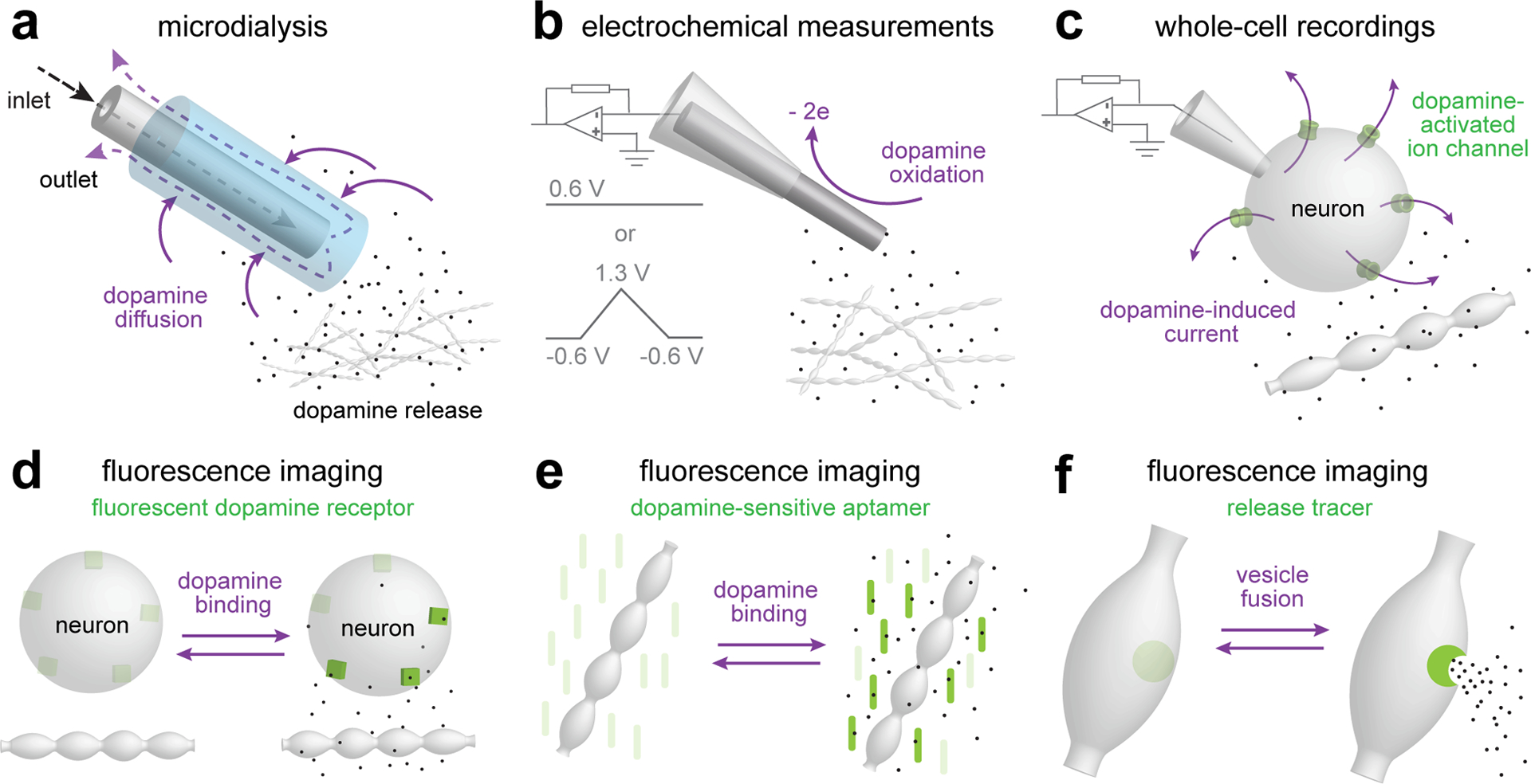
Dopamine is a prototypical neuromodulator that controls circuit function through G protein-coupled receptor signalling. Neuromodulators are volume transmitters, with release followed by diffusion for widespread receptor activation on many target cells. Yet, we are only beginning to understand the specific organization of dopamine transmission in space and time. Although some roles of dopamine are mediated by slow and diffuse signalling, recent studies suggest that certain dopamine functions necessitate spatiotemporal precision. Here, we review the literature describing dopamine signalling in the striatum, including its release mechanisms and receptor organization. We then propose the domain-overlap model, in which release and receptors are arranged relative to one another in micrometre-scale structures. This architecture is different from both point-to-point synaptic transmission and the widespread organization that is often proposed for neuromodulation. It enables the activation of receptor subsets that are within micrometre-scale domains of release sites during baseline activity and broader receptor activation with domain overlap when firing is synchronized across dopamine neuron populations. This signalling structure, together with the properties of dopamine release, may explain how switches in firing modes support broad and dynamic roles for dopamine and may lead to distinct pathway modulation.


Figures

Figure 1.
Figure 1.. Modes of chemical transmission
Overview of the fundamental modes of chemical transmission differing in release precision and organization of receptors. a. Endocrine cells release their transmitters, generally hormones, from the cell surface. The transmitters travel over long distances through the extracellular space and the blood stream to receptors residing far away from the release sites. Often, no specialized release site architecture is evident in these cells. b. Volume transmission relies on diffusion of transmitter in the extracellular space, and the receptors are only loosely coupled with the release sites. Often, specialized active zone-like release sites mediate neuromodulator secretion. A steep transmitter concentration gradient is built upon release, and the degree of receptor activation depends on their distance to these release sites. c. Synaptic transmission relies on tight spatial coupling between the active zone and receptor clusters, which are often aligned with one another at a subsynaptic scale. Signal transmission is confined to the synaptic cleft to ensure accuracy and efficient receptor activation.
Figure 2.
Figure 2.. Measurements of dopamine transmission
a. Microdialysis enables sampling of the chemical environment in the brain. A semipermeable probe inserted into the brain is perfused to exchange solutes with the surrounding tissue. The method can be used to quantitatively measure multiple neurotransmitters and molecules in vivo, with a detection threshold lower than 50 fg for dopamine. Microdialysis provides the best measurement for tonic dopamine levels, but temporal (minutes) and spatial (several hundred μm) resolution are too low to report subcellular organization of dopamine transmission or to detect fast dopamine transients. b. Electrochemical measurements rely on oxidation of dopamine at the surface of a carbon fiber electrode. Constant-potential amperometry, typically performed at 0.6 V for dopamine, provides the best temporal resolution (sub-millisecond, limited by the sampling frequency), but suffers from low chemical selectivity as any molecule that can be oxidized at the applied voltage will contribute to the signal. Fast-scan cyclic voltammetry (FSCV) improves chemical selectivity at the cost of temporal resolution (typically one data point per ~100 ms). Different molecules are oxidized at distinct voltages and can be distinguished by scanning across holding voltages (typically with a triangular wave ranging from −0.6 V to 1.3 V and a scan speed of 400–800 V/s). Because electrochemical measurements rely on subtraction of a reference current, they are suited to measure changes, but not for assessing baseline dopamine concentration. c. Whole-cell electrophysiology can be used to measure currents mediated by ion channels that are activated by dopamine. This method relies on natural coupling of GIRK2-channels to D2 receptors for somatodendritic dopamine release in the midbrain, on exogenous expression of GIRK2 channels to report striatal D2 receptor activation, or on introducing dopamine-sensitive ion channels called LGC-53. These whole-cell recordings have high temporal precision and report dopamine at the target cell. d-f. A range of fluorescent imaging techniques can be used to assess dopamine release. GRABDA and dLight (d) are genetically engineered dopamine receptors in which a circularly permuted GFP is inserted such that fluorescence increases upon dopamine binding,. The sensors exhibit good spatiotemporal resolution and can be used in vivo and in vitro. Although the sensors are engineered dopamine receptors, their expression pattern may not mimic that of endogenous dopamine receptors, and hence the signal may not report the spatial organization of dopamine transmission. Synthetic optical probes (e) are made by conjugating oligonucleotides to single-wall carbon nanotubes to gain dopamine-selectivity and -sensitivity of the infrared properties of these nanotubes. Key advantages are their resistance to photobleaching and their ability to report dopamine release with very high spatiotemporal resolution,. VMAT-pHluorin and FFNs (f) represent two tracing strategies for assessing vesicle fusion and content release, respectively, different from other methods that measure extracellular dopamine levels. VMAT-pHluorin contains a pH-sensitive fluorophore at the intraluminal side of the vesicular monoamine transporter 2 (VMAT2), and its fluorescence increases when the acidic vesicular lumen is neutralized upon fusion with the plasma membrane. FFNs are VMAT2 and/or DAT substrates and can be used to monitor dopamine vesicle fusion via dye release,. Both methods may permit detecting quantal events, but signal-to-noise ratios are in general relatively low, and translating the measurements to absolute dopamine levels is difficult.
Figure 3.
Figure 3.. Sparse dopamine release sites
a. Cellular (top) and molecular (bottom) organization of dopamine release sites. ~25% of dopamine varicosities contain functional release sites composed of active zone proteins. RIM and Munc13 are essential for action potential-triggered dopamine release, and mediate the coupling of release-ready vesicles to Ca2+ entry for fast release triggered by the fast Ca2+ sensor synaptotagmin-1,,,. The exact identities and distributions of Ca2+ channels in dopamine axons are not well understood, but they may be more broadly distributed than active zone proteins, and multiple different channel subtypes contribute to release,. Similarly, additional Ca2+ sensors are likely present, but their identities and roles are not known. b. Distribution of dopamine release sites and impact area of individual dopamine release events in the striatum. The sparsity of active zone-containing varicosities, the long-lasting depression of individual sites after a release event, and the rapid dilution of dopamine into the extracellular space suggest that, at any given time, only a small fraction of the space reaches high-enough dopamine levels for a sufficient amount of time for efficient receptor activation, and large striatal areas are not within reach of these varicosities during baseline activity. If dopamine receptors reside in this distant space, they are unlikely to be activated by single vesicular release events.
Figure 4.
Figure 4.. Dopamine neuron firing and release
a, b. Model of dopamine release of a single dopamine axon during tonic firing (black) and burst firing (purple). The amount of dopamine released from a single axon depends on how many sites release dopamine (red dots, active sites). Many sites do not release either because the initial release probability of the available sites is below 1 (black dots, inactive sites), or because sites are in a depressed state (blue dots, refractory sites). Neurons with a higher tonic firing frequency (a) will have more refractory sites and release less dopamine in response to each action potential. Neurons with a lower tonic firing frequency (b) will have less refractory sites and release more dopamine in response to each action potential. Burst firing leads to rapid depression of release from a single axon, with low-frequency neurons contributing more dopamine. Note that this speculative model presents the total amount of release from a single axon, which cannot currently be measured. Typical measurements using electrochemical methods (Fig. 2) reflect the average dopamine in a large area and from many neurons, not peak dopamine at release sites from a single axon. In addition, in vivo voltammetry may reveal a build-up of dopamine during rapid stimulation because dopamine reuptake is overwhelmed and because low frequency-measurements overestimate dopamine levels during the decay phase.
Figure 5.
Figure 5.. Dopamine receptor organization and activation
a, b. Working model of dopamine receptor organization with overview (a) and zoom-in on a varicosity that makes a synapse-like contact (b). Dopamine is released from non-synaptic and synaptic varicosities. The main dopamine receptors are segregated over two neuron subtypes, D1-MSNs and D2-MSNs. Both MSN types sense dopamine release from non-synaptic varicosities (a, left), and can receive synaptic-like inputs from dopamine axons with appositions between dopamine varicosities and GABAergic postsynaptic assemblies (a, right, and b). Dopamine receptors are widely distributed on MSNs, and may be present in clusters. Importantly, dopamine receptors are not found in the postsynaptic specializations with the currently available tools (b). Instead, these specializations may contain gephyrin and other proteins typically found at GABA-ergic synapses. Individual vesicular fusion events may activate both D1 and D2 receptors, and close-by receptors are more likely to be activated by dopamine than those farther away. The exact organization of dopamine receptors relative release sites is not known, but may strongly impact dopamine functions.
Figure 6.
Figure 6.. The domain-overlap model
a, b. Model of dopamine signaling domains during tonic and phasic release. In a given area of the striatum, tonic release (a) generates short-lived dopamine peaks that are confined to a small domain and only recruit proximal receptors (left). After a short time interval (Δt), a distinct set of release sites is active and targets a different subset of receptors (right). The synchrony of phasic release across many release sites in a given area (b) generates significant crosstalk between signaling domains. After a brief interval, dopamine spread overwhelms the DAT, and dopamine levels increase beyond the micrometer-sized domains of active sites. This leads to augmented dopamine dwell times and activation of new receptors residing farther away from release sites. This domain-overlap model may form a basis for recruiting small variable subsets of receptors during tonic activity (arising from small dopamine domains), and recruitment of larger numbers of distant receptors during phasic activity (arising from overlap of dopamine domains). Regional heterogeneity in release site distribution within the complex dopamine axonal arbor may influence receptor activation domains, and co-release could further shape the signaling of dopamine neurons. Differential distribution of distinct dopamine receptors or cell types at micrometer-scale distances may lead to distinct pathway modulation during tonic and phasic release, respectively.

References

    1. Grillner S & Robertson B The Basal Ganglia Over 500 Million Years. Curr. Biol 26, R1088–R1100 (2016). - PubMed
    1. Sawin ER, Ranganathan R & Horvitz HRC elegans Locomotory Rate Is Modulated by the Environment through a Dopaminergic Pathway and by Experience through a Serotonergic Pathway. Neuron 26, 619–631 (2000). - PubMed
    1. Surmeier DJ, Graves SM & Shen W Dopaminergic modulation of striatal networks in health and Parkinson’s disease. Curr. Opin. Neurobiol 29, 109–117 (2014). - PMC - PubMed
    1. Grace AA Dysregulation of the dopamine system in the pathophysiology of schizophrenia and depression. Nat. Rev. Neurosci 17, 524–532 (2016). - PMC - PubMed
    1. Gershman SJ & Uchida N Believing in dopamine. Nat. Rev. Neurosci 20, 703–714 (2019). - PMC - PubMed

Publication types

LinkOut - more resources