. 2021 Sep;76(9):2840-2854.
doi: 10.1111/all.14850. Epub 2021 May 10.

Silencing of SARS-CoV-2 with modified siRNA-peptide dendrimer formulation

Affiliations

Silencing of SARS-CoV-2 with modified siRNA-peptide dendrimer formulation

Musa Khaitov et al. Allergy. 2021 Sep.

Abstract

Background: First vaccines for prevention of Coronavirus disease 2019 (COVID-19) are becoming available but there is a huge and unmet need for specific forms of treatment. In this study we aimed to evaluate the anti-SARS-CoV-2 effect of siRNA both in vitro and in vivo.

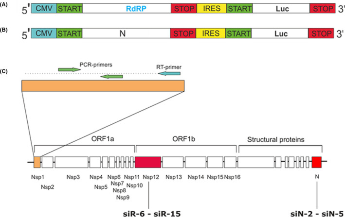
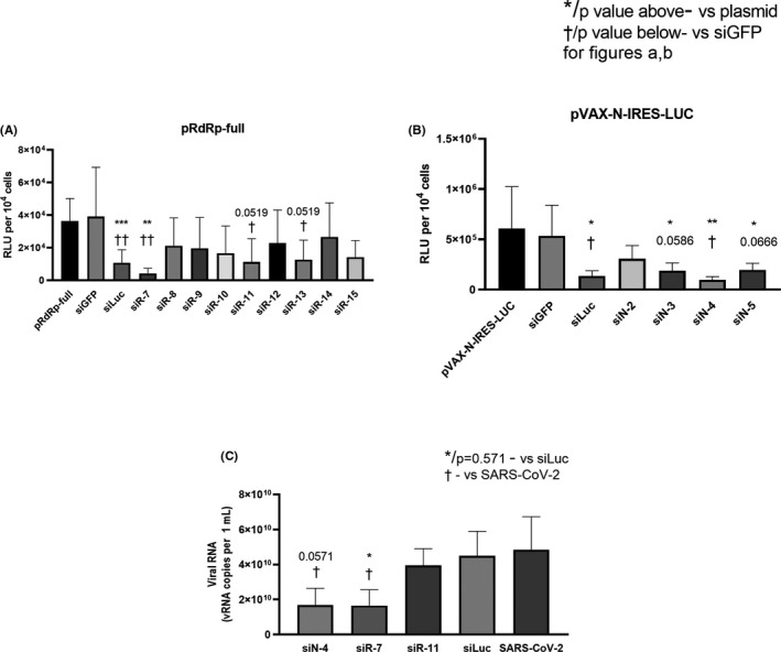
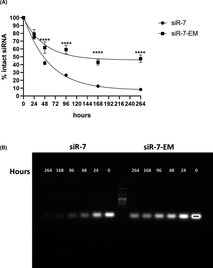
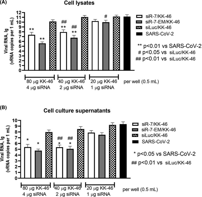
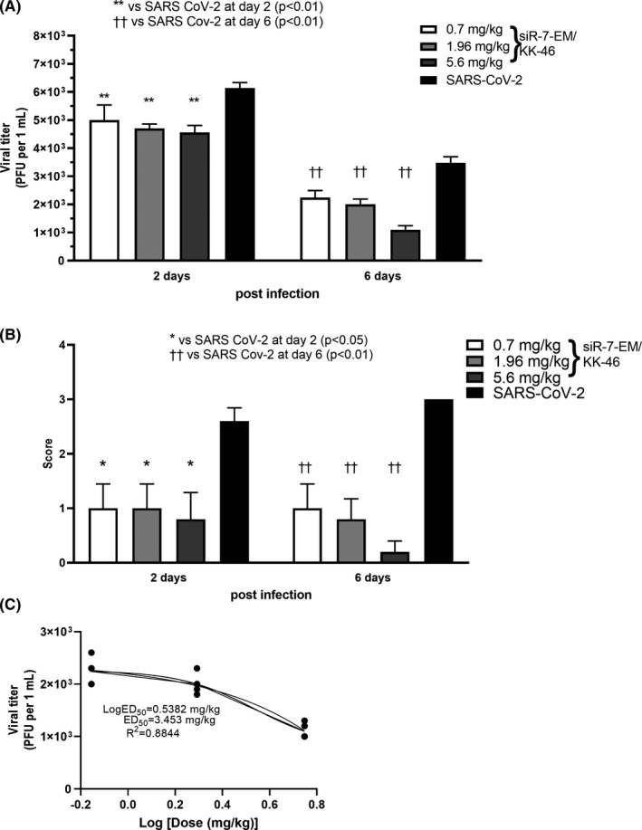
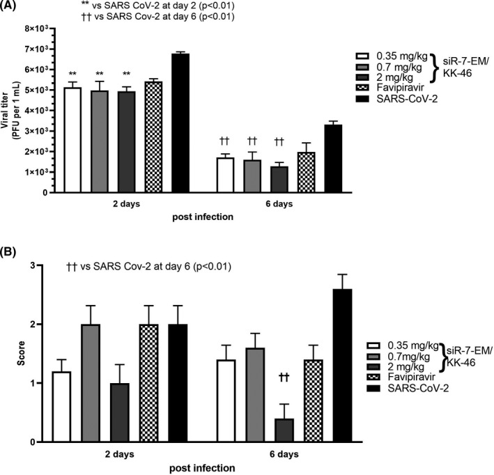
Methods: To identify the most effective molecule out of a panel of 15 in silico designed siRNAs, an in vitro screening system based on vectors expressing SARS-CoV-2 genes fused with the firefly luciferase reporter gene and SARS-CoV-2-infected cells was used. The most potent siRNA, siR-7, was modified by Locked nucleic acids (LNAs) to obtain siR-7-EM with increased stability and was formulated with the peptide dendrimer KK-46 for enhancing cellular uptake to allow topical application by inhalation of the final formulation - siR-7-EM/KK-46. Using the Syrian Hamster model for SARS-CoV-2 infection the antiviral capacity of siR-7-EM/KK-46 complex was evaluated.

Results: We identified the siRNA, siR-7, targeting SARS-CoV-2 RNA-dependent RNA polymerase (RdRp) as the most efficient siRNA inhibiting viral replication in vitro. Moreover, we showed that LNA-modification and complexation with the designed peptide dendrimer enhanced the antiviral capacity of siR-7 in vitro. We demonstrated significant reduction of virus titer and lung inflammation in animals exposed to inhalation of siR-7-EM/KK-46 in vivo.

Conclusions: Thus, we developed a therapeutic strategy for COVID-19 based on inhalation of a modified siRNA-peptide dendrimer formulation. The developed medication is intended for inhalation treatment of COVID-19 patients.

Keywords: COVID-19; LNA; SARS-CoV-2; peptide dendrimers; siRNA.


Conflict of interest statement

Rudolf Valenta has received research grants from the Austrian Science Fund (FWF), HVD Biotech, Vienna, Austria, Worg Pharmaceuticals, Hangzhou, China and Viravaxx, Vienna, Austria and serves as a consultant for Viravaxx. Veronica Skvortsova currently serves as head of the Federal Medico‐biological Agency of Russia (FMBA Russia). Musa Khaitov, Alexandra Nikonova, Ksenia Kozhikova, Ilya Kofiadi, Igor Shilovskiy, Valeriy Smirnov, Ivan Kozlov, Sergey Andreev, Olesya Koloskova and Ilya Sergeev are authors on a patent application related to this study. The other authors do not have any conflicts of interest to declare.

Figures

FIGURE 1
FIGURE 1
Plasmid constructs and design of siRNAs. Schematic representation of the bicistronic expression plasmid coding genes of firefly luciferase (Luc) and full size RdRp (pRdRp‐full) (A) or N (pVAX‐N‐IRES‐LUC) (B) genes of SARS‐CoV‐2. (C) Positions of the siRNA targeting SARS‐CoV‐2 RdRp (siR‐6‐siR‐15) and N (siN‐2‐siN‐5) genes. The RT‐PCR–amplified region at the most upstream region of ORF1 (NSP1‐leader protein) is marked
FIGURE 2
FIGURE 2
Properties of designed siRNA. (A, B) Inhibition of gene expression with synthetic siRNA. Hep‐2 cells were transfected with each of the plasmids coding SARS‐CoV‐2 genes fused with firefly luciferase gene (pRdRp‐full) (A) or pVAX‐N‐IRES‐LUC (B) followed by transfection with SARS‐CoV‐2‐specific or control siRNA. siLuc and siGFP were used as positive and negative controls, respectively. LipofectamineTM 3000 was used as vehicle for both pDNA and siRNA. After 24 h cells were harvested and luciferase activity was determined. Data are expressed as relative light units (RLU) per 104 cells. Footnotes: */† or adjusted p value above/below represents the difference compared to cells transfected with plasmid only/non‐specific siGFP, respectively. *†p < 0.05, **††p < 0.01, ***p < 0.001. (D) Inhibition of SARS‐CoV‐2 reproduction with synthetic siRNA. Vero E6 cells were transfected with siRNA/ LipofectamineTM 3000 complexes. Media with complexes were removed four hours after transfection and cells were infected with SARS‐CoV‐2 at MOI 0.0001. Viral load was determined by qRT‐PCR. The results are expressed as number of viral RNA copies per mL. Footnotes: * or p = 0.571 represent differences with cells infected with SARS‐CoV‐2 and treated with non‐specific siLuc. † represent differences with SARS‐CoV‐2 only infected cells.*†p < 0.05. Differences between multiple groups were estimated using a Kruskal–Wallis test followed by post‐hoc testing (if the Kruskal–Wallis was significant) using un‐paired Mann–Whitney U tests. Bars show means of four (A, B) and five (C) independent experiments ± SDs
FIGURE 3
FIGURE 3
Modified siRNA have increased resistance to nuclease degradation. A, The stability of unmodified siR‐7 (circles) and modified siR‐7‐EM (squares) in 50% mouse serum are compared over a period of 264 h at 37°C (N = 4). siRNA quantities at various time points were calculated by dividing the total counts of full‐length siRNA by the input starting material. B, For this purpose, aliquots of each sample (10 μg siRNA per lane) were analyzed by 1.5% agarose gel electrophoresis. Differences between multiple groups were analyzed using repeated measures one‐way ANOVA with Dunnetts's post hoc test. Bars show medians of four independent experiments+SEM. **** p < 0.0001
FIGURE 4
FIGURE 4
Inhibition of SARS‐CoV‐2 reproduction with unmodified or LNA‐modified siR‐7/peptide dendrimer KK‐46 complexes in vitro. Vero E6 cells were transfected with unmodified siR‐7 or LNA‐modified siR‐7‐EM‐peptide dendrimer KK‐46 complexes at three concentrations (x‐axes). Media with complexes were removed four hours after transfection and cells were infected with SARS‐CoV‐2 at MOI 0.0001. After 48 hours supernatants and cells were harvested. Viral load was determined by qRT‐PCR in cells lysates (A) and supernatants (B). The results are expressed as viral RNA copies per mL. Differences between multiple groups were estimated using a Kruskal–Wallis test followed by post‐hoc testing (if the Kruskal–Wallis was significant) using un‐paired Mann‐Whitney U tests. Bars show medians of five independent experiments +SDs. Footnotes: # represent differences with cells infected with SARS‐CoV‐2 and treated with non‐specific siLuc.*represent differences with SARS‐CoV‐2 only infected cells. *#p < 0.05, **##p < 0.01
FIGURE 5
FIGURE 5
Dose‐dependent inhibition of SARS‐CoV‐2 reproduction with modified siR‐7‐EM‐peptide dendrimer KK‐46 complexes in Syrian hamsters after repeated inhalation exposure. Syrian hamsters were infected with SARS‐CoV‐2 with a dose of 105 PFU/animal and treated with three doses of the modified siR‐7‐EM‐peptide dendrimer KK‐46 complexes (35, 98 and 289 μg siR‐7‐EM/animal, 0.7, 1.96 and 5.6 mg KK‐46/kg, respectively). Inhalations with complexes were repeated daily for six days. Two and six days after infection animals were sacrificed and determination of viral titer (A) and macroscopic evaluation plus scoring of histopathology (B) in the lung were performed. The results are expressed as plaque forming units (PFU) per mL (A) or scores (B) obtained for histological analysis of pathologic alterations in hamsters’ lung. (C) Dose‐response curve of SARS‐CoV‐2 levels in the lungs at day six after infection, where the ED50 estimation is ∼ 3.453 mg/kg. Differences between multiple groups were estimated using a Kruskal–Wallis test followed by post‐hoc testing (if the Kruskal–Wallis was significant) using un‐paired Mann‐Whitney U tests. Bars show medians of one experiment (five animals per group) + SDs. Data are provided for one experiment representative of two independent experiments with five hamsters /group. Footnotes: */† represent differences with hamsters infected with SARS‐CoV‐2 and assessed at day two/six after infection, respectively. **††p < 0.01, *p < 0.05
FIGURE 6
FIGURE 6
Effect of repeated low dose modified siR‐7‐EM‐peptide dendrimer KK‐46 complexes in Syrian hamsters. Syrian hamsters were infected with SARS‐CoV‐2 at a dose of 105 PFU/animal and exposed to different doses (0.175, 0.35 and 1.0 mg/kg of siR‐7‐EM/KK‐46 aerosol) twice a day with two hours interval. Two and six days after infection animals were sacrificed and viral titers (A) and macroscopic evaluation and scoring of the histopathology lesions (B) in the lung were analyzed. Orally administrated Favipiravir (1 h after infection, a dose of 1.2 mg/animal was administered twice a day, and then daily during 6 days after infection 0.4 mg/animal twice a day) served as positive control. The results are expressed as plaque forming units (PFU) per mL (A) or scores (B) obtained for histological analysis of pathologic lung alterations. Differences between multiple groups were estimated using a Kruskal–Wallis test followed by post‐hoc testing (if the Kruskal–Wallis was significant) using un‐paired Mann–Whitney U tests. Bars show medians of one experiment (five animals per group) + SDs. Data are from one experiment representative of two independent experiments with five hamsters/group are shown. Footnotes: */† represents difference with hamsters infected with SARS‐CoV‐2 and assessed at day two/six after infection, respectively. **††p < 0.01

Similar articles

  • Treatment of COVID-19 patients with a SARS-CoV-2-specific siRNA-peptide dendrimer formulation.
    Khaitov M, Nikonova A, Kofiadi I, Shilovskiy I, Smirnov V, Elisytina O, Maerle A, Shatilov A, Shatilova A, Andreev S, Sergeev I, Trofimov D, Latysheva T, Ilyna N, Martynov A, Rabdano S, Ruzanova E, Savelev N, Pletiukhina I, Safi A, Ratnikov V, Gorelov V, Kaschenko V, Kucherenko N, Umarova I, Moskaleva S, Fabrichnikov S, Zuev O, Pavlov N, Kruchko D, Berzin I, Goryachev D, Merkulov V, Shipulin G, Udin S, Trukhin V, Valenta R, Skvortsova V. Khaitov M, et al. Allergy. 2023 Jun;78(6):1639-1653. doi: 10.1111/all.15663. Epub 2023 Feb 14. Allergy. 2023. PMID: 36721963 Clinical Trial.
  • Designing an effective therapeutic siRNA to silence RdRp gene of SARS-CoV-2.
    Shawan MMAK, Sharma AR, Bhattacharya M, Mallik B, Akhter F, Shakil MS, Hossain MM, Banik S, Lee SS, Hasan MA, Chakraborty C. Shawan MMAK, et al. Infect Genet Evol. 2021 Sep;93:104951. doi: 10.1016/j.meegid.2021.104951. Epub 2021 Jun 2. Infect Genet Evol. 2021. PMID: 34089909 Free PMC article.
  • Suppression of SARS-CoV-2 Replication with Stabilized and Click-Chemistry Modified siRNAs.
    Traube FR, Stern M, Tölke AJ, Rudelius M, Mejías-Pérez E, Raddaoui N, Kümmerer BM, Douat C, Streshnev F, Albanese M, Wratil PR, Gärtner YV, Nainytė M, Giorgio G, Michalakis S, Schneider S, Streeck H, Müller M, Keppler OT, Carell T. Traube FR, et al. Angew Chem Int Ed Engl. 2022 Sep 19;61(38):e202204556. doi: 10.1002/anie.202204556. Epub 2022 Aug 12. Angew Chem Int Ed Engl. 2022. PMID: 35802496 Free PMC article.
  • Surface Engineered Dendrimers in siRNA Delivery and Gene Silencing.
    Tambe V, Thakkar S, Raval N, Sharma D, Kalia K, Tekade RK. Tambe V, et al. Curr Pharm Des. 2017;23(20):2952-2975. doi: 10.2174/1381612823666170314104619. Curr Pharm Des. 2017. PMID: 28292248 Review.
  • Non-viral vectors for RNA delivery.
    Yan Y, Liu XY, Lu A, Wang XY, Jiang LX, Wang JC. Yan Y, et al. J Control Release. 2022 Feb;342:241-279. doi: 10.1016/j.jconrel.2022.01.008. Epub 2022 Jan 10. J Control Release. 2022. PMID: 35016918 Free PMC article. Review.

Cited by

References

    1. Zhang JJ, Dong X, Cao YY, et al. Clinical characteristics of 140 patients infected with SARS‐CoV‐2 in Wuhan, China. Allergy 2020;75(7):1730‐1741. - PubMed
    1. Azkur AK, Akdis M, Azkur D, et al. Immune response to SARS‐CoV‐2 and mechanisms of immunopathological changes in COVID‐19. Allergy 2020;75(7):1564‐1581. - PMC - PubMed
    1. Tang D, Comish P, Kang R. The hallmarks of COVID‐19 disease. PLoS Pathog 2020;16(5):e1008536. - PMC - PubMed
    1. Morens DM, Folkers GK, Fauci AS. Emerging infections: a perpetual challenge. Lancet Infect Dis 2008;8(11):710‐719. - PMC - PubMed
    1. Morens DM, Breman JG, Calisher CH, et al. The origin of COVID‐19 and why it matters. Am J Trop Med Hyg 2020;103(3):955‐959. - PMC - PubMed