Review
. 2020:108:33-83.
doi: 10.1016/bs.aivir.2020.08.003. Epub 2020 Sep 23.

Structure-guided paradigm shifts in flavivirus assembly and maturation mechanisms

Affiliations
Review

Structure-guided paradigm shifts in flavivirus assembly and maturation mechanisms

Conrrad M R Nicholls et al. Adv Virus Res. 2020.

Abstract

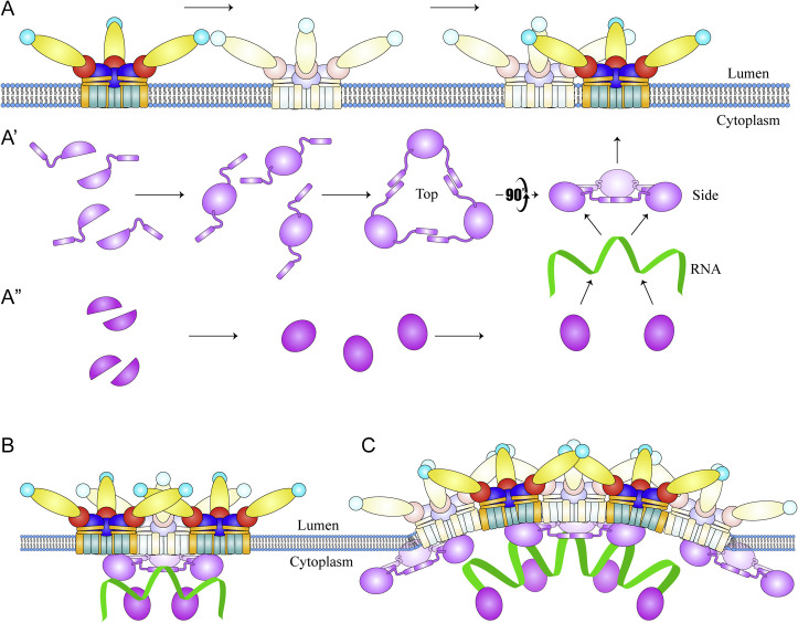
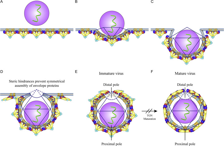
The flavivirus genus encompasses more than 75 unique viruses, including dengue virus which accounts for almost 390 million global infections annually. Flavivirus infection can result in a myriad of symptoms ranging from mild rash and flu-like symptoms, to severe encephalitis and even hemorrhagic fever. Efforts to combat the impact of these viruses have been hindered due to limited antiviral drug and vaccine development. However, the advancement of knowledge in the structural biology of flaviviruses over the last 25 years has produced unique perspectives for the identification of potential therapeutic targets. With particular emphasis on the assembly and maturation stages of the flavivirus life cycle, it is the goal of this review to comparatively analyze the structural similarities between flaviviruses to provide avenues for new research and innovation.

Keywords: Capsid; Dengue; Envelope; Flavivirus; Flavivirus assembly; Flavivirus maturation; Flavivirus structure; West Nile; Zika.


Figures

Unlabelled Image
Graphical abstract
Fig. 1
Fig. 1
Flavivirus life cycle: Flaviviruses initiate infection through a myriad of interactions with host cell receptors. (1) Once attached, the virus is internalized into the cell via clathrin-mediated endocytosis. (2) The E glycoproteins then undergo conformational changes to allow fusion between the viral envelope and the endosomal membrane, which ultimately leads to the release of the RNA into the cytoplasm. (3) The viral RNA genome is released and transported to the ER where it is translated. (4) Viral RNA replication occurs in virus-induced membrane invaginations known as replication complexes. These structures shield viral replication products and are spatially congruent with sites of virion assembly along the ER membrane. (5) Immature virions are assembled and bud into the ER lumen and are subsequently trafficked through the TGN. (6) The low pH of the TGN promotes virion maturation through glycoprotein conformational changes and subsequent cleavage of the pr domain from the prM protein. (7) After furin cleavage, the secretory vesicle containing the virion fuses with the plasma membrane, where the pr domain is released and the particle emerges into the extracellular environment as a fully mature and infectious virus.
Fig. 2
Fig. 2
Flavivirus genome and polyprotein domain structure: The ~ 11 kb flavivirus open reading frame genome (top) is translated into a single polyprotein along the ER membrane (bottom). This polyprotein is processed into ten functional proteins by both host and viral proteases (protease cleavage sites are labeled with arrows), generating three structural proteins (C, prM/M, and E) and seven non-structural proteins (NS1, NS2A, NS2B, NS3, NS4A, NS4B, NS5). As the name suggests, the structural proteins function to encapsulate and protect the viral RNA, forming an enveloped virion. In contrast, the non-structural proteins facilitate RNA genome replication and assist in cellular immuno-evasion. The amino acid length of each protein is indicated by the small number within each gene “box” (top).
Fig. 3
Fig. 3
Flavivirus capsid protein: (A) A single capsid monomer (ZIKV C protein—PDB ID: 6C44) with each helix (α1–α4) labeled within the structure. (B) Hydrophobic interactions (residues highlighted in green) between helices α2 and α2′ (top) and helices α4 and α4′ (bottom—rotated ninety-degrees horizontally) stabilizing the ZIKV capsid dimer. The helices of one capsid protomer within the dimer (α1–α4) are colored in purple, while the helices of the other capsid protomer (α1′–α4′) are colored in light gray. (C) Side view of the ZIKV capsid dimer. The positive exterior of the dimer is shown by the green (+) symbols, which has been hypothesized to interact with the viral RNA during assembly. The central hydrophobic cleft (HC), which has been shown to interact with lipids, is indicated in yellow. C-anchor is not present in this structure of C protein. For an understanding of the C protein structure with C-anchor included, readers are referred to Tan et al. (2020).
Fig. 4
Fig. 4
Mature and immature flavivirus glycoproteins: (A) Side view surface representation of the immature ZIKV (PDB ID: 5U4W) prM-E heterodimer trimeric spike (left). The prM protein is colored cyan, the E protein DI is colored red, DII is colored yellow, and DIII is colored blue. The E protein TMDs are colored orange while the prM/M TMDs are colored light blue. The viral membrane is indicated by the curved dotted line. On the right side of panel (A) is a top view ribbon representation of the trimeric spike. This structure is color coded the same as the surface structure on the left but includes a depiction of the fusion loop (green) and glycan loop (pink). (B) Side view surface representation of the Fab stabilized immature ZIKV (PDB ID: 6LNT) prM-E heterodimer inverted tripod described in Section 4 (Tan et al., 2020). In this structure, density was observed below the TMDs of prM and E protein (light blue and orange, respectively) that fit C protein (purple). The prM stem was also visualized (cyan). (C) Side view surface representation of the mature dimeric M-E heterodimers (PDB ID: 6C08) (far left). The E protein domains are color coded as follows: DI in red, DII in yellow, and DIII in blue. The E TMDs are colored orange while the M TMDs are colored light blue. The viral membrane is indicated by the curved dotted line. In the center of panel (C), a ribbon representation of the dimeric M-E heterodimer displays the glycan loop (pink) and the fusion loop (green). One heterodimer is color coded the same as the surface representation on the left, while the other heterodimer is colored gray. A 90-degree rotated top view of the dimeric heterodimer is present on the far-right side of panel (C), displaying the same color scheme as the ribbon structure in the center.
Fig. 5
Fig. 5
Mature and immature flavivirus structure: (A) Surface representation of the immature ZIKV (PDB ID: 5U4W) particle with prM and each E protein domain color coded as follows: prM (cyan), E protein domain I (red), E protein domain II (yellow), E protein domain III (blue). (B) Cross-section diagram of the immature flavivirus particle showing prM-E heterodimer trimeric spikes. The diagram is color coded to match panel A. Additionally, the prM TMDs (cylinders along the lipid bilayer) are colored in light blue, while the E protein TMDs are colored orange. (C) Cross-section diagram of the mature flavivirus particle showing dimeric M-E heterodimers. Color scheme is the same as panel (B). (D) Surface representation of the mature ZIKV (PDB ID: 6C08) particle with each E protein domain color coded the same as in panel (A), but also includes the fusion loop shown in green.
Fig. 6
Fig. 6
NS2A assisted assembly model—This model was originally proposed by Xie et al. (2019): The viral RNA (light green ribbon) is immediately captured by NS2A (blue dumbbells) upon its release from replication complexes (left), where it is transported to assembly sites (right). In parallel, other NS2A proteins recruit un-processed C-prM-E polyproteins and NS2B-3 proteases to the assembly site. Here, NS2B-3 (green) cleaves the C-prM-E polyprotein (purple, cyan and yellow) in tandem with host signalase (not shown). This facilitates in the dimerization of soluble C proteins, which are then able to receive the genomic RNA from NS2A, forming a NC. Meanwhile, the processed prM and E proteins are able to form heterodimers and subsequently form trimeric spikes on the luminal side of the membrane (color code similar to Fig. 3, Fig. 5). The NC then buds into the nascent viral envelope at the ER lumen.
Fig. 7
Fig. 7
Structural comparisons of flavivirus particles: Unique structural elements have been identified within immature flavivirus structures throughout the last 20 years (panels A–C). Meanwhile, the structural elements observed in mature flavivirus structures have remained somewhat constant, but the resolution has increased dramatically (Panel D). (A–D) Central section looking down the fivefold axis of the cryo-EM map. The central sections are contoured and colored radially from magenta (NC core), blue (capsid), green (TM) to yellow and red (ecto-domain). (A) Cross-section structure of the immature ZIKV particle at 9 Å, published by Prasad et al. (2017) (EMDB: 8505). Residual density (blue blob in red circle) underneath the prM-E spikes was shown to fit capsid protein. This was the first time any connections between the flavivirus NC and the envelope glycoproteins had been observed. (B) Cross-section structure of the antibody stabilized immature ZIKV particle at 8 Å, published by Tan et al. (2020) (EMDB: 0932). Density fitting C protein (red circle) was resolved to ~ 9.5 Å and showed that C protein retained its helix α5 (C-anchor). (C) Asymmetric reconstruction and cross-section structure of the immature KUNV particle at ~ 20 Å, published by Therkelson et al. (2018) (EMDB: 8983). Contact between the NC and the viral envelope was observed at the “proximal pole” (top), indicating an interaction during virus assembly/budding. A lack of density at the “distal pole” (bottom) indicated that symmetrical incorporation of the glycoproteins is sterically hindered at the bud neck (also see Fig. 8). (D) Cross-section structure of the mature ZIKV particle at 3.1 Å, published by Sevvana et al. (2018) (EMDB: 7543). No contacts were observed between the NC and envelope.
Fig. 8
Fig. 8
Capsid mediated assembly model—This model was originally proposed by Tan et al. (2020): All “A” panels occur in parallel. (A) Cleaved prM and E proteins oligomerize to form prM-E heterodimers, which in turn coalesce to form “inverted tripods.” These inverted tripods then associate to form trimeric spikes. A′ Un-processed C proteins retaining their helix α5 (C + α5) dimerize in the cytoplasm and subsequently form trigonal trimers through interactions between each helix α5. The positively charged exterior of these trimers then interacts with the genomic RNA, while the hydrophobic core of each dimer facilitates membrane interaction, which ultimately leads to binding with the prM-E TMDs. A” Fully processed C proteins (not containing helix α5) also dimerize and work in tandem with the C + α5 proteins to assist the condensation of genomic RNA. (B) The C + α5 trimers facilitate proper spacing between prM-E inverted tripods, which promotes trimeric spike formation. (C) Proper prM-E inverted tripod spacing and trimeric spike formation through C + α5-prM-E TMD interactions initiates membrane bending and drives viral budding, forming immature flavivirus particles. Color scheme is similar to Fig. 4, Fig. 5.
Fig. 9
Fig. 9
Asymmetric assembly model—This model was originally proposed by Therkelson et al., 2018: (A) Glycoproteins assemble along the ER membrane as prM-E heterodimer trimeric spikes. (B) The unstructured NC interacts with the trimeric spikes through their TMDs and initiates the budding process of an icosahedron. (C) As the NC pushes deeper into the ER membrane, more glycoproteins are wrapped into the virion envelope. (D) During the final stages of the budding process, the virion is unable to incorporate the final copies of the envelope glycoproteins due to the steric hinderance of the narrowing bud neck. (E) Highly distorted density at the distal pole of the immature virion due to the lack of glycoproteins suggests that icosahedral symmetry is incompletely fulfilled during budding. The NC also remains in close proximity with the envelope at the proximal pole after budding. (F) Viral maturation through the TGN produces dramatic conformational changes in the glycoproteins, allowing furin to cleave the pr peptide from the particle. As a result, the NC is shifted to the center of the particle, displaying no contact points with the envelope. Color scheme is similar to Fig. 3, Fig. 5.

Similar articles

Cited by

References

    1. Allison S.L. Synthesis and secretion of recombinant tick-borne encephalitis virus protein E in soluble and particulate form. J. Virol. 1995;69:5816–5820. - PMC - PubMed
    1. Allison S.L. Mutational evidence for an internal fusion peptide in flavivirus envelope protein. E. J. Virol. 2001;75:4268–4275. - PMC - PubMed
    1. Alsaleh K. The E glycoprotein plays an essential role in the high pathogenicity of 381 European -Mediterranean IS98 strain of West Nile virus. Virology. 2016;492:53–65. - PubMed
    1. Amberg S.M., Rice C.M. Mutagenesis of the Ns2b-Ns3-mediated cleavage site in the Flavivirus capsid protein demonstrates a requirement for coordinated processing. J. Virol. 1999;73:8083–8094. - PMC - PubMed
    1. Amberg S.M. Ns2b-3 proteinase-mediated processing in the yellow fever virus structural region: in vitro and in vivo studies. J. Virol. 1994;68:3794–3802. - PMC - PubMed

Substances

LinkOut - more resources