. 2021 Jan-Jun:296:100639.
doi: 10.1016/j.jbc.2021.100639. Epub 2021 Apr 8.

The multidomain architecture of a bacteriophage endolysin enables intramolecular synergism and regulation of bacterial lysis

Affiliations

The multidomain architecture of a bacteriophage endolysin enables intramolecular synergism and regulation of bacterial lysis

Frank Oechslin et al. J Biol Chem. 2021 Jan-Jun.

Abstract

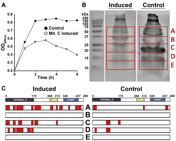
Endolysins are peptidoglycan hydrolases produced at the end of the bacteriophage (phage) replication cycle to lyse the host cell. Endolysins in Gram-positive phages come in a variety of multimodular forms that combine different catalytic and cell wall binding domains. However, the reason why phages adopt endolysins with such complex multidomain architecture is not well understood. In this study, we used the Streptococcus dysgalactiae phage endolysin PlySK1249 as a model to investigate the role of multidomain architecture in phage-induced bacterial lysis and lysis regulation. PlySK1249 consists of an amidase (Ami) domain that lyses bacterial cells, a nonbacteriolytic endopeptidase (CHAP) domain that acts as a dechaining enzyme, and a central LysM cell wall binding domain. We observed that the Ami and CHAP domains synergized for peptidoglycan digestion and bacteriolysis in the native enzyme or when expressed individually and reunified. The CHAP endopeptidase resolved complex polymers of stem-peptides to dimers and helped the Ami domain to digest peptidoglycan to completion. We also found that PlySK1249 was subject to proteolytic cleavage by host cell wall proteases both in vitro and after phage induction. Cleavage disconnected the different domains by hydrolyzing their linker regions, thus hindering their bacteriolytic cooperation and possibly modulating the lytic activity of the enzyme. PlySK1249 cleavage by cell-wall-associated proteases may represent another example of phage adaptation toward the use of existing bacterial regulation mechanism for their own advantage. In addition, understanding more thoroughly the multidomain interplay of PlySK1249 broadens our knowledge on the ideal architecture of therapeutic antibacterial endolysins.

Keywords: PlySK1249; bacteriophage; endolysin; intramolecular synergism; lysis regulation; proteolysis.


Conflict of interest statement

Conflict of interest The authors declare that they have no conflicts of interest with the contents of this article.

Figures

Figure 1
Figure 1
Characterization of the lytic activity of the PlySK1249 endolysin and its various truncated forms.A, parent PlySK1249 (full enzyme, aa 1–489); Ami (N-terminal amidase, aa 1–170); Ami_LysM (N-terminal amidase + LysM, aa 1–330); LysM_CHAP (LysM + C-terminal CHAP domain, aa 250–489). B, SDS-PAGE gel showing PlySK1249 and its various truncated constructions. The purified endolysin constructions were loaded (2 mg/ml) on NuPAGE 4 to 12% BisTris gels and stained with Coomassie blue. Molecular mass was determined with a prestained protein standard. C, cells of multiple bacterial species in the exponential growth phase were exposed to 1 μM of parent PlySK149 or its different truncated forms. Decrease in the turbidity of the cultures was measured at 600 nm after 30 min. No decrease in turbidity was observed in the absence of enzyme (not shown) or in the presence of LysM_CHAP. D, S. dysgalactiae cells in the exponential growth phase were also exposed to either 1 μM of Ami_LysM or LysM_CHAP constructs alone or in combination and the decrease in turbidity (at 600 nm) was followed over 1 h. E, viable counts from experiment shown in panel D. Counts were assessed by plating serial dilutions on nutrient agar. The experiments were repeated twice in triplicate and means ± standard deviations are shown.
Figure 2
Figure 2
Morphological aspects of cells lysed by the PlySK1249 endolysin and its various truncated forms.S. dysgalactiae in the exponential growth phase were observed after 1 h using phosphate buffer as a control (A), or LysM_CHAP at a final concentration of 3.5 μM (B). Although the CHAP domain did not impact directly on cell lysis, an effect on chain disruption was observed. The control population was composed mainly (90%) of chains between 6 and 37 cells long (144 cells observed in total) compared to the 60% of chains that were composed of two cells for the LysM_CHAP treatment (1044 cells observed in total). C, transmission electron microscopy of S. dysgalactiae cells treated with the native PlySK1249 endolysin or its various truncated versions. S. dysgalactiae cells in the exponential growth phase were treated with phosphate buffer for 1 h as a control, Plysk1249 for 15 min, Ami_LysM for 15 min, or LysM_CHAP for 1 h. Cells were then postfixed using glutaraldehyde and embedded in epoxy for ultrathin sectioning. Scale bars represent 500 nm. The inset in the LysM_CHAP figure highlights the nonlytic wall nibbling by the LysM_CHAP construct.
Figure 3
Figure 3
RP-HPLC chromatogram and LC-MS analysis of S. dysgalactiae peptidoglycan digested with PlySK1249 or its truncated catalytic domains.A, purified wall-peptidoglycan was digested overnight and glycans were sequentially precipitated before chromatography on a C18 Sephasil column. Equimolar concentrations (3.5 μM) of I) PlySK1249, II) Ami_LysM, III) LysM_CHAP, and IV) both Ami_LysM and LysM_CHAP were used and analyses were repeated three times for each, yielding the same results. B, relative abundances of the polymers observed in the native enzyme and amidase domain digestion experiments. The products of peptidoglycan digestion by the native enzyme and its truncated Ami domain were analyzed by LC-MS after a 5 kDa filtration. The masses of the precursors corresponding to the dimer, trimer, quadrimer, and heptamer of the AAAQKA monomer block were detected. All of these oligomers were detected in both samples, with the exception of the heptamers, which were only present in the amidase digestion. Polymer structures were deduced from masses after de novo peptide sequencing. C, representation of S. dysgalactiae peptidoglycan based on the masses and sequences observed. Arrows indicate the cleavage sites for the respective catalytic domains of the enzyme. The framed dotted-line area corresponds to the dimer block.
Figure 4
Figure 4
PlySK1259 proteolysis and its different truncated versions in the presence of trypsin or cell-wall-associated proteases. Forty μM of the parent enzyme (A) or the Ami_lysM (B) and LysM_CHAP (C) were incubated with 1 μg/ml of trypsin. Right panels, sample were migrated after different incubation times on NuPAGE 4 to 12% BisTris, stained with Coomassie blue. The subsequent degradation products obtained after ON digestion of the native enzyme (band 1 and 2) were sequenced and the identified peptides are indicated on the right of the panel. Left panels, digestion products were transferred to a nitrocellulose membrane for western blotting using Anti-6xHis antibody. D, 40 μM of the parent enzyme (I), Ami_lysM (II), or LysM_CHAP (III) were incubated overnight with cell wall protein extract from S. dysgalactiae SK1249. Samples were migrated on NuPAGE 4 to 12% BisTris and degradation was confirmed by western blotting using Anti-6xHis tag antibody.
Figure 5
Figure 5
Identification of the proteases involved in the PlySK1249 cleavage.A, proteins present in the total cell wall extract were serially precipitated using increasing concentrations of ammonium sulfate. Between each step, proteins were collected by centrifugation and then mixed with 40 μM of the LysM_CHAP construct and incubated overnight. Samples were then migrated on a NuPAGE 4 to 12% BisTris gel and degradation was confirmed by western blotting using Anti-6xHis tag antibody. B, the 55% to 80% ammonium sulfate precipitation fraction was further separated using size-exclusion chromatography. Fractions were tested for activity as described before. C, relative abundances after LC-MS analysis of the protease content present in the fractions B4, B6, B8, and B10. Proteases only observed in the active fractions B8 and B10 are highlighted in red (pepF, N, O, S, T).
Figure 6
Figure 6
PlySK1249-like endolysin proteolysis in vivo during prophage induction in strain S. agalactiae FSL-S3.A, the LambdaSa04-like prophage inserted in the S. agalactiae FSL-S3 strain was induced by adding 1 μg/ml of mitomycin at an OD600nm of 0.2 and lysis of the culture was followed for 6 h (full circle noninduced, empty circle mitomycin C-induced culture). B, the supernatant of the culture was then concentrated 5000× time and migrated onto a 15% SDS gel. A total of five bands were cut from the gel, covering a molecular weight range of 75 to 10 kDa. C, extracted bands from the gel were analyzed by LC-MS for the presence of the PlySK1249-like endolysin in both induced and uninduced cultures. Peptides detected that matched with the PlySK1249-like amino acid sequences are highlighted in red.
Figure 7
Figure 7
Effect of proteolytic cleavage on PlySK1249 lytic activity and impact of endolysin truncation on diffusion.A, S. dysgalactiae cells in the exponential growth phase were exposed to 3.5 μM of PlySK1249 or 3.5 μM of PlySK1249 pretreated with 1 μg/ml of trypsin during 30 min. Decrease in bacterial cell turbidity was measured at 600 nm during 30 min. B, PlySK1249, Ami, and Ami_LysM (40 μM) diffusion across a layer of soft agar containing heat-inactivated S. dysgalactiae cells with formation of a lysis halo. C and D, diffusion of the amidase domain was compared with the Ami_LysM construct and the native enzyme at a concentration of 40 μM and on different streptococcal species (C) or at different concentrations for the Ami and Ami_LysM constructs (D). Each experiment was repeated three times. Means ± standard deviations are shown.

Similar articles

Cited by

References

    1. Young R. Phage lysis: Three steps, three choices, one outcome. J. Microbiol. 2014;52:243–258. - PMC - PubMed
    1. Nelson D.C., Schmelcher M., Rodriguez-Rubio L., Klumpp J., Pritchard D.G., Dong S., Donovan D.M. Endolysins as antimicrobials. Adv. Virus Res. 2012;83:299–365. - PubMed
    1. Fischetti V.A. Bacteriophage lysins as effective antibacterials. Curr. Opin. Microbiol. 2008;11:393–400. - PMC - PubMed
    1. Fischetti V.A. Bacteriophage endolysins: A novel anti-infective to control gram-positive pathogens. Int. J. Med. Microbiol. 2010;300:357–362. - PMC - PubMed
    1. Schleifer K.H., Kandler O. Peptidoglycan types of bacterial cell walls and their taxonomic implications. Bacteriol. Rev. 1972;36:407–477. - PMC - PubMed

Publication types

MeSH terms

Supplementary concepts