A Novel Role of Claudin-5 in Prevention of Mitochondrial Fission Against Ischemic/Hypoxic Stress in Cardiomyocytes
- PMID: 33838228
- PMCID: PMC8497653
- DOI: 10.1016/j.cjca.2021.03.021
A Novel Role of Claudin-5 in Prevention of Mitochondrial Fission Against Ischemic/Hypoxic Stress in Cardiomyocytes
Abstract
Background: Downregulation of claudin-5 in the heart is associated with the end-stage heart failure. However, the underlying mechanism ofclaudin-5 is unclear. Here we investigated the molecular actions of claudin-5 in perspective of mitochondria in cardiomyocytes to better understand the role of claudin-5 in cardioprotection during ischemia.
Methods: Myocardial ischemia/reperfusion (I/R; 30 min/24 h) and hypoxia/reoxygenation (H/R; 24 h/4 h) were used in this study. Confocal microscopy and transmission electron microscope (TEM) were used to observe mitochondrial morphology.
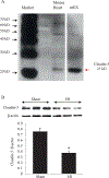
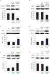
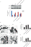
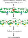
Results: Claudin-5 was detected in murine heart tissue and neonatal rat cardiomyocytes (NRCM). Its protein level was severely decreased after myocardial I/R or H/R. Confocal microscopy showedclaudin-5 presented in the mitochondria of NRCM. H/R-induced claudin-5 downregulation was accompanied by mitochondrial fragmentation. The mitofusin 2 (Mfn2) expressionwas dramatically decreased while the dynamin-related protein (Drp) 1 expression was significantly increased after H/R. The TEM indicatedH/R-induced mitochondrial swelling and fission. Adenoviral claudin-5 overexpression reversed these structural disintegration of mitochondria. The mitochondria-centered intrinsic pathway of apoptosis triggered by H/R and indicated by the cytochrome c and cleaved caspase 3 in the cytoplasm of NRCMs was also reduced by overexpressing claudin-5. Claudin-5 overexpression in mouse heart also significantly decreased cleaved caspase 3 and the infarct size in ischemic heart with improved systolic function.
Conclusion: We demonstrated for the first time the presence of claudin-5 in the mitochondria in cardiomyocytes and provided the firm evidence for the cardioprotective role of claudin-5 in the preservation of mitochondrial dynamics and cell fate against hypoxia- or ischemia-induced stress.
Copyright © 2021 Canadian Cardiovascular Society. All rights reserved.
Conflict of interest statement
Figures


Similar articles
-
Sevoflurane postconditioning attenuates cardiomyocytes hypoxia/reoxygenation injury via PI3K/AKT pathway mediated HIF-1α to regulate the mitochondrial dynamic balance.BMC Cardiovasc Disord. 2024 May 29;24(1):280. doi: 10.1186/s12872-024-03868-1. BMC Cardiovasc Disord. 2024. PMID: 38811893 Free PMC article.
-
Tongmai formula improves cardiac function via regulating mitochondrial quality control in the myocardium with ischemia/reperfusion injury.Biomed Pharmacother. 2020 Dec;132:110897. doi: 10.1016/j.biopha.2020.110897. Epub 2020 Oct 28. Biomed Pharmacother. 2020. PMID: 33113431
-
TBC1D15-Drp1 interaction-mediated mitochondrial homeostasis confers cardioprotection against myocardial ischemia/reperfusion injury.Metabolism. 2022 Sep;134:155239. doi: 10.1016/j.metabol.2022.155239. Epub 2022 Jun 6. Metabolism. 2022. PMID: 35680100
-
Enhancement of Mitochondrial Homeostasis: A Novel Approach to Attenuate Hypoxic Myocardial Injury.Int J Med Sci. 2024 Oct 28;21(15):2897-2911. doi: 10.7150/ijms.103986. eCollection 2024. Int J Med Sci. 2024. PMID: 39628681 Free PMC article. Review.
-
Mitochondrial Stress as a Central Player in the Pathogenesis of Hypoxia-Related Myocardial Dysfunction: New Insights.Int J Med Sci. 2024 Sep 23;21(13):2502-2509. doi: 10.7150/ijms.99359. eCollection 2024. Int J Med Sci. 2024. PMID: 39439461 Free PMC article. Review.
Cited by
-
Suppression of RBFox2 by Multiple MiRNAs in Pressure Overload-Induced Heart Failure.Int J Mol Sci. 2023 Jan 9;24(2):1283. doi: 10.3390/ijms24021283. Int J Mol Sci. 2023. PMID: 36674797 Free PMC article.
-
Epigenetics of Homocystinuria, Hydrogen Sulfide, and Circadian Clock Ablation in Cardiovascular-Renal Disease.Curr Issues Mol Biol. 2024 Dec 5;46(12):13783-13797. doi: 10.3390/cimb46120824. Curr Issues Mol Biol. 2024. PMID: 39727952 Free PMC article. Review.
-
Claudin-10 overexpression suppresses human clear cell renal cell carcinoma growth and metastasis by regulating ATP5O and causing mitochondrial dysfunction.Int J Biol Sci. 2022 Mar 6;18(6):2329-2344. doi: 10.7150/ijbs.70105. eCollection 2022. Int J Biol Sci. 2022. PMID: 35414767 Free PMC article.
-
Interaction Between Rumen Microbiota and Epithelial Mitochondrial Dynamics in Tibetan Sheep: Elucidating the Mechanism of Rumen Epithelial Energy Metabolism.BioTech (Basel). 2025 Jun 5;14(2):43. doi: 10.3390/biotech14020043. BioTech (Basel). 2025. PMID: 40558392 Free PMC article.
-
Mitochondrial apoptosis in response to cardiac ischemia-reperfusion injury.J Transl Med. 2025 Jan 28;23(1):125. doi: 10.1186/s12967-025-06136-8. J Transl Med. 2025. PMID: 39875870 Free PMC article. Review.
References
-
- Taddei A, Giampietro C, Conti A, et al. Endothelial adherens junctions control tight junctions by VE-cadherin-mediated upregulation of claudin-5. Nature cell biology. 2008;10:923–934. - PubMed
-
- Huang P, Zhou CM, Qin H, et al. Cerebralcare Granule(R) attenuates blood-brain barrier disruption after middle cerebral artery occlusion in rats. Experimental neurology. 2012;237:453–463. - PubMed
-
- Kashiwamura Y, Sano Y, Abe M, et al. Hydrocortisone enhances the function of the blood-nerve barrier through the up-regulation of claudin-5. Neurochemical research. 2011;36:849–855. - PubMed
-
- Zhu H, Wang Z, Xing Y, et al. Baicalin reduces the permeability of the blood-brain barrier during hypoxia in vitro by increasing the expression of tight junction proteins in brain microvascular endothelial cells. Journal of ethnopharmacology. 2012;141:714–720. - PubMed
Publication types
MeSH terms
Substances
Grants and funding
LinkOut - more resources
Full Text Sources
Other Literature Sources
Research Materials
Miscellaneous

