Review
. 2021 Mar;86(3):248-261.
doi: 10.1134/S0006297921030020.

Structure and Expression of Large (+)RNA Genomes of Viruses of Higher Eukaryotes

Affiliations
Review

Structure and Expression of Large (+)RNA Genomes of Viruses of Higher Eukaryotes

Alexey A Agranovsky. Biochemistry (Mosc). 2021 Mar.

Abstract

Viral positive-sense RNA genomes evolve rapidly due to the high mutation rates during replication and RNA recombination, which allowing the viruses to acquire and modify genes for their adaptation. The size of RNA genome is limited by several factors, including low fidelity of RNA polymerases and packaging constraints. However, the 12-kb size limit is exceeded in the two groups of eukaryotic (+)RNA viruses - animal nidoviruses and plant closteroviruses. These virus groups have several traits in common. Their genomes contain 5'-proximal genes that are expressed via ribosomal frameshifting and encode one or two papain-like protease domains, membrane-binding domain(s), methyltransferase, RNA helicase, and RNA polymerase. In addition, some nidoviruses (i.e., coronaviruses) contain replication-associated domains, such as proofreading exonuclease, putative primase, nucleotidyltransferase, and endonuclease. In both nidoviruses and closteroviruses, the 3'-terminal part of the genome contains genes for structural and accessory proteins expressed via a nested set of coterminal subgenomic RNAs. Coronaviruses and closteroviruses have evolved to form flexuous helically symmetrical nucleocapsids as a mean to resolve packaging constraints. Since phylogenetic reconstructions of the RNA polymerase domains indicate only a marginal relationship between the nidoviruses and closteroviruses, their similar properties likely have evolved convergently, along with the increase in the genome size.

Keywords: SARS-CoV; closteroviruses; evolution; gene expression; nidoviruses; viral positive-sense RNA genomes.


Conflict of interest statement

The author declares no conflict of interest. This article does not contain description of studies with the involvement of humans or animal subjects.

Figures
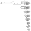
Fig. 1.
Fig. 1.
Genomic RNA and sgRNAs of SARS-CoVs: L, leader sequence; -1 RFs, -1 ribosomal frameshifting signal. Encoded proteins and protein domains: PLP, papain-like cysteine protease; MP, (main) serine protease; POL, RNA polymerase; HEL, RNA helicase; S, spike glycoprotein; GP, accessory proteins and outer membrane glycoprotein; M, matrix protein; N, nucleocapsid protein. Genes unavailable for translation in each type of mRNA are shown as shaded boxes. Arrows indicate 3′-ends of RNAs. Drawn approximately to scale.
Fig. 2.
Fig. 2.
Transcription and replication of coronaviral RNA. a) Discontinuous transcription. (–)RNA synthesis on the genomic (+)RNA may stop on the transcription regulatory signal (B-TRS, light box), with the following transition of the nascent (–)strand to the 5′-leader, annealing of B-TRS to L-TRS, and copying of the leader sequence. Anti-sgRNAs serve as templates to produce sgRNAs. b) Continuous replication. Free viral nucleocapsid protein N0 and cell helicase DDX1 attach to B-TRSs, allowing replicase to ignore the stop signals and to synthesize the full-length (–)RNA, which serves as a template for producing progeny (+)RNAs. The RNA 3′-ends are shown by arrowheads. Coding and noncoding sequences are drawn not to scale.
Fig. 3.
Fig. 3.
Structure of the SARS-CoV-1 replicative pp1ab. Vertical dotted line indicates the boundary between 1a and 1b in the pp1ab protein. The domains for the nonstructural proteins nsp1-16 are shown as filled boxes. Designations: PLP, papain-like protease (in cis cleavage sites are indicated by curved arrows); MP, main chymotrypsin-like protease (in trans cleavage sites are shown by arrowheads); TM, transmembrane domain; Pr, putative primase; POL, RNA polymerase; Z, zinc-binding domain; HEL, RNA helicase; Exo, 3′-5′ exonuclease; Mtr, N7-guanine methyltransferase; NU, nidoviral uridylate-specific endoribonuclease; MT, 2′-O-ribose methyltransferase.
Fig. 4.
Fig. 4.
Genomic RNA and sgRNAs of beet yellows closterovirus (BYV). Genes unavailable for translation in each type of mRNA are shown as shaded boxes; +1 RFs, +1 ribosomal frameshifting signal for the translation of ORFs 1a and 1b; arrows and dotted lines indicate cleavage sites in pp1a; Mem, membrane-binding domain; p6, small hydrophobic protein; HSP70h, homolog of HSP70 family heat-shock proteins; p64, 64-kDa protein; CPm, minor capsid protein; CP, major capsid protein.

References

    1. Koonin E. V., Dolja V. V. Evolution and taxonomy of positive-strand RNA viruses: implications of comparative analysis of amino acid sequences. Crit. Rev. Biochem. Mol. Biol. 1993;28:375–430. doi: 10.3109/10409239309078440. - DOI - PubMed
    1. Steinhauer D. A., Holland J. Rapid evolution of RNA viruses. Annu. Rev. Microbiol. 1987;41:409–431. doi: 10.1146/annurev.mi.41.100187.002205. - DOI - PubMed
    1. Drake J. W. Rates of spontaneous mutation among RNA viruses. Proc. Natl. Acad. Sci. USA. 1993;90:4171–4175. doi: 10.1073/pnas.90.9.4171. - DOI - PMC - PubMed
    1. Holmes E. C. Error thresholds and the constraints to RNA virus evolution. Trends Microbiol. 2003;11:543–546. doi: 10.1016/j.tim.2003.10.006. - DOI - PMC - PubMed
    1. Dolja V. V., Karasev A. V., Koonin E. V. Molecular biology and evolution of closteroviruses: sophisticated build-up of large RNA genomes. Annu. Rev. Phytopathol. 1994;32:261–285. doi: 10.1146/annurev.py.32.090194.001401. - DOI